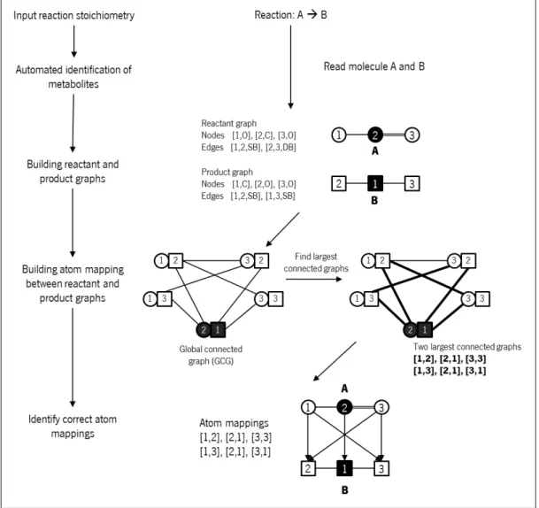
A critical evaluation of automatic atom mapping algorithms and tools
94
0
0
Texto
Imagem
+7
Documentos relacionados