Abstract—These Passive anterior-posterior (A-P) stability of the knee, measured in terms of joint laxity, is considered important for good clinical outcome following knee arthroplasty. In vitro and in vivo studies measured the laxity at
selected joint positions in the intact and replaced knees. However, analyzing the effects of surgical techniques on the joint stability is difficult to implement experimentally.
In the present study a mathematical model of the knee with unconstrained unicompartmental arthroplasty was used to study relative translations of the bones during a simulated A-P laxity test over 0-120o flexion. The knee ligaments were modeled as bundles of non-linear elastic fibers. Anatomical data, material properties of ligaments, geometries of the prosthetic components and guidelines for component placement on the bones were taken from literature. The model calculations for tibial translations resulting from ±130 N A-P forces were compared with the experimental measurements of Lo et al. (2010), reported as mean of 14 cadaver knees. Further, the effects of component placement on the bones were also studied.
The model calculations agreed in general with the experimental measurements showing similar patterns during flexion. The joint laxity first increased from 0o to about 45o flexion and decreased thereafter. An increase in the A-P force resulted in uniform increase in laxity over flexion. A change of 1 mm in the placement of femoral component affected the laxity by nearly 3 mm near extension. However, this effect of change varied significantly with flexion. Such effects can alter the joint kinematics and may be clinically significant. The analysis has clinical relevance .
Index Terms—Component Placement, Knee Mechanics, Knee Stability, Unicompartmental Knee Arthroplasty.
I. INTRODUCTION
ASSIVE A-P stability of the knee is estimated from laxity tests where known magnitudes of A-P forces are applied on the tibia at fixed flexion angles and corresponding tibial translations are recorded. Such translations of the tibia in the absence active muscle forces are considered important clinical measure to assess knee function after joint replacement [1], [2]. In the sagittal plane, primary restrain to anterior tibial translation is the anterior cruciate ligament, while primary restrain to the posterior tibial translation is the posterior cruciate ligament.
Manuscript received March 23, 2011; revised April 11, 2011.
A. Imran is with Ajman University of Science and Technology, Ajman, U.A.E. (phone: 971-50-2850131; fax: 971-6-7438888; e-mail: [email protected] or [email protected]).
Unicompartmental arthroplasty with unconstrained prosthetic surfaces depends on the knee ligaments for A-P stability [3]. Therefore, appropriate ligament balancing during surgery can play significant role in determining kinematics of the replaced knee [1], [3]. Incorrect placement of the prosthetic components can affect the ligament lengths and result in altered kinematics of the joint.
In vitro and in vivo experimental studies have analyzed the passive knee stability in the intact and replaced knees at selected flexion angles and external loads [4], [5]. However, analyzing the effects of surgical techniques such as placement of the prosthetic components is difficult to implement experimentally. An understanding of these effects, particular for minimally invasive surgery, is important as they can influence patient outcome [6]–[9]. Theoretical models validated with the experimental observations are useful tools to study such effects [9], [10].
In the present study a sagittal plane mathematical model of the knee is used to analyze the passive stability of the joint in terms of anterior and posterior translations of the tibia after unicompartmental replacement with intact ligaments. The model is also used to analyze the effects on the A-P translations resulting from incorrect placement of the prosthetic components.
II. METHODS
The knee with intact cruciate and collateral ligaments and unconstrained prosthetic components was modeled in the sagittal plane. The ligaments were modeled as bundles of non-linear elastic fibers [11]. A flat tibial and a circular femoral component were used. Anatomical data, attachments of the ligaments and their material properties as well as geometries of the prosthetic components and surgical guidelines for their attachment on the bones were taken from literature [3], [11], [12]. The components were attached to the bones such that no fiber of any ligament was significantly stretched at 0 or 90o flexions. This position of the prosthetic components was defined as the correct position (or correct placement). The knee motion was defined during 0–120o flexion at 5o interval.
At each flexion position, an A-P laxity test was simulated by applying a known anterior (+) or posterior (-) force on the tibia while the femur was held fixed and the resulting tibial translations were noted. The model calculations were compared with in vitro experimental measurements from Lo
et. al. [4]. Further, the femoral component was positioned 1 mm proximal and 1 mm distal to its correct position. The A-P simulation was repeated and total A-A-P translations of the tibia were noted.
Passive Anterior-Posterior Knee Stability After
Unconstrained Unicompartmental Arthroplasty
Ahmed Imran, Member, IAENG
P
Proceedings of the World Congress on Engineering 2011 Vol III WCE 2011, July 6 - 8, 2011, London, U.K.
ISBN: 978-988-19251-5-2
ISSN: 2078-0958 (Print); ISSN: 2078-0966 (Online)
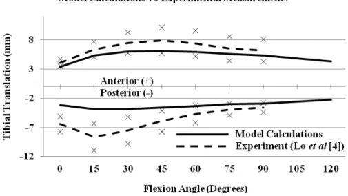
Fig. 1. A-P tibial translation under ±130 N A-P force over the flexion range. Model calculations (continuous lines) are compared with the experimental measurements of Lo et al. [4] (dashed lines correspond to the mean values (at 15o interval) and ‘×’ show standard deviation).
Fig. 2. Model calculations for anterior tibial translation corresponding to 50, 100, 150 and 200 N anterior force.
Fig. 3. Model calculations for total A-P translation under ±150 N force, comparing correct placement with 1mm proximal or 1 mm distal placement of the femoral component.
Proceedings of the World Congress on Engineering 2011 Vol III WCE 2011, July 6 - 8, 2011, London, U.K.
ISBN: 978-988-19251-5-2
ISSN: 2078-0958 (Print); ISSN: 2078-0966 (Online)
III. RESULTS
Figure 1 shows a comparison between A-P tibial translations under ±130 N force calculated from the model during 0–120o flexion and those reported by Lo et. al. [4] given as mean values from 14 intact cadaver knees during 0–90o flexion (at 15o interval).
Figure 2 gives anterior tibial translations as calculated from the model corresponding to 50, 100, 150 and 200 N anterior force on the tibia.
Figure 3 shows a comparison of the total A-P translations for the correct placement and for incorrect placement of the femoral component by 1 mm proximal and 1 mm distal to its correct position.
IV. ANALYSIS
From figure 1, the patterns of A-P tibial translations calculated from the model show reasonable agreement with those from the experiment. The model calculations for anterior translations are similar to the mean experimental values and are within the reported standard deviation. The model calculations for posterior translations for angles greater than 45o are similar to the mean experimental values and are within the reported standard deviation. For 0–45o flexion positions, the model calculations for the posterior translation underestimate the experiment.
The anterior translation at any load (figure 2), first increases with flexion angle from 0 to about 45o and then decrease in higher flexion. Similarly, the posterior translation first increases from 0 to about 30o and then decreases in higher flexion. These patterns of variation with flexion are in agreement with the experimental measurements (figure 1).
Figure 2 shows that the tibial translations increased uniformly with the magnitude of the external applied load.
Figure 3 gives the effects over the flexion range of a femoral component placed 1 mm proximal or 1 mm distal to the correct position. Total A-P translations under ±150 N forces are plotted over the flexion range. For each incorrect placement, the variations in translation were flexion dependent, except at 90o. Also, the patterns of change in the tibial translation reversed beyond 90o flexion. For example, the tibial translation due to a proximal placement resulted in 2.9 mm increase at 0o and 1 mm decrease at 120o flexion. On the contrary, the tibial translation due to a distal placement resulted in 2.9 mm decrease at 0o and 1.1 mm increase at 120o flexion. There was no change at 90o. Such variations altered the joint kinematics and, therefore, may be clinically significant for the patient outcome.
V. CONCLUSION
Reasonable agreement between the model calculations and experimental measurements shows that unconstrained prosthetic components with intact ligaments can reproduce the patterns of A-P translations similar to those observed in the intact knee. However, the model simulations also show that the surgical placement of prosthetic components can
affect the joint kinematics significantly and variably over the flexion range. This may influence the patient outcome. The analysis has clinical relevance.
REFERENCES
[1] Y. Ishii, H. Noguchi, Y. Matsuda, M. Takeda, S. Walker and R. Komistek, “ Effect of knee laxity on in vivo kinematics of meniscal-bearing knee prostheses,” The Knee, vol. 14, pp. 269–274, 2007. [2] D. Jones, C. Locke, J. Pennington and J. Theis, “The effect of sagittal
laxity on function after posterior cruciate-retaining total knee arthroplasty,” J Arthroplasty, vol. 21, pp. 719–723, 2006.
[3] J. Goodfellow and J. O’Connor, “The mechanics of the knee and prosthesis design,” J Bone Jt Surg (Br), vol. 60-B, pp. 358–369, 1978.
[4] J. H. Lo, O. Müller, T. Dilger, N. Wülker, M. Wünschel, “Translational and rotational knee joint stability in anterior and posterior cruciate-retaining knee arthroplasty,” Knee, doi:10.1016, 2010.
[5] Y. Ishii, H. Noguchi, M. Takeda, H. Kiga and S. Toyabe, “Effect of voluntary soft tissue tension and articular conformity after total knee arthroplasty on in vivo anteroposterior displacement,” The Knee, vol. 18, pp. 11–14, 2011.
[6] D. Fisher, M. Watts and K. Davis, “Implant position in knee surgery: a comparison of minimally invasive, open unicompartmental, and total knee arthroplasty,” J Arthroplasty, vol. 18-7(S-1), pp.2–8, 2003. [7] A. Gulati, R. Chau, D. Simpson, C. Dodd, H. Gill and D. Murray,
“Influence of component alignment on outcome for unicompartmental knee replacement,” The Knee, vol. 16, pp. 196–9, 2009.
[8] P. Muller, C. Pellengahr, M. Witt, J. Kircher, H. Refior and V. Jansson, “Influence of minimally invasive surgery on implant positioning and the functional outcome for medial unicompartmental knee arthroplasty,” J Arthroplasty, vol. 19(3), pp. 296–301, 2004. [9] A. Imran, “Unicompartmental knee arthroplasty (UKA): effects of
component placement on joint mechanics studied with a mathematical model,” in Proc. IFMBE (31), 2010, pp. 616–619.
[10] A. Imran, “Knee laxity after unicompartmental joint replacement: A planar mathematical analysis,” presented at the First Middle East Conference on Biomedical Engineering (MECBME’11), 22-25 Feb. 2011, Sharjah, U.A.E.
[11] A. Zavatsky and J. O’Connor, “A model of human knee ligaments in the sagittal plane: Part 1. Response to passive flexion,” J Engng in Med., vol. 206 (H), pp. 125–134, 1992.
[12] T.W. Lu and J. O’Connor, “Fiber recruitment and shape changes of knee ligaments during motion,” J Engineering in Med., vol. 210(H), pp. 71–79, 1996.
Proceedings of the World Congress on Engineering 2011 Vol III WCE 2011, July 6 - 8, 2011, London, U.K.
ISBN: 978-988-19251-5-2
ISSN: 2078-0958 (Print); ISSN: 2078-0966 (Online)