Study of loss-on-ignition anomalies found in ashes from combustion of
iron-rich coal
R.E. Vandenberghe
a,*, V.G. de Resende
a, G.M. da Costa
b, E. De Grave
a aDepartment of Physics and Astronomy, University of Ghent, B-9000 Gent, BelgiumbChemistry Department, Federal University of Ouro Preto, 35400-000 Ouro Preto (MG), Brazil
a r t i c l e
i n f o
Article history:
Received 3 September 2009
Received in revised form 19 January 2010 Accepted 20 January 2010
Available online 30 January 2010
Keywords: Fly ash Bottom ash Loss on ignition Mössbauer spectroscopy
a b s t r a c t
Samples of a fly and a bottom ash, each before and after ignition at 960°C, have been studied by X-ray diffraction, Mössbauer spectroscopy, carbon analysis, and thermogravimetric analysis with the aim to explain an observed negative loss on ignition. Ashes after ignition contain more maghemite resulting from oxidation of newly formed magnetite. Moreover, the fly ash that already contained magnetite exhib-ited an increase of hematite after ignition. Hercynite present in both ashes transforms to hematite and magnetite after the ignition. All these oxidation processes are responsible for a weight gain which may compensate the loss due to the burning of the remaining carbon in the ashes. Also,a-Fe is formed after ignition which may have originated from wustite.
Ó2010 Elsevier Ltd. All rights reserved.
1. Introduction
From environmental, economical, and fundamental points of view fly ashes and bottom ashes from coal combustion have been the subject of various studies with respect to their mineralogical composition, their morphology, and their carbon content [1–4]. Particularly, the latter is important, because the remaining carbon content is not only a measure for the efficiency of the combustion process, but is also a necessary parameter for further application of these ashes. Indeed, those ashes are mainly used as additives for cement and concrete and have to meet certain standards. The remaining carbon is usually measured by the loss on ignition (LOI), i.e., the relative loss of mass after heating the ash during a short period at high temperatures (750–960°C). The LOI for fly ashes used for cement additions must be usually less than 5%, whereas fly ashes of poorer quality (>5% LOI) are then often used in the brick industry.
Although mostly used as a routine measurement, LOI is not al-ways a direct appropriate measure for the remaining carbon con-tent in the ashes. Natural occurring or added limestone (mostly calcite) or dolomite, and the presence of volatile organic com-pounds, all have a serious impact on the LOI, leading to overesti-mated unburned carbon contents[5,6]. On the other hand, when the ashes contain a substantial amount of iron minerals, it might
be possible that the weight after ignition changes to a large extent in an opposite way by modification of the Fe-containing compo-nents. For instance, oxidation of magnetite to hematite can take place, which would result in a gain of weight.
At the Electrabel-Rodenhuize electric generating plant (Destel-donk, Belgium) the quality of the fly and bottom ashes are moni-tored daily by measuring the LOI after burning the ashes for 30 min at 960°C. After coal from a new origin was used LOI values for both the fly ash and bottom ash strongly deviated from the usual expected values and even became negative. It was readily suggested that the increased iron content in the new coal played a crucial role in these unexpected LOI results. In order to unravel this problem, a study applying a combination of various techniques was performed to gain information about the character and the behaviour of the Fe-bearing materials in both ashes before and after ignition.
2. Experimental
Samples of fly ash (F1 and F2) and bottom ash (B1 and B2), each before and after ignition, respectively, were investigated. X-ray fractograms (XRD) were recorded on a XRD-6000 Shimadzu dif-fractometer equipped with a Fe tube and a graphite monochromator. Mössbauer spectra were collected at 295, 140, and 80 K with spectrometers operating at constant acceleration mode with triangular reference signals and using57Co(Rh) sources.
The spectrometers were calibrated by collecting the spectrum of standard hematite or of a metallic iron foil at room temperature.
0016-2361/$ - see front matterÓ2010 Elsevier Ltd. All rights reserved. doi:10.1016/j.fuel.2010.01.022
* Corresponding author. Address: University of Ghent, Department of Physics and Astronomy, Division NUMAT, Proeftuinstraat 86, B-9000 Gent, Belgium. Fax: +32 9 264 6697.
E-mail address:[email protected](R.E. Vandenberghe).
Contents lists available atScienceDirect
Fuel
Spectra were analysed using Lorentzian line profiles and consider-ing distributions of hyperfine fields where necessary. Isomer shifts are referenced with respect to
a
-Fe at room temperature. The car-bon content in the ashes was measured by the flash combustion method with an accuracy of ±2%. Thermogravimetric measure-ments between 25 and 1000°C and under synthetic air (100 ml/min) were carried out on a Du Pont SDT 2960 apparatus applying a heating rate of 5°C/min.
3. Results and discussion
3.1. Fly and bottom ashes before ignition
In the XRD patterns of the fly and bottom ashes before ignition (samples F1 and B1, respectively) characteristic diffraction peaks of quartz (SiO2), mullite (Al6Si2O13), hematite (
a
-Fe2O3), hercynite(FeAl2O4) and magnetite/maghemite (Fe3O4/
c
-Fe2O3) areunambig-uously identified (Fig. 1). In the bottom ash, some additional peaks are observed which can be attributed to plagioclase. The mineral-ogy of sample F1 is typical for the so-called class F fly ashes (ASTM C618) that are obtained from burning high-rank coals[7].
Concerning the iron-bearing compounds, the Mössbauer spec-trum at 295 K of the ash F1 shows the two sextets of (oxidized) magnetite, the sextet of weakly ferromagnetic (WF) hematite, and a sextet with zero quadrupole shift 2
e
Q, pointing to a spinelphase which is most likely maghemite, although the presence of other spinel ferrites cannot be excluded. Therefore, this component will be denoted as a maghemite/ferrite phase. The asymmetrical line shape in the latter is probably due to small-particle morphol-ogy as usually observed in such systems and in some cases also from mixed ferrite compounds[8,9]. It is worth to remark at this point that the relative areas (RA) for the magnetite and maghe-mite/ferrite sextets are not at all accurate due to the strong overlap and possible asymmetric lines for magnetite. Moreover, the broad-lined inner sextet has been fitted with a hyperfine field distribu-tion, which might show some fluctuations in the intensity proba-bilities. Anyway, this inner sextet is not arising from goethite as claimed by Hinckley et al.[1]in their ashes because well crystal-lized goethite, being magnetic at room temperature, can only be formed under extreme hydrothermal conditions and is not at all stable at such high temperatures.
The paramagnetic part of the spectrum consists of Fe3+and Fe2+ doublets. Although many doublets of each kind can be expected, the spectrum could only be significantly fitted with one Fe2+and
one Fe3+ doublet. The latter doublet, with d0.32 mm/s and DEQ0.99 mm/s, is mainly representing an average spectrum for
the mullite Fe3+doublets. At room temperature, this doublet might
also partly represent some superparamagnetic maghemite/ferrite because at lower temperatures the intensity of the Fe3+ doublet
has somewhat decreased. The doublet with d1.06 mm/s and DEQ2.28 mm/s is an average for the various Fe2+doublets
repre-senting the presence of ferrous ions in the structure of hercynite. The Mössbauer spectrum of hercynite at room temperature shows almost complete overlap of the different Fe2+doublets[10].
There-fore, no quantitative estimation of the Fe2+cation distribution is
possible from the 295 K spectrum.
The 295 K Mössbauer spectrum of the bottom ash (sample B1) is similar to the spectrum obtained for the fly ash (sample F1) with the exception that no clear sextets of magnetite were observed. Also, the Fe2+ doublet contribution is much more pronounced in
comparison with the fly ash spectrum.
In these 295 K Mössbauer spectra it is difficult to resolve quan-titatively the various sextets due to the expected asymmetrical line shapes arising from small-particle morphology. Spectra were col-lected at 140 K to determine more accurately the amounts of iron in the various compounds. The latter temperature reduces the asymmetry in the line shape for hematite and maghemite/spinel on the one hand and is situated above the Verwey transition (130 K for pure magnetite) for the magnetite on the other hand,
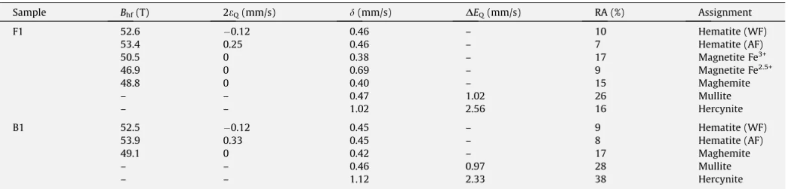
because the Mössbauer spectrum of magnetite below its Verwey-transition temperature is very complicated[11]. The 140 K spectra of the fly and the bottom ash are shown inFig. 2and the hyperfine parameters obtained from the fitted spectra are listed inTable 1.
10 20 30 40 50 60 70
0 20 40 60 80 100 0 20 40 60 80 100
Pl Pl
B1
2θ (°)
Mg Mu
Mg Mg
Mg
Mu Mu H Q Mu He Mu
He M u Mu
Mu
Q Q
Q
H H
H
H
Intensity (counts)
F1
Fig. 1.X-ray diffraction patterns of the fly and bottom ashes before ignition (samples F1 and B1, respectively). Mu: mullite; He: hercynite; Q: quartz; H: hematite; Mg: magnetite/maghemite and Pl: plagioclase.
-10 -5 0 5 10
97.0 97.5 98.0 98.5 99.0 99.5 100.0 98.5 99.0 99.5 100.0
T
ransm
is
si
o
n
(
%
)
B1
Velocity (mm/s)
F1
For the Mössbauer spectrum of sample F1 the same components as in the 295 K spectrum were considered, but, a sextet due to antifer-romagnetic (AF) hematite phase has additionally been taken into account. The coexistence of both magnetic hematite phases (WF and AF) might be explained by either small-particle morphology [12,13]or by the presence of two hematite fractions with an ex-treme difference in particle sizes or with a distinct difference in aluminium substitution.
Although no significant amount of magnetite is present in the bottom ash (B1), the Mössbauer spectrum was also measured at 140 K (Fig. 2) to compare with those of the fly ash (F1). The Mössbauer spectrum was fitted using two sextets due to WF and AF hematite, a sextet accounting for maghemite/ferrite/(magne-tite), an Fe3+doublet and an Fe2+ doublet (Table 1). Concerning
the relative area parameter (RA), the values were found, within experimental error, similar to the ones obtained from the spectrum collected at 295 K, indicating that sample B1 does not show super-paramagnetic effects due to small-particle morphology of the iron-bearing oxides.
In order to provide more accurate hyperfine parameters and to obtain a better insight in the iron distribution of the different phases, both samples F1 and B1 were subjected to a magnetic sep-aration resulting in a magnetic and a non-magnetic fraction. An intermediate magnetic fraction was also obtained, however, the obtained Mössbauer results will not be discussed here because the spectra are very similar to the one of the total fraction.
The Mössbauer spectrum at 295 K of the non-magnetic fraction of sample F1 showed only the contribution of the Fe3+and Fe2+
doublets, no magnetic sextets were observed in the spectrum. However, by lowering the temperature to 140 K (Fig. 3a) the spec-trum shows, in addition to the Fe3+and Fe2+doublets, three weak
sextets, one due to WF hematite and two from magnetite. This re-sult corroborates the Mössbauer rere-sults obtained from the total fraction of sample F1 in which small-particle morphology resulted similarly in a slight decrease of the Fe3+doublet at lower
temper-ature. The spectrum at 295 K of the magnetic fraction of sample F1 shows four components: a WF hematite sextet, the two magnetite sextets, and a weak Fe3+doublet (Fig. 3b). The broad-lined sextet
due to maghemite/ferrite is not clearly observed in this fraction, but might be somewhat hidden by the inner magnetite sextet.
The spectrum collected at 80 K of the non-magnetic fraction of sample B1 (Fig. 3c) does not show any sextet contribution, con-firming the absence of superparamagnetic effects in the bottom ash sample. The Mössbauer spectrum was fitted with four Fe2+
doublets characteristic for hercynite, a weak Fe3+doublet that is
considered as an average for the three mullite doublets, and an-other Fe3+doublet with quadrupole splitting of 0.98 mm/s and
iso-mer shift of 0.69 mm/s (Table 2). The identification of the precise
nature of the latter iron phase on the basis of the adjusted hyper-fine parameters is not possible because of the strong overlap be-tween the various doublet lines in the central part of the spectrum. However, it is not unreasonable to suggest that the dou-blet may be due to wustite (Fe1xO) that may indeed be present in
a small amount in the bottom ash. The Mössbauer spectrum of the
Table 1
Hyperfine parameters derived from the Mössbauer spectra at 140 K of the fly (F1) and bottom (B1) ash before ignition.
Sample Bhf(T) 2eQ(mm/s) d(mm/s) DEQ(mm/s) RA (%) Assignment
F1 52.6 0.12 0.46 – 10 Hematite (WF)
53.4 0.25 0.46 – 7 Hematite (AF)
50.5 0 0.38 – 17 Magnetite Fe3+
46.9 0 0.69 – 9 Magnetite Fe2.5+
48.8 0 0.40 – 15 Maghemite
– – 0.47 1.02 26 Mullite
– – 1.02 2.56 16 Hercynite
B1 52.5 0.12 0.45 – 9 Hematite (WF)
53.9 0.33 0.45 – 8 Hematite (AF)
49.1 0 0.42 – 17 Maghemite
– – 0.46 0.97 28 Mullite
– – 1.12 2.33 38 Hercynite
Bhf: hyperfine field at maximum of the distribution; 2eQ: quadrupole shifts;DEQ: quadrupole splitting;d: isomer shifts; RA: relative spectral areas. The values of isomer shifts are with reference to metallic iron at room temperature.
-2 0 2 4
95 96 97 98 99 100 96 97 98 99 100
-10 -5 0 5 10
-10 -5 0 5 10
99.0 99.2 99.4 99.6 99.8 100.0 100.2
(c)
Transmission (%)
Velocity (mm/s)
(b)
Transmission (%)
Velocity (mm/s)
(a)
Transmission (%)
Velocity (mm/s)
magnetic fraction of sample B1 is very similar to the total fraction spectrum.
3.2. Fly and bottom ash after ignition
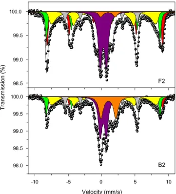
The Mössbauer spectrum at 140 K of the fly ash after ignition (sample F2) shows also the two hematite sextets (WF and AF phases), the distributed maghemite/ferrite sextet and the mullite doublet (Fig. 4). However, magnetite is absent and the Fe2+
/hercy-nite doublet has nearly completely disappeared. On the other hand, the characteristic sextet of
a
-Fe is clearly observed.Considering the relative area values of the subspectra of the var-ious components observed in the fly ash before (Table 1, sample F1) and after ignition (Table 3, sample F2), one can readily observe that besides the formation of metallic iron, the contribution of hematite as well as maghemite/(ferrite) increased considerably after ignition. The contribution of the Fe3+doublet did not change,
confirming the previous assignment of this doublet to mullite, which is indeed stable up to very high temperatures. On the con-trary, magnetite has completely vanished whereas the doublet attributed to Fe2+/hercynite is also strongly reduced. The formation of
a
-Fe after ignition in air could be explained by the following reaction:4FeO!Fe3O4þFe
which could, for instance, be the final result of the decomposition of siderite (FeCO3)[14]. However, siderite is only present in the coal
and transforms during combustion to hematite or magnetite. The latter is usually formed in the higher temperature regions of the fur-nace, which is the reason for being solely present in the fly ash. Moreover, the hyperfine parameters of the siderite doublet are quite different of those observed for the Fe2+ doublets in the
Mössbauer spectra. As already suggested, one of the doublets appearing in the spectra of the non-magnetic fraction of the bottom ash sample before ignition could be assigned to wustite, the pres-ence of this phase resulting in the formation of
a
-Fe after ignition. Therefore, it is not excluded that wustite may also be present in the fly ash before ignition, although its spectral contribution could not be directly resolved in the Mössbauer spectrum of the non-mag-netic fraction of the sample due to the overlap with the inner lines of the sextets (Fig. 3a).From the changes in the relative areas of the subspectra, it can be suggested that magnetite and hematite are formed from oxida-tion of hercynite. In order to verify this oxidaoxida-tion process, a
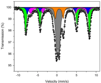
syn-thetic hercynite sample was subjected to a heat treatment at 960°C for 30 min in air, which are the same conditions used in the routine LOI measurements. The Mössbauer spectrum at 295 K of the obtained product is shown inFig. 5. The spectrum is com-posed of a WF hematite sextet, two magnetite sextets, an Fe3+
dou-blet with hyperfine parameters typical for iron in the structure of corundum (
a
-Al2O3), and a weak Fe2+/hercynite doublet. The latterone is most probably the result of the non-complete transforma-tion of the hercynite during the short heating. From this experi-ment, it is clear that magnetite and hematite are the main iron-bearing oxidation products from hercynite, which can be roughly represented by the following reaction
Table 2
Hyperfine parameters derived from the Mössbauer spectra at 140 K of the non-magnetic fraction and at 295 K of the magnetic fraction of the fly (F1) ash sample, and at 80 K of the non-magnetic fraction of the bottom (B1) ash sample.
Temperature (K) Sample Bhf(T) 2eQ(mm/s) d(mm/s) DEQ(mm/s) Assignment
140 F1 non-magnetic fraction 52.6* 0.12* 0.46* – Hematite (WF)
49.4 0 0.38* – Magnetite Fe3+
43.4 0 0.69* – Magnetite Fe2.5+
– – 0.48 0.97 Mullite
– – 1.17 2.11 Hercynite
295 F1 magnetic fraction 51.1 0.15 0.37 – Hematite (WF)
48.6 0 0.28 – Magnetite Fe3+
45.4 0 0.59 – Magnetite Fe2.5+
– – 0.40 0.91 Mullite
80 B1 non-magnetic fraction – – 0.45 0.72 Mullite
– – 1.13 3.07 Hercynite
– – 1.15 2.57 Hercynite
– – 1.10 1.52 Hercynite
– – 1.15 2.08 Hercynite
– – 0.69 0.98 Iron phase
Bhf: hyperfine field at maximum of the distribution; 2eQ: quadrupole shifts;DEQ: quadrupole splitting;d: isomer shifts. The values of isomer shifts are with reference to metallic iron at room temperature.
*Fixed parameters.
-10 -5 0 5 10
98.0 98.5 99.0 99.5 100.0 98.5 99.0 99.5 100.0
B2
Velocity (mm/s)
Transmission (%)
F2
5FeAl2O4þO2!
a
-Fe2O3þ5Al2O3þFe3O4The presence of maghemite instead of magnetite in the burned ashes might be explained by the newly formed magnetite from both hercynite and wustite, which afterwards oxidizes to maghe-mite according to
2Fe3O4þ 1
2O2!3
c
-Fe2O3On the other hand, the magnetite that was already present in the fly ash oxidizes completely to hematite after ignition.
The spectrum at 140 K of the bottom ash after ignition (B2) shows two sextets of hematite, a maghemite/ferrite sextet, a Fe3+/mullite doublet, a Fe2+/hercynite doublet, and in addition, a sextet of
a
-Fe. Comparing the relative areas of the bottom ash be-fore and after ignition (Tables 1 and 3, respectively), it can be ob-served that the hematite and maghemite contribution increases at the expense of the hercynite phase after ignition. It is not ex-cluded that the remaining doublet attributed to hercynite may also represent other Fe2+-containing components such as glasses.Final-ly, as suggested
a
-Fe is formed from the wustite after the ignition. The observed negative LOI can now be explained by the differ-ent oxidation processes involving the formation of magnetite, maghemite, and hematite, which is responsible for a weight gain and compensates the loss due to the burning of the remaining car-bon in both samples fly (F1) and bottom (B1) ash before ignition. The unburned carbon has been directly measured by carbon anal-ysis. The carbon content was found to be 2.31 wt.% for sample F1 and 1.09 wt.% for sample B1.The various processes resulting in weight changes can normally be observed from thermogravimetric analysis (TGA). The TGA curve and the corresponding derivative curve of sample F1 are rep-resented inFig. 6. A minor weight loss (0.2%) that could account for the release of adsorbed water is observed between 25 and 280°C. At higher temperatures, in the range 500–700°C with a maximum at 618°C, a loss in weight is clearly noticed. This weight loss can be
mainly ascribed to release of the remaining unburned carbon in the sample as CO2. At temperatures beyond 700°C, a gain in weight
takes place and is still not completed at temperatures up to 1000°C. The weight gain is clearly associated with the
aforemen-tioned oxidation processes resulting in the formation of hematite, magnetite, and maghemite. Precise results on the amount of un-burned carbon in the fly ash sample cannot be derived from the TGA analysis, but it is nevertheless clear that the weight gain can compensate the loss due to unburned carbon.
Table 3
Hyperfine parameters derived from the Mössbauer spectra at 140 K of the fly (F2) and bottom (B2) ash after ignition.
Sample Bhf(T) 2eQ(mm/s) d(mm/s) DEQ(mm/s) RA (%) Assignment
F2 52.7 0.15 0.45 – 17 Hematite (WF)
53.6 0.31 0.45 – 21 Hematite (AF)
48.5 0 0.43 – 24 Maghemite
33.5 0 0.07 – 8 a-Fe
– – 0.38 0.94 27 Mullite
– – 1.08 2.16 3 Hercynite
B2 52.7 0.16 0.45 – 13 Hematite (WF)
53.5 0.36 0.45 – 10 Hematite (AF)
48.8 0 0.47 – 23 Maghemite
33.5 0 0.07 – 9 a-Fe
– – 0.37 0.91 27 Mullite
– – 1.14 2.23 18 Hercynite
Bhf: hyperfine field at maximum of the distribution; 2eQ: quadrupole shifts;DEQ: quadrupole splitting;d: isomer shifts; RA: relative spectral areas. The values of isomer shifts are with reference to metallic iron at room temperature.
-10 -5 0 5 10
95 96 97 98 99 100
Transmission (%)
Velocity (mm/s)
Fig. 5. Mössbauer spectra at 295 K of a pure hercynite sample heated in air at 960°C during 30 min. WF hematite (green); magnetite Fe3+(blue); magnetite Fe2.5+ (magenta); Fe3+/Al
2O3(gray); and Fe2+/hercynite (orange). (For interpretation of the references to colour in this figure legend, the reader is referred to the web version of this article.)
97 98 99 100
99.7 %
98.1 %
99.8 %
Temperature (°C)
Der
iv
. w
eight (%/°C)
200 400 600 800 1000
0.02 0.01 0.00 -0.01 -0.02 -0.03 -0.04 -0.05 -0.06 -0.07 -0.08
618 °C
W
eight loss (%)
0
4. Conclusions
From the present study on a fly and a bottom ash, before and after ignition, it follows that a hercynite phase is present which transforms to hematite and magnetite. In the short time of the ignition experiment at 960°C, the magnetite is not converted to
hematite but oxidizes rather (probably afterwards) to maghemite. The magnetite which was already present in the fly ash is con-verted completely to hematite after ignition. All those oxidation processes lead to a weight gain that can offset the weight loss due to the burning of the remaining carbon. Therefore, negative LOI values cannot be excluded in this type of iron-rich ashes when the ignition is performed at relatively high temperatures (960°C).
In order to avoid such anomalies, LOI should only be measured after ignition at moderate temperatures (750°C) at which, at
least, hercynite remains stable[15].
Acknowledgement
This work was supported by FWO-Flanders. Prof. Peter Van den haute (Geology Dept. UGent) is acknowledged for the use of his magnetic separator. The authors are grateful to Jozef Janssens
(Chemistry Department, University of Antwerp) for carrying out the TGA measurements.
References
[1] Hinckley CC, Smith GV, Twardowska H, Saporoschenko M, Shiley RH, Griffen RA. Fuel 1980;59:161–5.
[2] Huffman GP, Huggins FE, Dunmyre GR. Fuel 1981;60:585–97. [3] Hower JC, Robl TL, Thomas GA. Fuel 1999;78:701–12. [4] Vassilev SV, Vassileva CG. Energy Fuels 2005;19:1084–98. [5] Brown RC, Dykstra J. Fuel 1995;74:570–4.
[6] Fan M, Brown RC. Energy Fuels 2001;15:1414–7. [7] American Society for Testing and Materials (ASTM) C618.
[8] Gomes S, François M, Abdelmoula M, Refait Ph, Pellissier C, Evrard O. Cem Concr Res 1999;29:1705–11.
[9] Bayukov OA, Anshits NN, Balaev AD, Sharonova OM, Rabchevskii EV, Petrov MI, et al. Inorg Mater 2005;41:50–9.
[10] Larsson L, O’Neill HSTC, Annersten H. Eur J Mineral 1994;6:39–51. [11] Vandenberghe RE, De grave E. In: Long GJ, Grandjean F, editors. Mössbauer
spectroscopy applied to inorganic chemistry, vol. 3. New York: Plenum Press; 1989. p. 59–182.
[12] De Grave E, Chambaere D, Bowen LHJ. J Magn Magn Mater 1983;30:349–54. [13] Van San E, De Grave E, Vandenberghe RE, Datas L, Barron V, Rousset A. Phys
Chem Miner 2001;28:488–97.
[14] Gallagher PK, West KW, Warne SStJ. Thermochim Acta 1981;50:41–7. [15] de Resende VG, Peigney A, De Grave E, Laurent Ch. Thermochim Acta