Unis, Gisela et al.
Acute pulmonary histoplasmosis in the state of Rio Grande do Sul, Brazil
Original Article
Acute pulmonary histoplasmosis in the state of Rio
Grande do Sul, Brazil
GISELA UNIS, ELIANE WURDIG ROESCH, LUIZ CARLOS SEVERO
*Study carried out in the Santa Casa Hospital Mycology Laboratory, in the city of Porto Alegre – RS
Correspondence to: Luiz Carlos Severo, Laboratório de Micologia, Hospital Santa Rita, Santa Casa-Complexo Hospitalar. Rua Annes Dias, 285. CEP 90020-090. Porto Alegre - RS
Phone #: 55 51 3214 8435. E-mail: [email protected]
Submitted: 15 May 2004. Accepted, after review: 22 September 2004. J Bras Pneumol 2005; 31(1): 52-9.
Background: Acute pulmonary histoplasmosis is a respiratory infection occurring when an otherwise healthy individual inhales a large quantity of fungal propagules. Length of exposure determines disease severity. An epidemic is influenced by factors affecting the growth and transmission of Histoplasma capsulatum var. capsulatum in nature.
Objective: To identify epidemiological and clinical aspects of patients with acute pulmonary histoplasmosis in the state of Rio Grande do Sul (RS) and compare these aspects with those of other cluster outbreaks reported in Brazil.
Method: The charts of 212 patients diagnosed with histoplasmosis over a 25-year period (1977-2002) were obtained from the archives of the Laboratório de Micologia from Complexo Hospitalar Santa Casa (Santa Casa Hospital Mycology Laboratory), in the city of Porto Alegre (RS). In reviewing these patient charts, we identified and included in the study cases of acute pulmonary histoplasmosis in which there was a positive culture and/or histopathological findings consistent with the diagnosis. Outbreaks were defined as one confirmed case or positive immunodifusion Histoplasma capsulatum with compatible clinical history. All reported Brazilian outbreaks were reviewed.
Results: Of the 212 patient charts reviewed, 18 (8.5%) were selected for inclusion in the study. Among those 18 patients, ages ranged from 8 to 63 years (median, 35.4), and 67% were male. Epidemiological histories were suggestive of the disease in 11 patients (61%). The most common disease type, seen in 17 patients (95%), was primary acute pulmonary histoplasmosis, and there was a predominance of isolated cases.
Conclusion: The identification of isolated cases and the presence of cluster outbreaks demonstrate the abundance of H. capsulatum in the soil and, together with the occurrence of all forms of the disease, confirms the assumption that Rio Grande do Sul is a hyperendemic region for histoplasmosis.
Jornal Brasileiro de Pneumologia 31(1) - Jan/Fev de 2005
INTRODUCTION
Human infection with Histoplasma capsulatum var. capsulatum (H. capsulatum) is benign and regressive(1). Clinical manifestations of the disease
are dependent on the immunological status and a n a t o m y o f t h e h o s t , a s w e l l a s o n t h e concentration of fungal propagules inhaled. Cellular immunodeficiency predisposes to the disseminated form of the disease, which is potentially fatal if not treated. Structural changes in the lung parenchyma (emphysema) propitiates colonization of the airways when there is prolonged exposure to the fungus, and, via an allergic mechanism, leads to the chronic form of the disease, accompanied by pulmonary fibrosis(2). However, in healthy hosts,
acute pulmonary histoplasmosis only occurs when a substantial quantity of fungal propagules is inhaled(3).
The objective of the present study was to identify epidemiological and clinical aspects of patients diagnosed with acute pulmonary histoplasmosis over a 25-year period in the state of Rio Grande do Sul (RS), Brazil and to briefly review this presentation of the mycosis in Brazil.
METHODS
Diagnosis of acute pulmonary histoplasmosis and inclusion in the study were based on the following criteria: residence in the state of Rio Grande do Sul; clinical evidence of histoplasmosis ( c u l t u r e p o s i t i v e f o r H . c a p s u l a t u m) , histopathological findings showing fungal elements consistent with H. capsulatum or immunodiffusion test results revealing H or M bands; culture or histopathological evidence of H. capsulatum located exclusively in the lungs of patients presenting no structural defects in lung anatomy. Materials used for diagnosis were sputum, bronchoalveolar lavage fluid or lung biopsy sample. The smears on the slides were stained with silver methenamine according to the Grocott-Gomori technique. Cultures were grown on Sabouraud agar (DIFCO, Detroit, MI, USA), 1% chloramphenicol (União Química Farmacêutica Nacional S.A., São Paulo, Brazil), and Mycosel® (BBL) media, processed
in a model FVL, series 636 Class II biosafety laminar flow hood (Trox do Brasil Ltda., São Paulo, Brazil), and incubated at 25ºC. Cultures testing positive for H. capsulatum were confirmed by the m i c r o m o r p h o l o g i c a l a s p e c t ( t u b e r c u l a t e
macroconidia), and thermal dimorphism was characterized through conversion to the yeast phase on brain heart infusion (BHI) agar at 37ºC. Outbreaks were defined as one confirmed case o r p o s i t i v e m y c o l o g i c a l b l o o d c u l t u r e (immunodiffusion) with compatible clinical history. Patient charts were obtained from the archives of the Laboratório de Micologia do Complexo Hospitalar Santa Casa (Santa Casa Hospital Mycology Laboratory), in the city of Porto Alegre. These charts were reviewed regarding age, gender, race, epidemiological history, origin of referral, signs and symptoms, concomitant or predisposing condition, mycological blood culture, treatment and evolution. All reported Brazilian outbreaks were reviewed.
RESULTS
The charts of 212 patients diagnosed with histoplasmosis over a 25-year period (1977-2002) were obtained from the archives of the laboratory. Of the 212 patient charts, 18 (8.5%) presented acute pulmonary histoplasmosis. Among those 18 patients, ages ranged from 8 to 63 years (median, 35.4; mean, 34.5), and 12 (67%) were male. Epidemiological histories were suggestive of exposure to a fungal ecological niche in 11 patients (61%), of which 8 had been exposed to chicken feces and 3 to bat feces. The interval between exposure and symptom development ranged from 7 days to 11 months. The most common disease type, seen in 17 patients (95%), was primary acute pulmonary histoplasmosis, with acute development of respiratory and systemic symptoms, in combination with diffuse micronodular infiltrate seen on the chest X-ray. One patient (Case 17) presented an uncommon radiological pattern with nodular opacities, 3 presented bilateral mediastinal adenopathy, and 7 were diagnosed in groups: 3 (Cases 6, 7 and 8) belonging to the same family; a couple (Cases 12 and 13); and 2 brothers (Cases 1 and 2). The rest were isolated cases. One patient presented the recurrent form of the disease due to exposure to the same hen house where he had been infected a year ago.
Unis, Gisela et al.
Acute pulmonary histoplasmosis in the state of Rio Grande do Sul, Brazil
methenamine silver stain showed small, oval yeast-like cells suggestive of H. capsulatum and budding in the center of the caseous necrosis (Figure 1B). The remaining 5 patients were group cases, among which 3 were diagnosed through immunodiffusion assay. Culture was performed in 6 patients and was positive in 3.
Ten patients (56%) presented spontaneous cure, and 8 (44%) were treated with ketoconazole or itraconazole for a period that ranged from two months to one year and presented favorable evolution (Table 1). Among these 18 patients, 4 (22%) had been treated for tuberculosis prior to diagnosis.
Cases of acute pulmonary histoplasmosis from a recent cluster outbreak in the state of Rio Grande do Sul are shown below:
Case 6, 34-year-old white male:
Epidemiological history: exposure to chicken feces brought from the city of São Gabriel (RS).
Symptomatology: dyspnea caused by physical effort, ventilatory-dependent chest pain, dry cough, fever, prostration and sudden weight loss. I m a g i n g : i l l - d e f i n e d n o d u l a r l e s i o n s , predominantly in the lower halves of the lungs, seen on the chest X-ray.
Mycological evaluation: mycological blood culture with immunodiffusion was positive for H. capsulatum and the M band was present; histopathology of the lingula segment (obtained through lung biopsy) stained with hematoxylin and eosin showed granuloma with hemorrhagic necrosis and vasculitis in the lung parenchyma; and Grocott-Gomori methenamine silver staining
revealed small, budding yeast-like organisms suggestive of H. capsulatum.
T r e a t m e n t a n d e v o l u t i o n : o u t p a t i e n t observation with no antifungal therapy; subsequent spontaneous regression of symptoms and eventual clinical cure.
Case 7, 29-year-old white female:
Epidemiological history: exposure to chicken feces brought from the city of São Gabriel (RS).
Symptomatology: dry cough, dyspnea, chest pain, fever, cephalgia, prostration and sudden weight loss.
Imaging: both lungs presenting diffuse micronodular infiltrate on the chest X-ray.
Mycological evaluation: mycological blood culture with immunodiffusion negative for H. capsulatum; histopathological examination not performed.
T r e a t m e n t a n d e v o l u t i o n : o u t p a t i e n t observation with no antifungal therapy; subsequent spontaneous regression of symptoms and eventual clinical cure.
Case 8, 18-year-old white female:
Epidemiological history: exposure to chicken feces brought from the city of São Gabriel (RS).
Symptomatology: cough with purulent expectoration, dyspnea, fever, prostration, sudden body aches and sore throat.
Imaging: both lungs presenting diffuse interstitial infiltrate on the chest X-ray.
Mycological evaluation: not performed. T r e a t m e n t a n d e v o l u t i o n : o u t p a t i e n t observation with no antifungal therapy; subsequent
Figure 1 – A. Tuberculoid granuloma with caseous necrosis, hematoxylin-eosin staining (x100); B. Presence
55
Jor
n
al
B
ra
sil
ei
ro
de
Pn
eu
m
ol
ogi
a 3
1
(1
)
J
an
/F
ev
de
2
0
0
5
TABLE 1
Cases of acute pulmonary histoplasmosis diagnosed in the Laboratório de Micologia do Complexo Hospitalar Santa Casa (Santa Casa Hospital Mycology Laboratory), in the city of Porto Alegre, from 1997 to 2002
Case Symptoms Radiological study of the chest Diagnosis Specific
Age T R S H&E GMS Cult IDh treatment
Evol-Gender ution
1* 24, M 60 + + Bilaterally disseminated micronodular infiltrate TGCN + NP NP No Cure
2* 35, M 60 + + Bilaterally disseminated micronodules NP NP NP +M No Cure
3 8, M 90 + + Diffuse micronodular interstitial pulmonary infiltrate and hilar TGCN + - NP No Cure 4 23, M 21 + + Nodules and micronodules disseminated in both lungs and TGCN + + +M No Cure
probable enlargement of interlobular and paratracheal lymph nodes
5 46, M 15 + + Numerous nodules of acinar or lobular extension, disseminated in both lungs TGCN + + +M No Cure 6* 34, M 16 + + Nodular bilateral lesions of acinar or lobular extension TGCN + NP +M No Cure
7* 29, F 12 + + Diffuse bilateral micronodular infiltrate NP NP NP - No Cure
8* 18, F 27 + + Diffuse bilateral interstitial infiltrat NP NP NP NP No Cure
9 51, M 7 + + Diffuse bilateral pulmonary infiltrate TGCN + NP - Keto 6 m Cure
10 59, M 120 + + Interstitial infiltrate with granular pattern, bilateral interlobar TGCN + + NP Keto 6 m Cure lymph node enlargement
11 54, F 180 + + Thin reticular diffuse pulmonary infiltrate, bilateral, symmetric TGCN + NP - Keto Cure 12* 23, F 14 + + Limited bilateral infiltrative lesions, some of them nodular, NP NP NP +M Keto 2 m Cure
with possible bilateral enlargement of interlobular lymph nodes
13* 23, M 14 + + Multiple ill-defined nodular lesions, micronodules and striation, disseminated NP NP NP +M & H Keto 2 m Cure bilaterally and symmetrically. Slight bilateral enlargement of the
mediastino-pulmonary lymph nodes
14 38, M 45 + + Symmetrically bilateral, diffuse reticulonodular pulmonary infiltrate TGCN + NP - Keto 15d Itra 6 m Cure
15 39, F 730 + - Diffuse bilateral pulmonary infiltrate TGCN + NP - Itra 12 m Cure
Reinfection
16 31, F 150 + + Micronodular and nodular infiltrate predominatly TGCN + NP +M No Good
in the lower two-thirds of the lungs
17 63, M 330 + + Chest CT-proven diffuse bilateral pulmonary infiltrate with granular TGCN + - - Itra Cure zones and some central nodular opacities.
18 40, M 15 + + Bilateral micronodular lesions predominantly in the more caudal areas of both TGCN + - NP No Cure lungs, some confluent, possible paratracheal adenopathy
T: time (months); R: respiratory; S: systemic; CT: computed tomography; H&E: hematoxylin-eosin; GMS: Grocott-Gomori methenamine silver. Cult: culture; IDh: immunodiffusion for histoplasmosis;
Unis, Gisela et al.
Acute pulmonary histoplasmosis in the state of Rio Grande do Sul, Brazil
spontaneous regression of symptoms and eventual clinical cure.
These three cases occurred simultaneously. The individuals lived in an apartment in the urban area of the city of Porto Alegre (RS). They used chicken feces from the city of São Gabriel as an organic fertilizer for the soil in their apartment flowerbox. The next case, also a case of acute pulmonary histoplasmosis, was an isolated case.
Case 17, 63-year-old white male: Epidemiological history: not referred.
Symptomatology: anterior chest pain, dyspnea upon significant exertion, cough with purulent expectoration, anorexia and a 9-kg weight loss in 11 months.
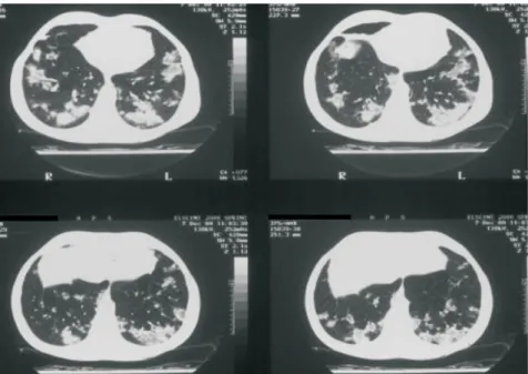
Imaging: bilateral diffuse micronodular infiltrate, with granular areas and some central nodular opacities, on the computed tomography scan of the chest. There was no evidence of lymph node enlargement in the mediastinum (Figure 2).
Mycological evaluation: mycological blood culture with immunodiffusion negative for H. capsulatum; histopathology of lingula sample (obtained through lung biopsy) stained with hematoxylin and eosin showing tuberculoid granuloma with caseous necrosis and fibrosis in the lung parenchyma; the Grocott-Gomori methenamine silver staining revealing numerous
small, round or oval budding yeast-like cells suggestive of H. capsulatum; culture was negative. Treatment and evolution: itraconazole 200 mg/ day, resulting in regression of the symptoms and clinical cure.
This unusual radiological pattern is suggestive of massive inoculation in a non-endemic area, causing a severe inflammatory reaction(1). Culture
is positive in only 15% of cases of the self-limited forms(4).
Up until 1978, only five cluster outbreaks, involving a total of 50 patients and one dog, had been reported in Brazil(7). Subsequently, it was
reported that thirteen cluster outbreaks (involving 102 patients) occurred in the state of Rio de Janeiro(8-12), one each occurred in the states of
Paraíba(13), Amazonas(14), Minas Gerais (15), and
Bahia(16), and two occurred in the state of Rio Grande
do Sul(17,18) (Chart 2). In addition to the two cluster
outbreaks documented in the literature(17,18), a new
outbreak was reported during the present study period (Cases 6, 7 and 8).
DISCUSSION
Acute pulmonary histoplasmosis is characterized by the development of respiratory symptoms from one to three weeks after substantial exposure to H. capsulatum(2). Duration of exposure determines
Figure 2 – Bilateral diffuse pulmonary infiltrate with granular areas and some central
Jornal Brasileiro de Pneumologia 31(1) - Jan/Fev de 2005
disease severity: short exposure times (20 minutes or less) produce mild symptoms, whereas a 50- to 60-hour exposure time results in severe illness(5).
There are three radiological patterns that appear after massive inoculation: nodular pneumonia, micronodular infiltrate and miliary pattern. The first one is the least common, and is suggestive of a pronounced inflammatory reaction, located on the periphery of the endemic area, in hosts with no previous infection. The second pattern is the most frequent, and the third one is suggestive of re-exposure of previously infected hosts who maintained a high level of immunity(1). The course
of the disease is self-limited, with spontaneous regression of the symptoms(3). Whether the host
has been previously infected or not differentiates the findings. Chart 1 shows the principal differences between primary infection and reinfection.
Clinical presence of cough, fever, dyspnea and asthenia in a previously healthy individual may, in most cases, be associated with epidemiologically relevant episodes(1). In 11 patients (61%), onset of
the disease can be related to a specific activity (such as the exposure to chicken or bat feces observed in the representative cluster outbreak). Due to the simultaneous occurrence of the cases and the facility of identifying the source of exposure, cluster outbreaks are more easily recognized than are isolated cases. In the latter, significant clinical suspicion or, preferably, routine mycological investigation including mycological blood culture (immunodiffusion), fungus-specific staining of the histological sections, and fungi culture of the tissue samples obtained through biopsy, is needed(2).
Systemic mycoses, especially histoplasmosis, mimic tuberculosis in terms of clinical, radiological and histopathological aspects since they are granulomatous diseases. Diagnostic differentiation demands special stains as well isolation of the etiologic agent in culture. The presence of tuberculoid granuloma with caseous necrosis on tissue sections from lung biopsies, stained with hematoxylin and eosin, misleads the physician into the diagnostic hypothesis of tuberculosis. In fact, the term tuberculoid comes from the word tubercle, meaning small and round in shape, and not from tuberculosis, the disease. In the present study, 22% of patients were subject to the complications from and deleterious effects of a treatment for tuberculosis, as well as to delayed diagnosis of the disease. A diagnostic routine that includes the Grocott-Gomori methenamine silver stain technique or immunodiffusion for histoplasmosis, or both, in patients with a presumptive diagnosis of tuberculosis reveals unsuspected cases(6).
Many factors indicate that Rio Grande do Sul is a hyperendemic region for histoplasmosis, as are other regions of the country. Such factors include the types of histoplasmosis cases found, the fact that the acute pulmonary form was seen, the presence of isolated cases and cluster outbreaks, isolation of the etiologic agent in the
soil( 1 9 ), the strong cutaneous reactivity to
histoplasmin(20), the large number of cases of the
disseminated form in patients with acquired immunodeficiency syndrome(21), the presence of
the acute disseminated disease in infants(22), and
the presence of the chronic pulmonary form(21).
CHART 1
Goodwin & Des Prez classification
PRIMARY REINFECTION
Incubation period 10-18 days 3-7 days
Disease severity More severe Less severe
Geographical location Periphery of the endemic area Highly endemic area
Population of the endemic area Immigrants Natives
Radiological findings Bronchopneumonia Miliary nodulation
Hilar adenopathy No adenopathy
Possible pleural involvement No pleural involvement
Late calcification Usual None
58 U
ni
s,
G
is
e
la
e
t
a
l.
A
cut
e
p
ul
m
o
na
ry
h
is
to
p
la
sm
o
sis
i
n
th
e
st
a
te
o
f
R
io
G
ra
nd
e
d
o
Sul
,
B
ra
zi
l
CHART 2
Year Reference city State Source of infection Activity Cases Sero- Histo- Isolation in soil logy logy
1958 Paraíba do Sul RJ Cave with bats Leisure/study 13 * 0 1 NP Paula, 1959(23) 1959 Santa Teresa RJ Water reservoir Leisure 7 ND ND NP Paula, Aidé, 1979(9) 1966 Ubatuba SP House ceiling Cleaning 8 6 7 (+) Bat feces Fava Netto et al., 1967(24) 1967 Brasília DF Cave with bats Leisure 14 8 13 (+) Soil Schmidt et al., 1973(25) 1971/73 Ubatuba SP Cave with bats Leisure 10 10 9 NP Fava Netto et al., 1976(26)
1972 Vassouras RJ Cave with bats Prospecting 5 ND ND NP Paula, Aidé, 1979(9)
1975 Rio de Janeiro RJ Cave with bats Leisure 5 0 5 NP Rêgo et al., 1976(27) 1978 Angra dos Reis RJ Cave Leisure 8 ND 8 NP Paula, Aidé, 1985(10) 1978 Canoas RS Hollow tree trunk with bats Guano harvest 2* 1 ND NP Severo et al., 1981(17) 1981 Rio do Ouro RJ Cave Leisure 10** ND ND (+) Soil Paula, Aidé, 1985(10) 1981 São Gonçalo RJ Abandoned mine Leisure 10 8 ND (+) Soil and feces Wanke, 1985(8) 1981 São Gonçalo RJ Abandoned mine Study 4 ** 4 ND (+) Soil and feces Wanke, 1985(8) 1981 Itaipava-Petrópolis RJ Abandoned mines Leisure 10 9 ND (+) Soil and feces Wanke, 1985(8)
1982 São Gonçalo RJ Cave with bats Leisure 6** 5 ND NP Verbicário et al., 1993(11) 1982 Itaipava-Petrópolis RJ Abandoned mines Leisure 5 2 ND (+) Soil Wanke, 1985(8)
1982 Niterói RJ Abandoned mine Leisure 6 ** 5 ND (+) Soil Wanke, 1985(8) 1984 PendotibaNiterói RJ Water pipe Study 17 8 17 (+) Soil Paula, Aidé, 1985(10)
1984 Tinguá RJ Hen house Cleaning 12 ND ND ND Paula, Aidé, 1985(10)
1986 Borborema PB Chimney with bats Cleaning 6 4 5 (+) Bat feces Fernandes et al.,1989(13)
1993 Taquari RS Hen house Cleaning 2 2 ND NP Severo et al., 1993(18)
1993 Manaus AM Cave with guano Leisure 8 8 ND NP Suzaki et al., 1995(14)
1997 Pedro Leopoldo MG Cave with bats Leisure 4 3 ND NP Cury et al., 2001(15) 2000 Jequié BA Basement with bats Cleaning 4* 4 ND NP Martins et al., 2000(16) 2003 Niterói RJ Out-of-use oven Cleaning 5 5 ND NP Martins et al., 2003(12)
Total 181
ND: no data; NP: not performed
Jornal Brasileiro de Pneumologia 31(1) - Jan/Fev de 2005
REFERÊNCIAS
1. Goodwin RA, Loyd JE, Des Prez RM. Histoplasmosis in normal hosts. Medicine (Baltimore). 1981;60:231-66. 2. Chung KJ, Bennett JE. Histoplasmosis. In: Kwon-Chung KJ, Bennett JE. Medical mycology. Philadelphia: Lea & Febiger; 1992. p.248-79.
3. Goodwin RA, Des Prez RM. Histoplasmosis, state of the art. Am Rev Respir Dis. 1978;117:929-56. 4. Wheat LJ, Kauffman CA. Histoplasmosis. Infect Dis Clin
N Am. 2003;17:1-19.
5. Larsh HW. Histoplasmosis. In: di Salvo AF. Occupational mycoses. Philadelphia: Lea & Febiger; 1983. p.29-42. 6. Unis G. Histoplasmose no teste terapêutico para t u b e r c u l o s e . B u s c a d e c a s o s p e l a i m u n o d i f u s ã o [dissertação]. Porto Alegre: Universidade Federal do Rio Grande do Sul; 2000.
7. Londero AT, Ramos CD. The status of histoplasmosis in Brazil. Mycopathologia. 1978;3:153-6.
8. Wanke B. Histoplasmose. Estudo epidemiológico, c l í n i c o e e x p e r i m e n t a l [ t e s e ] . R i o d e J a n e i r o : Universidade Federal do Rio de Janeiro; 1985. 9. Paula A, Aidé MA. Histoplasmose no Brasil. JBM.
1979;37:67-81.
1 0 . Paula A, Aidé MA. As microepidemias de histoplasmose do Estado do Rio de Janeiro. JBM. 1985;49:18-28. 11 . Verbicário LPS, Andrade E, Alvim MAM, Andrade CRM,
Gabetto JM, Pinho RR, et al. Histoplasmose aguda: reavaliação de seis casos 10 anos após a microepidemia. Arq Bras Med. 1993;67:361-4.
1 2 . Martins EML, Marchiori E, Damato SD, Pozes AS, Silva ACG, Dalston M. Histoplasmose pulmonar aguda: relato de uma microepidemia. Radiol Bras. 2003;36:147-51. 1 3 . Fe r n a n d e s F O, C o s t a W, W a n k e B , O l i ve i ra R Z . Microepidemia de histoplasmose capsulata. Clínica e epidemiologia do primeiro surto ocorrido no Estado da Paraíba. CCS. 1989;11:189-99.
1 4 . Suzaki A, Kimura M, Kimura S, Shimada K, Miyaji M, K a u f m a n L . A n o u t b r e a k o f a c u t e p u l m o n a r y histoplasmosis among travelers to a bat-inhabited cave in Brazil. Kansenshogaku Zasshi: 1995;69:444-9. 1 5 . Cury GC, Diniz Filho A, Costa e Cruz AG, Hobaika ABS.
Surto de histoplasmose em Pedro Leopoldo, Minas Gerais, Brasil. Rev Soc Bras Med Trop. 2001;34:483-6.
1 6 . Martins ACP, Neves MLC, Lopes AA, Santos NNQ, Araújo NN, Pereira KM. Histoplasmosis presenting a s a c u t e r e s p i r a t o r y d i s t r e s s s y n d r o m e a f t e r exposure to bat feces in a home basement. Braz J Infect Dis. 2000;4:103-6.
1 7 . Severo LC, Picon PD, Londero AT, Rubião HFºJ. Histoplasmose aguda. Relato de dois casos. Rev AMRIGS. 1981;25:64-7.
1 8 . S e ve r o L C , R i z z o n C F C , R o e s c h E W, Po r t o N S . Histoplasmose pulmonar aguda: episódio em casal de adultos. Rev AMRIGS. 1993;37:281-3.
1 9 . Severo LC, Petrillo VF, Camargo JJ, Geyer GR, Porto N S . A c u t e p u l m o n a r y h i s t o p l a s m o s i s a n d f i r s t isolation of Histoplasma capsulatum from soil of Rio Grande do Sul, Brasil. Rev Inst Med Trop S Paulo. 1986;28:51-5.
2 0 . Zembrzuski MM, Bassanesi MC, Wagner LC, Severo LC. I n q u é r i t o i n t r a d é r m i c o c o m h i s t o p l a s m i n a e paracoccidioidina em duas regiões do Rio Grande do Sul. Rev Soc Bras Med Trop. 1996;28:1-3.
21. Severo LC, Oliveira FM, Irion K, Porto NS, Londero AT. Histoplasmosis in Rio Grande do Sul, Brazil: a 21-year experience. Rev Inst Med Trop S Paulo. 2001;43:183-7. 22. Severo LC, Zardo IB, Roesch EW, Hartmann AA. Acute disseminated histoplasmosis in infancy in Brazil: report of a case and review. Rev Iberoamer Micol. 1998;15:48-50. 2 3 . Paula A. Microepidemia de histoplasmose. Rev Serv
Nac Tuberc. 1959;3:11-20.
24. Fava Netto C, Andrade e Silva U, Chammas F, Lacaz CS. Histoplasmose epidêmica. Estudo clínico, radiológico, micológico e imunológico de surto ocorrido no Estado de São Paulo. Rev Inst Med Trop S Paulo. 1967;9:222-32. 2 5 . Schmidt S, Machado OP, Galvão AB. Microepidemia
de histoplasmose na zona rural de Brasília, DF. 2. Estudos epidemiológico e parasitológico da fonte de infecção. Rev Soc Bras Med Trop. 1973;7:107-15. 26. Fava Netto C, Almeida Neto JM, Guerra MAG, Costa EO.
Histoplasmose epidêmica. Novos surtos ocorridos no litoral norte do Estado de São Paulo. Inquérito epidemiológico com histoplasmina e paracoccidioidina. Rev Inst Med Trop S Paulo. 1976;18:108-12.