Pedro Arêde Rei
Licenciado
On the track of β-lactam resistance:
Studies on the regulation of
methicillin-resistance in Staphylococcus aureus
Dissertação para obtenção do Grau de Doutor em Biologia
Orientador: Doutor Duarte Carvalho Oliveira, Assistant
Researcher, CREM, Faculdade de
Ciências e Tecnologia, Universidade Nova
de Lisboa
Co-orientador: Doutora Hermínia de Lencastre, Full
Professor, ITQB, Universidade Nova de
Lisboa
Presidente: Profª. Doutora Maria João Romão Arguentes: Prof. Doutor S. Mobashery
Profª. Doutora B. Berger-Bächi
Vogais: Prof. Doutor Arsénio M. Fialho Prof. Doutor Adriano O. Henriques Prof. Doutor Carlos São José Profª. Doutora Marta Aires de Sousa
i
Pedro Arêde Rei
Licenciado
On the track of β-lactam resistance: Studies on the regulation of
methicillin-resistance in Staphylococcus aureus
© Pedro Arede
ISBN: 978-989-20-3273-3
A Faculdade de Ciências e Tecnologia e a Universidade Nova de Lisboa têm o direito, perpétuo e sem limites geográficos, de arquivar e publicar esta dissertação através de exemplares impressos reproduzidos em papel ou de forma digital, ou por qualquer outro meio conhecido ou que venha a ser inventado, e de a divulgar através de repositórios científicos e de admitir a sua cópia e distribuição com objectivos educacionais ou de investigação, não comerciais, desde que seja dado crédito ao autor e editor.
iii
Acknowledgments
First of all, I would like to thank Duarte Oliveira, my supervisor, for accepting me in his laboratory, giving me the opportunity to perform the PhD and believing in my work capacity. I must also thank him for guiding me so patiently in the first years of training at the bench, for having accompanied me with enthusiasm during the work leading to this PhD. Above all, I would like to thank him for all the unconditional support, friendship, incentive, persistence and the enthusiastic scientific discussions as well as having shared with me his passion for science.
To my co-supervisor, professor Hermínia de Lencastre, Head of the Molecular Genetics laboratory at Instituto de Tecnologia Química e Biológica, Universidade Nova de Lisboa, where I spent the first years of my scientific career as a graduate student. I thank her for the supervision during this PhD, the criticism, the discussion and sharing scientific ideas.
To Professor Xavier Gomis-Rϋth, Head of Proteolysis Laboratory, at Molecular Biology Institute of Barcelona, in Barcelona, Spain, where I performed part of the experimental work presented in this thesis, for accepting me in his lab and for his supervision. I am very grateful to him for the excellent conditions, experimental support and also for the all knowledge that he and all of his team shared with me.
Special thanks to Tiago Botelho, who accompanied me in my stay in Barcelona, for teaching me some of the laboratory skills I know today. To him and to Soraya Hernandez, Mar Lopez, Nuria Cerda, Tibisay Guevara and Theodoros Goulas for all the good moments and also for making me feel at home.
To my former lab. colleagues, Ana Portelinha and Joana Ministro for their availability, friendship and also for the good moments.
I thank my colleagues, past and present, from CREM, Isabel Correia, Raquel Portela, Lia Godinho, Mário Ferreira, Renato Pires, Carolina Alves, Elizabete Valério, Marco Coelho, Pedro Almeida, Carla Gonçalves, Márcia Rato and Márcia Palma, for the helpful work environment, the constant sharing of ideas and also for all the good moments.
I also thank my colleagues, from the laboratory of Molecular Genetics at ITQB, Catarina Milheiriço, Rita Sobral, Sandro Pereira, Susana Gardete, Nuno Faria,Teresa Conceição, Nelson Frazão, Sónia Nunes, Alexandra Simões and Isilda Gueifão, for their inconditional help at the beginning of my scientific journey, and also for their friendship and good work environment.
iv A special thanks to Rui Almeida and Ana Teresa Lopes, from REQUINTE, for their inexhaustible help in the use of the necessary apparatus to protein purification.
To Centro de Recursos Microbiológicos (CREM), for providing me the excellent conditions to perform my experimental work.
My acknowledgement to Fundação para a Ciência e a Tecnologia (FCT) for the financial support for the accomplishment of this Doctoral Project (SFRH/BD/38316/2007) and for the financial support that allowed me to attend scientific meetings which gave me the opportunity to exchange scientific information with other researchers.
To all my friends whom, unfortunately, I cannot name, I thank the endless friendship and constant support that make life so interesting.
Finally I would like to thank my Family, for all their trust, incentive, for supporting me in every moment, for always being by my side and for giving me the strength to go further…. To them I dedicate this thesis.
v
Abstract
Methicillin-resistant Staphylococcus aureus (MRSA) is an important human pathogen, causing a wide range of infections. MRSA has not only developed an intrinsic resistance to all β-lactams, but has also acquired resistance to virtually all classes of antimicrobial agents. The characteristic MRSA phenotype is conferred by the presence of mecA gene which is regulated by a sensor-inducer (MecR1) and a repressor (MecI). However, mecA induction by its cognate sensor/inducer is very inefficient and, therefore, it is believed that optimal expression of β-lactam resistance in MRSA requires a non-functional MecI system. Surprisingly, no correlation was found between the presence of functional MecR1-MecI and the level of β-lactam resistance in a representative collection of epidemic MRSA strains, suggesting the existence of other mecA regulators.
In these studies, we show that the mecA regulatory locus is not a two-component system but, actually, it is a three-component system containing besides mecR1-mecI, the previously unidentified anti-repressor mecR2. The crystal structure of MecR2 reveals a three-domain architecture, with an N-terminal DNA-binding-like domain, an intermediate scaffold domain, and a C-terminal dimerization domain, important to the functional dimeric oligomerization state. MecR2 disturbs the binding of the repressor MecI to the mecA promoter, which leads to its proteolytic inactivation independently from MecR1, presumably by non-specific cytoplasmatic proteases. Our data also demonstrates that in the presence of functional mecR1-mecI genes, mecR2 is essential for a robust induction of mecA transcription and, consequently, for the optimal expression of resistance phenotype in MRSA. These observations point to a revision of the current model for the transcriptional control of the mecA gene.
vii
Resumo
Os Staphylococcus aureus resistentes à meticilina (MRSA, do inglês “methicillin-resistant
Staphylococcus aureus”) são um importante agente patogénico, que em certas circunstâncias podem
causar infecções. Para além de terem desenvolvido resistência a todos os antibióticos β-lactâmicos, os MRSA são também frequentemente resistentes a outras classes de agentes antimicrobianos. O fenótipo caracteristico dos MRSA deve-se à presença do gene mecA, que é regulado por um sensor/transdutor (MecR1) e um repressor (MecI). No entanto, como a indução do gene mecA através do MecR1 é muito ineficiente, pensa-se que os MRSA só conseguem expressar elevados níveis de resistência aos β-lactâmicos se o sistema MecR1-MecI não estiver funcional. Curiosamente, dados recentes demonstram ausência de relação entre a expressão das proteínas MecR1-MecI funcionais e o nível de resistência aos β-lactâmicos numa colecção de estirpes MRSA epidémicas.
Os resultados aqui apresentados mostram que o locus que regula a transcrição do gene mecA contém três genes reguladores (mecR1-mecI-mecR2). A resolução da estrutura tridimensional do MecR2 revela que esta proteína é constituída por três domínios, um semelhante a domínios com capacidade de ligação ao ADN, um intermédio e um de dimerização. Os nossos resultados indicam que o MecR2 desestabiliza a ligação do repressor à região operadora do gene mecA, e que a inativação do MecI ocorre independente do MecR1, sendo efectuada possivelmente por proteases nativas. Este estudo demostra ainda que nas estirpes MRSA, que contêm os genes mecR1-mecI funcionais, o gene mecR2 é essencial para induzir eficazmente a transcrição do mecA. Em conjunto, estes dados revelam que o actual modelo que descreve o controlo da transcrição do gene mecA, deve ser repensado.
Palavras-chave: MRSA; resistência aos β-lactâmicos; regulação do gene mecA; Anti-repressor MecR2; repressor MecI
ix
Table of Contents
ACKNOWLEDGMENTS ... III
ABSTRACT ... V
RESUMO ... VII
TABLE OF CONTENTS ... IX
FIGURE INDEX ... XIII
TABLE INDEX ... XV
ABBREVIATIONS ...XVII
LIST OF PUBLICATIONS ... XIX
THESIS OUTLINE ... XXI
CHAPTER I... 1
GENERAL INTRODUCTION ... 1
1.1 Staphylococcus aureus as a human pathogen ... 3
1.1.1 General features ... 3
1.1.2 Colonization and infection ... 4
1.1.3 Antibiotic resistance ... 5
1.1.3.1 Resistance to β-lactam antibiotics ... 7
1.1.4 Epidemiology and evolution of MRSA ... 8
1.1.4.1 Hospital-acquired MRSA (HA-MRSA) ... 9
1.1.4.2 Community-acquired MRSA (CA-MRSA) ... 10
x
1.1.4.4. mecA gene origin ... 13
1.2. Regulation of gene expression in Prokaryotes ... 14
1.2.1 Initiation of transcription ... 15
1.2.2 Promoter recognition ... 16
1.2.3 The molecular mechanisms regulating transcription initiation ... 18
1.2.3.1 Activators vs repressors ... 18
1.2.3.2 The helix-turn-helix domain ... 18
1.3. Regulation of β-lactam resistance in S. aureus ... 19
1.3.1 Cell-wall biosynthesis: the cellular target of β-lactams ... 19
1.3.1.1 PBPs ... 20
1.3.1.2 PBP2a ... 21
1.3.1.3 Factors affecting methicillin resistance ... 22
1.3.1.4 Heterogeneity ... 23
1.3.2 bla-system ... 24
1.3.3 mec-system ... 27
1.4 References... 31
CHAPTER II ... 51
THE ROLE OF MECR2 IN THE REGULATION OF METHICILLINRESISTANCE IN MRSA . 51
2.1 Abstract ... 53 2.2 Introduction ... 53
2.3 Results and Discussion ... 55
2.3.1 The mecA cognate regulatory locus is a three-component system ... 55
2.3.2 mecR2 is involved in the optimal expression of β-lactam resistance ... 59
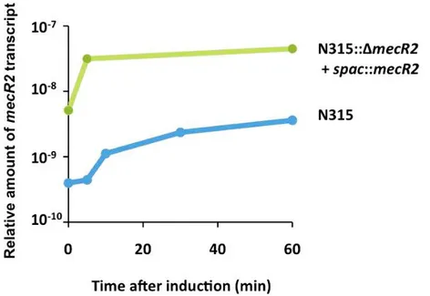
2.3.3 mecR2 is required for the full induction of mecA transcription ... 64
2.3.4 mecR2 transcription analysis ... 66
2.3.5 mecR2 is essential for the optimal expression of β-lactam resistance in strains with functional mecI-mecR1 regulatory locus. ... 68
2.3.6 mecR2 function is not dependent on mecR1 neither on the β-lactamase locus ... 70
xi
2.3.8 MecR2 interferes with the binding of MecI to the mecA promoter ... 72
2.3.9 MecR2 promotes the proteolytic cleavage of MecI ... 76
2.4 MecR2, the missing link in the signal-transduction mechanism of mecA expression ... 77
2.5 Concluding remarks ... 78
2.6 Materials and Methods ... 80
2.6.1 Bacterial strains and growth conditions ... 80
2.6.2 DNA manipulations ... 80
2.6.3 Construction of recombinant S. aureus strains ... 85
2.6.4 Transcription analysis ... 87
2.6.5 Bacterial two-hybrid assays ... 88
2.6.6 Electrophoretic mobility shift assays (EMSA) ... 89
2.6.7 Western blotting ... 90
2.7 Acknowledgments ... 90 2.8 References... 91
CHAPTER III ... 95
STRUCTUREFUNCTION STUDIES OF THE ANTIREPRESSOR MECR2 ... 95
3.1 Abstract ... 97 3.2 Introduction ... 97
3.3 Results and Discussion ... 98
3.3.1 Large-scale recombinant heterologous overexpression and purification of MecR2 ... 98
3.3.2 Biological activity of purified MecR2 ... 99
3.3.3 MecR2 establishes a non-obligate triggered transient interaction with MecI ... 99
3.3.5 MecR2 is a functional dimer ... 103
3.3.6 Structural similarities ... 105
3.3.7 MecR2 has a non-functional ligand-binding cleft ... 107
3.3.8 MecR2 has a non-functional DNA-binding domain ... 108
3.3.9 Functional implications of MecR2 ... 108
xii
3.3.11 Site-directed mutagenesis of MecR2 ... 111
3.4 Materials and Methods ... 113
3.4.1 Recombinant overexpression and purification ... 113
3.4.2 Biological activity of purified MecR2 ... 113
3.4.3 Site-directed mutagenesis of MecR2 ... 114
3.4.4 In vitro MecR2::MecI crosslinking ... 114
3.4.5 Bio-layer interferometry ... 116
3.4.6 Crystallization and structure analysis ... 117
3.5 Miscellaneous ... 118 3.6 Acknowledgments ... 118 3.7 References... 119
CHAPTER IV ... 123
CONCLUDING REMARKS & FUTURE PERSPECTIVES ... 123
4.1 Conclusion ... 125 4.2 Future Perspectives ... 129 4.3 References... 131
xiii
Figure Index
Figure 1.1 - Scanning Electron Microscopy of S. aureus N315. ... 3
Figure 1.2 - The bacterial cell with the 4 main antibiotic targets; cell-wall synthesis, nucleic acid synthesis, protein synthesis, and folic acid metabolism. ... 6
Figure 1.3 - Schematic representation of some β-lactam antibiotics. ... 8
Figure 1.4 - Schematic representation of the RNA polymerase core with the isoforms present in E.coli. 16 Figure 1.5 - The RNA polymerase-promoter interactions. ... 17
Figure 1.6 - The peptidoglycan structure of S. aureus. ... 20
Figure 1.7 - Schematic representation of BlaR1 membrane protein. ... 26
Figure 1.8 - Overall view of BlaI bound to DNA. ... 26
Figure 1.9 - Schematic representation of the β-lactamase and mecA operons. The levels of the aminoacid identity of the regulatory proteins are shown. ... 28
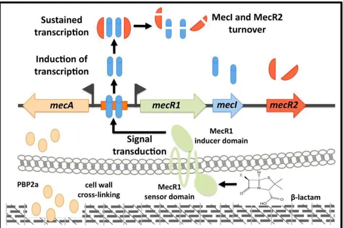
Figure 1.10 - Schematic representation of the mechanisms involved in the regulation of the mecA and blaZ genes expression in S. aureus. ... 29
Figure 2.1 - The mecA regulatory locus is a three-component system. ... 57
Figure 2.2 - Multiple sequence alignment between the repressor of the xylose operon (XylR) and the anti-repressor MecR2 found in staphylococci. ... 57
Figure 2.3 - Transcriptional analysis of mecR2. ... 59
Figure 2.4 - mecR2 interferes with the mecI-mediated repression of β-lactam resistance. ... 60
Figure 2.5 - Insertion-deletion strategies used for the reconstruction of the mecA regulatory locus in the chromosome of strain COL and deletion of mecR2 from the chromosome of strain N315. ... 61
Figure 2.6 - Reconstruction of the mecA regulatory locus in prototype strain COL. ... 62
Figure 2.7 - Role of mecR2 on the optimal expression of β-lactam resistance. ... 63
Figure 2.8 - Effect of mecR2 on the induction of mecA transcription. ... 65
Figure 2.9 - Effect of mecR2 on the induction of mecA transcription in strain COL. ... 66
Figure 2.10 - mecR2 transcription analysis. ... 67
Figure 2.11 - mecR2 is essential for the optimal expression of β-lactam resistance in strains with functional mecI-mecR1 regulatory locus. ... 69
Figure 2.12 - The mecR2 function is not dependent of mecR1 neither of the β-lactamase locus and does not interfere with the function of β-lactamase regulatory genes. ... 71
Figure 2.13 - MecR2 interacts directly with MecI, interfering with the binding of MecI to the mecA promoter and fostering the proteolysis of MecI. ... 75
Figure 2.14 - Control experiments for the electrophoretic mobility shift assays. ... 76
Figure 2.15 - Model for the mecA induction by MecR1-MecI-MecR2. ... 79
Figure 3.1 - Binding of MecR2 to MecI. ... 100
xiv Figure 3.3 - Ligand-binding cleft and quaternary structure of MecR2. ... 105 Figure 3.4 - MecR2 interacts with MecI. ... 110 Figure 3.5 - Biological activity of purified MecR2 and mutagenized MecR2 variants. ... 112 Figure 4.1 - Model depicting the transcriptional induction of mecA in the presence of β-lactams mediated by the regulator proteins MecR1-MecI-MecR2. ... 128
xv
Table Index
Table 2.1 - Strains used in the study of chapter II ... 81
Table 2.2 - Plasmids used in the study of chapter II ... 83
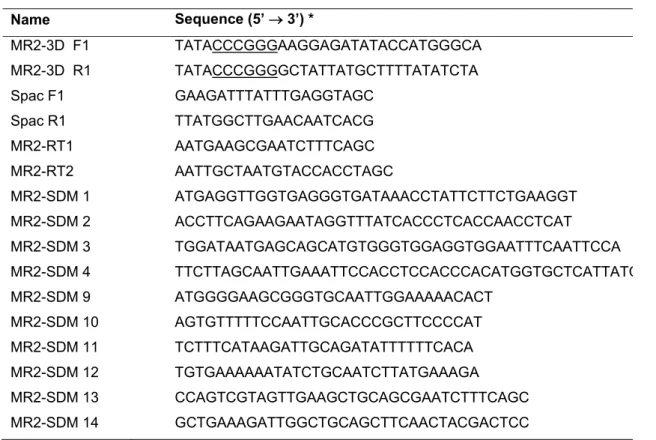
Table 2.3 - Primers used in the study of chapter II ... 84
Table 2.4 - Strains and plasmids used in the bacterial two-hybrid assays ... 89
Table 3.1 - Crystallographic data ... 106
Table 3.2 - Strains and plasmids used in the study of chapter III ... 115
xvii
Abbreviations
ACME Arginine catabolic mobile element
aux Auxiliary genes
agr accessory gene regulator BLAST Basic local alignment search tool BTH Bacterial two-hybrid
CA-MRSA Community-associated methicillin-resistant Staphylococcus aureus
cat Chloramphenicol resistance gene
CC Clonal complex
CDD C-terminal dimerization domain CoNS Coagulase negative staphylococci
DBD DNA-binding domain
DEPC Diethyl pyrocarbonate-treated water DHFA Dihydrofolic acid
DNA Deoxyribonucleic acid
EDTA Ethylenediaminetetraacetic acid EMSA Electrophoretic mobility shift assay
ery Erythromycin resistance gene
fem Factors essential for methicillin resistance
GlcNAc N-Acetylglucosamine
HA-MRSA Hospital-acquired methicillin-resistant Staphylococcus aureus HMW High-molecular weight
LMW Low-molecular weight
HTH Helix-turn-helix
IPTG Isopropyl β-D-1-thiogalactopyranoside
IS Insertion sequence
ISD Intermediate scaffold domain KD Dissociation constant KDa Kilodalton LA Luria agar LB Luria broth MBP Maltose-binding protein MGT Glycosyltransferases
MIC Minimum inhibitory concentration MOPS Morpholine propanesulfonic acid mRNA Messenger ribonucleic acid
xviii MRSA Methicillin-resistant Staphylococcus aureus
MSSA Methicillin sensitive Staphylococcus aureus MurNAc N-Acetylmuramic acid
NDD N-terminal DNA-binding-like domain
OD Optical density
ORF Open reading frame
PAP Population analysis profiles PBP Penicillin binding protein PMSF Phenylmethanesulfonylfluoride PVL Panton-Valentine leukocidin qRT-PCR Quantitative Real-time RT-PCR
RNA Ribonucleic acid
RNAP RNA polymerase
RNase Ribonuclease
ROK Repressors, open-reading frames, and kinases RT-PCR Reverse-transcriptase PCR
SCCmec Staphylococcal cassette chromosome mec
SDS-PAGE Sodium dodecyl sulfate polyacrylamide gel electrophoresis
ST Sequence type
TBE Tris-borate EDTA buffer
TCRS Two-component regulatory systems
TE Tris-EDTA buffer
tet Tetracycline resistance gene
TEV Tobacco etch virus THFA Tetrahydrofolic acid
TM Transmembrane segments
Tris Tris(hydroxymethyl)aminomethane TSA Trypticase soy agar
TSB Trypticase soy broth
van Vancomycin resistance gene
VISA Vancomycin-intermediate Staphylococcus aureus VRSA Vancomycin-resistant Staphylococcus aureus
xix
List of Publications
This thesis is based on two articles:
Arêde, P., C. Milheirico, H. de Lencastre, and D.C. Oliveira. The anti-repressor MecR2 promotes the proteolysis of the mecA repressor and enables optimal expression of β-lactam resistance in MRSA. PLoS Pathog, 2012. 8(7): p. e1002816.
Arêde, P. Botelho, T. Guevara, T. Usón, I, Oliveira, DC. Gomis-Rüth, FX. Structure-function studies of the staphylococcal methicillin resistance anti-repressor, MecR2. 2012. Submitted.
xxi
Thesis Outline
This Ph.D. Thesis is organized into four chapters. It includes a general introduction, one published manuscript and one manuscript in preparation.
Chapter I includes a general description of the mechanisms involved in methicillin resistance in S.
aureus. In this Chapter, special attention was given to the mode of action of this class of antimicrobial
agents, their targets, and the strategies developed by the bacteria to acquire resistance against these drugs, specially the resistance mechanism mediated by mecA gene.
Chapter II is entitled “The role of mecR2 in the regulation of methicillin-resistance in MRSA.” and includes a published manuscript. This Chapter describes the genetic and biochemical studies carried out in order to elucidate the role of the previous unidentified mecR2 gene on regulation mecA transcription in MRSA strains with a fully functional mec regulatory system.
Chapter III is entitled “Structure-function studies of the anti-repressor, MecR2” and includes a manuscript in preparation. This chapter describes the three-dimensional structure of the anti-repressor MecR2, as well as further evidences regarding MecR2::MecI interaction. It is also shown that the main function of the anti-repressor MecR2 is its binding to the MecI repressor, disrupting the interaction of MecI to the promoter mecA.
Chapter IV provides a general conclusion of results and includes a model, which illustrates the induction of mecA by its three cognate regulators (mecR1-mecI-mecR2) and an outlook for future research.
1
CHAPTER I
3
1.1 Staphylococcus aureus
as a human pathogen
1.1.1 General features
Staphylococcus aureus are a gram-positive cocci and microscopically are observed as individual
organisms, in pairs, in irregular, or in grapelike clusters. The term Staphylococcus is derived from the Greek term staphyle, meaning "a bunch of grapes." S. aureus are non-motile, non–spore-forming bacteria and their colonies are usually large (6-8 mm in diameter), smooth, and translucent (see Figure 1.1). S.
aureus were first recognized by Koch in 1878 and Pasteur in 1879 [1] but their more detailed
characterization only came some years later with Ogston [2] and Rosenbach [3], who described two pigmented colony types of staphylococci and proposed the appropriate nomenclature: Staphylococcus
aureus (golden-like) and Staphylococcus albus (white). The latter species is now named Staphylococcus
epidermidis. The colonies of most strains of S. aureus are pigmented, ranging from cream-yellow to
orange, which is due to the presence of triterpenoid (C30) carotenoids, rather than the more typical C40 carotenoids [4, 5]. S. aureus is part of the human flora, mainly in the axillae, the inguinal and perineal areas, and the anterior nares, whereas, S. epidermidis is ubiquitous in skin [6].
Figure 1.1 - Scanning Electron Microscopy of S. aureus N315. Adapted from [7].
At a biochemical level, S. aureus is a facultative anaerobe, which can grow by aerobic respiration or lactic acid fermentation of glucose. It is catalase positive, and can survive in NaCl concentrations of up to 15 percent. While most staphylococci are coagulase-negative, S. aureus is coagulase positive [8] and can grow between 10ºC and 45ºC. The G +C content of S. aureus DNA is within the range of 30 to 38
4 mol percent, being staphylococci one of the members of the low G +C group of the gram-positive bacterial phylogenetic group [9].
The staphylococcal cell is surrounded by a mesh-like structure 20-40 nm thick, called peptidoglycan, which is composed of a series of short glycan chains of c.a. 20 alternating N-acetyl-muramic-acid and β-1,4-N-acetylglucosamine residues [10]. Peptidoglycan is an essential and specific component of the bacterial cell wall found on the outside of the cytoplasmic membrane of almost all bacteria [11, 12]. Its main function is to preserve cell integrity and it also contributes to the maintenance of a defined cell shape and serves as a scaffold for anchoring other cell envelope components such as proteins and teichoic acids [13]. S. aureus-specific pentaglycine interpeptide cross-bridges are assembled in the cytoplasm by auxiliary genes femX [14], femA [15], and femB [16], which attach the glycine residues to the L-lysine residue of the stem peptide, in a sequential manner [17]: FemX adds the first glycine, FemA adds the 2nd and 3rd, whereas, the FemB adds the 4th and 5th. FemA and FemB are not interchangeable, meaning that inactivation of one of these genes results in cell walls that contain mono-or triglycine cross-bridges, respectively.
1.1.2 Colonization and infection
The capacity to asymptomatically colonize healthy individuals is a biological property of S.
aureus, with approximately 30% of humans being asymptomatic nasal carriers of S. aureus [18]. Indeed,
S. aureus carriers have a higher risk of acquiring S. aureus related infections and are an important
dissemination vehicle of S. aureus, either by direct contact (i.e. skin-to-skin contact with a carrier or infected individual) or by contact with contaminated surfaces or objects [19-21].
Due to its capacity to cause opportunistic infections, S. aureus should always be considered a potential pathogen which, in certain circumstances, may cause a variety of suppurative (pus-forming) infections and toxinoses in humans [19]. Examples are superficial skin lesions such as boils, styes and furunculosis or more serious infections such as pneumonia, mastitis, phlebitis, meningitis, mastitis, phlebitis and urinary tract infections and even deep-seated infections, such as osteomyelitis and endocarditis. Moreover, S. aureus is a major cause of hospital acquired (nosocomial) infection of surgical wounds and infections associated with indwelling medical devices [18]. S. aureus is also responsible for food poisoning by releasing enterotoxins into food, and for the toxic shock syndrome by releasing superantigens into the blood stream [22-24].
The pathogenicity of S. aureus is a complex process involving a diverse array of extracellular and cell wall components that are coordinately expressed during different stages of infection; i.e. colonization, escape from the host defense, growth and cell division, and bacterial dispersion. The adaptive response is highly coordinated and is modulated by regulatory elements via signal transduction pathways [19].
5 Genomic analysis has revealed two major families of global regulators in S. aureus: two-component regulatory systems (TCRS) [25, 26] and the SarA homologs, a family of proteins homologous to SarA [25, 27], of which the best-characterized regulators of virulence factors are the accessory gene regulator (agr) and the staphylococcal accessory regulator (SarA), respectively. The agr locus consists of two divergent transcripts, RNAII and RNAIII, driven by two distinct promoters, P2 and P3, respectively [28]. RNAIII is the effector of the agr response that involves up-regulation of genes involved in exoprotein synthesis and down-regulation of genes encoding surface proteins [29, 30]. Unlike agr, the sar locus activates the synthesis of both extracellular (e.g. hemolysins) and cell wall-associated proteins (e.g. fibronectin-binding protein) [31]. These extracellular proteins can be divided into two groups depending on when they are expressed in cells growing in rich medium: proteins that are expressed only when cell densities are low and proteins exclusively expressed at high cell densities. By virtue of their proteolytic activities and toxin effects on host cells, the exotoxins synthesized during the post-exponential phase facilitate the local invasion and hematogenous dissemination phases of S. aureus infections [32].
The virulence factors of S. aureus can be classified in three different groups: (i) those which are involved in the attachment to the host cells, such as collagen-binding protein, coagulase or fibronectin-binding proteins A and B; (ii) factors involved in the invasion of host cells and consequently tissue penetration, such as α-toxin and hemolysins; and (iii) virulence factors involved in evasion of host defences such as protein A, lipase and toxic shock syndrome toxin 1, [33-37]. The coordinated regulation of virulence determinants during the exponential and post exponential phases has a decisive contribution to the development of S. aureus infections.
1.1.3 Antibiotic resistance
S. aureus has been a stumbling block for anti-microbial chemotherapy able to develop resistance
to all therapeutic agents deployed in clinical practice. Antibiotics, which literally means agents “against life”, are molecules that prevent microbes, both bacteria and fungi, from growing. One of the first documented identification of an antibiotic compound dates from 1929 by Alexander Fleming, who observed that a culture plate of S. aureus had been contaminated by a blue-green mould (Penincillinium
notatum) and that colonies of S. aureus adjacent to the mould could not grow. Then, Fleming grew the
mould in a pure culture and found that it produced a substance that killed a number of disease-causing bacteria. He named the substance penicillin and realised that his discovery might have therapeutic value if the antibiotic could be produced in large quantity [38].
Antibiotics can be classified according to their mode of action, cellular target, and main clinically relevant mechanism of resistance. According to the physiological cellular target, antibiotics are often grouped into four major classes (see Figure 1.2): cell wall synthesis, protein synthesis, nucleic acid synthesis, or folic acid metabolism inhibitors. The cell wall synthesis inhibitors, such as the β-lactams and the glycopeptides are the most representative and widely used class of antibiotics against S. aureus
6 infections. The β-lactams (e.g. penicillins and cephalosporins) bind covalently to PBPs, and also inhibit the reticulation of the pentapeptidic chains of the peptidoglycan precursors. The glycopeptides (vancomycin and teicoplanin), also target cell wall synthesis. These later antibiotics bind the D-Ala-D-Ala termini of peptidoglycan precursors, blocking the transpeptidation reaction carried out by PBPs. Although glycopeptides are the last resort to treat methicillin resistant S. aureus (MRSA) infections, strains with reduced susceptibility to these antibiotics have already been reported [39, 40]. Vancomycin intermediate S. aureus (VISA) strains are characterized by a thickening of the cell wall, which is believed to reduce the ability of vancomycin to diffuse into the division septum of the cell, whereas, the vancomycin resistant S.
aureus (VRSA) modify peptidoglycan precursors ending in D-Ala-D-Ala to D-Ala-D-Lac instead, with low
affinity for glycopeptides [41-43].
Figure 1.2 - The bacterial cell with the 4 main antibiotic targets; cell-wall synthesis, nucleic acid synthesis, protein synthesis, and folic acid metabolism. Examples of the most representative and most used antibiotics are shown, as well as their specific targets. mRNA, messenger RNA; tRNA, transfer RNA; PABA, p-aminobenzoic acid; DHFA, dihydrofolic acid; THFA, tetrahydrofolic acid. Adapted from [54].
Another class of antibiotics are the inhibitors of protein synthesis such as the macrolides, lincosamides and streptogramins, which target the 50S subunit of the ribosome. On the other hand, tetracyclines and aminoglycosides selectively block the 30S subunit of the ribosome. This antimicrobial class also inhibit the elongation step of the protein synthesis and prevent the association of
aminoacyl-Folic acid metabolism
Sulphonamides Trimethoprim 50S 50S 30S 30S DHFA THF PAB A mRNA DNA ribossomes Cell wall Periplasm Cell-wall synthesis Β-lactamics Cycloserine Glycopeptides Bacitricin
Protein synthesis (tRNA)
Mupirocin DNA-directed RNApolymerase Rifampicin DNA gyrase Quilononas 30S inhibitors Tetracycline Spectinomycin aminoglycosides 50S inhibitors Macrolides Chloranphenicol Clindamycin
Folic acid metabolism
Sulphonamides Trimethoprim 50S 50S 30S 30S DHFA THF PAB A mRNA DNA ribossomes Cell wall Periplasm Cell-wall synthesis Β-lactamics Cycloserine Glycopeptides Bacitricin
Protein synthesis (tRNA)
Mupirocin DNA-directed RNApolymerase Rifampicin DNA gyrase Quilononas 30S inhibitors Tetracycline Spectinomycin aminoglycosides 50S inhibitors Macrolides Chloranphenicol Clindamycin
7 trRNA to the receptor site on the mRNA-ribosome complex. Currently, most S. aureus strains are fully resistant to this antibiotic class, either by the active efflux of the macrolides and tetracyclines to outside of the cell, or by modification of the target (ribosomal methylation) [44-46].
The inhibition of the nucleic acids synthesis is accomplished by drugs belonging to the quinolones and fluoroquinolones family. This antibiotic class binds to enzymes involved in DNA coiling (e.g. topoisomerase IV and DNA girase), to DNA polymerase and inhibits the chromosomal replication. Mutations in grlA, the gene encoding topoisomerase IV subunit A, are the main resistance mechanism in S. aureus [47-50].
Finally the inhibitors of acid folic synthesis class, such as the sulfamides and diaminopyridines, selectively bind to enzymes involved in the synthesis of acid folic [51, 52]. Bacterial resistance to sulfamides reported in S. aureus is mainly due to chromosomal point mutations leading to an increased production of p-aminobenzoic acid [53].
1.1.3.1 Resistance to β-lactam antibiotics
β-lactam antibiotics are the most widely used class of antimicrobial agents, mainly because they have broad spectrum, have low toxicity and have low cost. These agents are characterized by a four-membered β-Lactam ring and target the bacterial enzymes involved in the last steps of cell wall synthesis, the so-called penicillin-binding proteins (PBPs) [55, 56] (see Figure 1.3). β-lactams mimic the D-Ala-D-Ala dipeptide, particularly regarding the distribution of three electrostatic-negative wells, and act as suicide inhibitors. The active site serine attacks the carbonyl of the β-lactam ring, resulting in the opening of the ring and formation of a covalent acyl-enzyme complex. This complex is hydrolysed very slowly, thus effectively preventing further reactions [57, 58]. This antibiotic class encloses a large number of drugs which can be divided into several groups according to their chemical structure, such as: penicillins derivatives (penicillin G, cloxacillin and ampicillin-like agents); cephalosporins (have a 3,6-dihydro-2H-1,3-thiazine ring fused to the β-lactam ring as in cefoxitin); carbapenems (contain a β-lactam ring fused to a five-membered ring as in imipinem); monobactams (with a second thiazole ring not fused to the β-lactam ring) and β-lactamase inhibitors ( e.g. clavulanic acid combined with pecicillin) [59, 60]. The mechanism of penicillin resistance is due to the production of a plasmid borne β-lactamase enzyme encoded by the blaZ gene [61, 62]. Penicillinase-resistant penicillins, such as methicillin, were then developed to treat those infections, with apparently success, but shortly after MRSA strains began to arise and spread. MRSA have spread first in hospital settings and then, within community, in parallel to the earlier emergence and spread of penicillin-resistant S. aureus [54, 63]. Nowadays, most S. aureus strains are resistant to natural penicillins, as well as to aminopenicillins and antipseudomonal penicillins [54, 64, 65]. This mechanism of resistance is not due to β-lactamase production but rather to the expression of an
additiona antibiotic the β-lac antibiotic rapidly h caused infection has also al penicillin cs [66]. Figure 1.3 -ctam ring is m s. Adapted fro Like PBPs, hydrolysed by
1.1.4 Epid
Before the i by S. aure ns [69]. Curre o become an binding pro - Schematic marked. R rep om [67]. β-lactamase y these enzydemiology
ntroduction eus exceedeently, this org important co
otein (PBP2a
representati presents the
es also have ymes into bio
y and evol
of antibiotics ed 80% and ganism is a l ommunity- a a), which ion of some chemical sub the ability to ologically inaution of M
s into clinica d over 70% eading caus nd livestock-has an extr β-lactam an bstitute group o bind β-lacta ctive metaboMRSA
al practice, th of the infe se of infection -associated p remely low ntibiotics. The that confers ams, but in t olites [68]. he rate of m ected patient ns in hospita pathogen [19 reactivity w e characterist different prothis case the
mortality due ts developed als in many c 9, 70-73]. 8 with β-lactam tic structure of perties to the e antibiotic is to infections d metastatic countries and 8 m f e s s c d
9 S. aureus strains were fully susceptible to penicillin G when it was initially introduced in early 1940s, until the appearance of the first penicillin-resistant S. aureus, in 1942 [74, 75]. Penicillin-resistant S.
aureus were uncommon at the beginning, appearing only in healthcare settings, but in a very few years
an increasing number of resistant isolates were detected and became increasingly prevalent in the community as well [76]. During the 1950s others antibiotics with a broad spectrum of activity like, tetracycline, streptomycin and chloramphenicol started to be widely used against a variety of different bacteria, and also against S. aureus isolates positive for the β-lactamase [77].
In 1961, methicillin, the first semi synthetic penicillinase-resistant penicillin, was introduced into clinical practice, specifically for the treatment of penicillin-resistant S. aureus infections. However, its introduction was rapidly followed by reports of methicillin-resistant isolates, and the outcome of infections caused by MRSA were worse than the outcome of those that resulted from methicillin-sensitive strains [78, 79]. In the early 1980s and 1990s methicillin-resistant clones started also to be reported in the community - Community-associated MRSA (CA-MRSA), in individuals with no prior hospital exposure [80, 81]. CA-MRSA isolates carry a distinct molecular makeup and lack the multidrug resistance pattern usually harboured by nosocomial MRSA strains [82-85].
Another landmark of antimicrobial chemotherapy was the emergence of S. aureus clinical isolates with reduced susceptibility to vancomycin (the so-called intermediate resistance or VISA) in 1997 in Japan [86]. Shortly after, other cases were reported in other countries [40, 87], as well as infections caused by highly vancomycin-resistant S. aureus (VRSA) that have remained relatively rare [88, 89].
1.1.4.1 Hospital-acquired MRSA (HA-MRSA)
MRSA has been recognized as a major problem both in hospital and community in many countries and data collected by the SENTRY Antimicrobial Surveillance Program indicates average prevalences of MRSA in hospitals from different regions worldwide, as follows: 23% in Europe, 36% in USA, 29% in Latin America, 23% in Australia and 67% in Japan [90-93]. Data from the European Antimicrobial Resistance Surveillance Network (EARSS) have shown that the prevalence of MRSA in European countries is not uniform, varying widely between the Northern and Southern countries [94]. While the MRSA prevalence either in Scandinavian countries or Netherlands is extremely low, (below 1% and 5%, respectively), the minimum rate of MRSA prevalence in Southern countries is 25% reaching 50% in some of these countries [95].
Nosocomial MRSA is remarkable for its pattern of spread, being associated with just a few genetic lineages [96]. Historically, two hypothesis for the evolution of MRSA have been proposed: (i) the single-clone theory described by Kreiswirth and colleagues, which suggests that all MRSA single-clones have the same MSSA ancestor that acquired the PBP2a [97], and (ii) the multi-clone theory, which suggests that PBP2a has been acquired several times by different MSSA lineages [96]. Recent data from several studies supports the multi-clone theory [96, 98-100].
10 Crisostomo et al [101] have studied a collection of MSSA and MRSA isolates recovered in Denmark and the U.K. in the 1960s, and showed a close relationship between the MRSA clones and the prevalent epidemic MSSA lineages in the 1960s, which suggests that those epidemic MSSA lineages were the first recipients of the SCCmec element, the polymorphic chromosomal cassette that harbours the mecA gene coding for PBP2a. MRSA and MSSA lineages shared phenotypic and genetic properties, including phage group, antibiotype, pulsed-field gel electrophoresis pattern, spaA type and multilocus sequence-type (ST). For instance, MSSA isolates belonging to ST250 group were proposed to represent the progeny of a strain that served as one of the first S. aureus recipients of the methicillin-resistance determinant in Europe [101, 102]. The early MRSA were referred to as Archaic clone. Two different studies carried out by Enright et al and Gomes et al have corroborated these findings and further clarified the origins of early MRSA clones [96, 100]. Robinson and colleagues have investigated the frequency of SCCmec transfer, using a collection of 147 MRSA strains from different countries, demonstrating that the acquisition of SCCmec element has occurred at least 20 times and that its acquisition was four times more common than its replacement [103]. Furthermore, SCCmec type IV is twice more predominant when compared with other SCCmec, which might mean that most of the MRSA clones emerged through the acquisition of this SCCmec element. This success could be related with its small size, facilitating its transfer among staphylococci [103, 104].
Interestingly, S. aureus strains associated with pandemic MRSA lineages were easier transformed by a mecA-containing plasmid than other S. aureus lineages, suggesting that the genetic background is important for the stability and maintenance of SCCmec [105]. Additionally, some S. aureus lineages are not able to integrate the SCCmec element into their chromosome, apparently due to their specific attBSCC sequence (designated orfX) which varies among the different S. aureus lineages [106]. However, the main S. aureus lineages share the ability to become MRSA [107, 108].
The prevalence of nosocomial MRSA clones can shift over time, either in one region or in a specific hospital [109-111]. For instance, in a Portuguese hospital between 1996 and 2005 the ST239-MRSA-III (Brazilian clone) was replaced by the ST22-MRSA-IV (EMRSA-15) clone [112]. Moreover, sporadic isolates (MRSA strains from single patients) and minor clones (MRSA strains from a single hospital) have been reported additionally to the major HA-MRSA clones [113].
1.1.4.2 Community-acquired MRSA (CA-MRSA)
Since the 1990s, the epidemiology of MRSA has changed because infections are no longer confined to the hospital settings, and have also started to appear in healthy community individuals without established risk factors for the acquisition of MRSA. Several CA-MRSA clones have spread worldwide, not only in the community but, in most recent years, also in healthcare facilities as well, replacing the classic HA-MRSA clones in some hospitals [64, 114, 115]. CA-MRSA was firstly reported in Western Australia in 1993 from indigenous populations with no previous contacts with the nosocomial setting [81].
11 CA-MRSA prevalence varies substantially among countries, with high prevalence rates in the USA (USA300 clone), Australia (mainly the ST30 clone) and Europe (mainly the ST80 clone) [116], whereas prevalence rates are low in Scandinavia countries, Switzerland or Netherlands [91, 117]. Outbreaks of CA-MRSA infections have been described in specific populations groups, such as prison inmates [118], military recruits [119], native Americans [120] and competitive sports participants [121]. CA-MRSA has mainly been isolated from skin and soft tissues, such as abcesses, cellulitis, folliculitis and impetigo [120, 122-124]. In addition, severe necrotising pneumonia caused by CA-MRSA has already been described [123] .
When comparing CA-MRSA with HA-MRSA, several differences have been found, mainly in predominant SCCmec types, growth rates, and distribution of antibiotic resistance and toxin genes [116, 125]. The majority of CA-MRSA isolates harbour the SCCmec type IV, type V or type VII [103, 126, 127]. However, SCCmec types I, II, and III can also be found in some CA-MRSA isolates [31, 128]. It has been proposed that CA-MRSA are associated with several specific lineages of S. aureus, by the acquisition of SCCmec elements, mainly SCCmec type IV, by MSSA strains [126, 129]. In fact, detailed molecular characterization of CA-MRSA has shown that the genetic background of CA-MRSA strains are distinct from the predominant HA-MRSA clones within defined geographic regions, suggesting that CA-MRSA did not emerge from local HA-MRSA [116]. Moreover, the larger clonal diversity of CA-MRSA compared to HA-MRSA suggests that more S. aureus lineages have the ability to evolve to CA-MRSA strains than to HA-MRSA [96, 126, 130]. Many studies have reported that specific genetic backgrounds together with PVL (which is a specific S. aureus exotoxin) and SCCmec types IV or V, are genetic markers for CA-MRSA [129, 131, 132]. The existence of pvl positive MSSA strains in the community which share the same genetic background than the pvl positive CA-MRSA corroborates this hypothesis [100, 133]. However, other studies reporting on PVL-positive CA-MRSA which harbour SCCmec elements other than type IV or V [134], and on PVL-negative CA-MRSA have contradict that those genetic markers are specific for CA-MRSA strains [135-137].
Until recently, it was believed that dissemination of PVL-positive CA-MRSA clones was restricted to continents, i.e. the ST1 and ST8 clones in USA, the ST80 clone in Europe and the ST30 clone in Australia [116]. However, at the present, the five major PVL-positive CA-MRSA clones appear to be disseminating worldwide: the ST1 clone has been also observed in Asia, USA and Europe; the ST8 clone in USA and Europe; the ST30 clone in South America and Europe; and the ST59 and ST80 clones in Asia, Europe, USA and Middle-East [71, 133, 136, 138-142]. In addition to these major CA-MRSA clones, several minor PVL-positive CA-MRSA clones have been also reported worldwide [143-145]. CA-MRSA clones, such as USA 300, are often more virulent than the HA-MRSA clones, and it has been proposed that not only PVL is involved in skin or soft-tissue infections, but others genes such as hla (which encodes for an α-hemolysine involved in pathogenesis of pneumonia) or the mobile genetic element found in USA300 (called arginine catabolic mobile element (ACME)), may be also involved. ACME seems to be essential and contributes to the growth and survival of this clone, playing an important role in virulence as
12 well [146, 147]. The observation that these CA-MRSA clones have already spread worldwide, even in healthcare facilities is worrying, since PVL-positivity is related to high mortality and morbidity.
In addition to the MRSA emergence in the community, MRSA has also emerged in the farm environment [148]. In 2003, a new MRSA clone isolated from pigs and pig farmers in The Netherlands, which was not related to HA-MRSA or CA-MRSA, was reported [149] and since then MRSA clones have been isolated from other animals, such as, pets and horses [150-152]. One interesting characteristic related with this clone, is the presence of a new DNA methylation enzyme, making these strains non-typeable by pulsed-field gel electrophoresis (PFGE) with restriction endonuclease SmaI. This clone is characterized by ST398 and carries a SCCmec type V [149, 153]. The same clone has also been reported in USA, in Asia and in other European countries, such as: Italy, Portugal, Germany or France [151, 154-157].
Transmission of MRSA between animals and humans has been previously described, mainly associated with colonized companion animals, horses, and persons who take care of them [150, 158]. Therefore, the surveillance of MRSA clones from animal origin should be considered in order to understand the transmission routes and reservoirs of these clones and to re-define control measures that favour the prevention of infections caused by them [150]. Currently, little information exists concerning the genetic determinants or metabolic changes responsible for the enhanced epidemicity of the farm-associated MRSA strains, and consequently, few strategies exist that might control the spread of these pathogens [149, 159].
1.1.4.3 SCCmec element
The acquisition of the SCCmec element, which harbours the central determinant of methicillin resistance, the mecA gene, is the genetic event required for S. aureus to become a MRSA. This mobile element inserts into the S. aureus chromosome at a specific site (attBSCC), which is located closely to the chromosomal replication origin, at the 3’ end of an open reading frame (ORF) with unknown function (orfX) [160]. The genetic origin of SCCmec is still unknown. However, it has been speculated that SCCmec element was acquired by S. aureus from coagulase negative staphylococci (CoNS) [161, 162]. A study published by Wielders et al seems to support this idea, since from a neonate, who had not been previously in contact with MRSA, an epidemic MSSA and a S. epidermidis resistant strain and shortly after an isogenic MRSA strain were isolated. Moreover, the SCCmec element found in MRSA strain appeared to be identical to the one carried by the S. epidermidis isolate. This finding documents the in vivo transmission, by horizontal transfer, of the mecA gene between the two staphylococcal species [163]. In addition, Tsubakishita et al have recently shaded some light on this matter, reporting a plasmid in Macrococcus caseolyticus carrying a transposon containing an unusual form of the mec gene complex which is located in the same operon than blaZ, revealing a potential mechanism of the generation of a
13 new SCCmec-like element in those species [164]. Moreover, the same authors in another recent study observed a similar sequence between the mecA locus of the Staphylococcus fleurettii chromosome and the mecA-containing region (~12 kbp long) of SCCmec in S. aureus, which suggests that the mec gene complex found in MRSA might have been assembled in this species [165].
So far, eleven main types of SCCmec elements (SCCmec type I to type XI) and several variants, which range in size from 20.9 to 66.9 kb, have been described and characterized [166-168]. This classification is based on the mec complex class and type of ccr complex [126, 169-173]. The mec gene complex is composed of the mecA gene, its regulatory elements mecI-mecR1, and insertion sequences which can be located upstream or downstream of mecA [174]. Several SCCmec types carry insertion sequences upstream and downstream of mecA gene (for instance, SCCmec types I, and IV carry insertion sequence IS1272, whereas SCCmec V harbours the insertion sequence IS431), that truncate completely the mecI repressor as well as the most part of mecR1, leading to a de-repression of the structural mecA gene [169, 174, 175]. The cassette chromosome recombinase (ccr) genes, which belong to the invertase/resolvase class, and allow the integration and/or excision of SCCmec from the S. aureus genome [172]. The ccr gene complex can be constituted by two genes (ccrA and ccrB) or by a single gene (ccrC) [171, 176]. Besides the mec complex and the ccr genes, the remaining parts of the SCCmec are called the J (“joining”) regions (J1 to J3). The J1 region is located between the chromosomal right junction and the ccr genes, while the J2 region corresponds to the region between ccr genes and the mec complex and the region spanning from the mec complex to orfX is called J3 [169, 171, 174, 177-180]. Several variants of the SCCmec type I to type IV are defined by differences in the J regions [178, 180-182]. The J regions are no essential components to the cassette, although in some cases they harbour additional antibiotic determinants, particularly in SCCmec type II and III. In fact, the SCCmec elements are important reservoirs of non-β-lactam resistant genes. SCCmec types I, IV, V, VI, VII only contain the resistant gene mecA, while SCCmec types II, III and VIII, contain other resistance determinants to multiple classes of antibiotics (e.g. aminoglycosides and macrolides) due to the presence of additional resistance genes integrated in genetic mobile elements like plasmids (e.g. pUB110, pI258 and pT181) and transposons (Tn554 and ΨTn554). Additionally, the SCCmec element also contain several ORFs with unknown function, as well as genes coding for virulence factors that are involved in infections (pls or clf genes).
Several characteristics are common at all types of SCCmec that had been describes so far, (i) the ccr gene complex [176]; (ii) the structural mecA gene and its regulatory locus; and (iii) the typical flanking nucleotide sequences, which are inverted and directed repeats located at both ends of the SCCmec [183].
14 The mecA gene is not exclusive to MRSA strains, and can be also found in methicillin-resistant coagulase-negative staphylococci [184, 185]. Based on DNA and amino acid sequences homologies, it has been proposed that the mecA gene of S. aureus may have evolved from a fusion event between a β-lactamase gene and a PBP gene [186].
Couto et al have shown that a close homologue of the S. aureus mecA gene was ubiquitous in epidemiologically unrelated isolates of S. sciuri [187]. The introduction of the mecA homologue from S.
sciuri strain, which was methicillin susceptible, in a MSSA strain conferred resistance to β-lactams and
allowed growth and continuous synthesis of peptidoglycan in the presence of high β-lactam antibiotic concentrations [188]. Furthermore, comparing the transpeptidase and transglycosylase domains of the mecA gene in S. sciuri and MRSA, they share 96% and 80% of similarity, respectively. Antignac et al have provided further evidence for the proposition that the genetic resistance determinant mecA present in MRSA strains has evolved from S. sciuri, by reconstructing the methicillin resistant phenotype in S.
aureus strain COL (lacking the SCCmec), using the homologous mecA S. sciuri (pbpD gene) [189] . The
authors demonstrated that S. aureus transductants were able to produce large amounts of the 84-KDa S.
sciuri PBP 4 and exhibited properties typical of those of wild type strain, including broad-spectrum,
high-level, and homogeneous resistance to structurally different β-lactams [187]. Recently another divergent mecA homologue was found out in a MRSA clone isolated from human and bovine populations and characterized by a novel staphylococcal SCCmec (SCCmec type-XI) [190].
1.2. Regulation of gene expression in Prokaryotes
Adaptation to predictable environmental changes is dependent to a large extent on the ability of an organism’s proteins and RNAs to be regulated at the level of gene expression [191]. Changes in gene regulation might contribute to morphological diversity. Jacob and co-workers [192] in the 1970s, developed the concept that changes in patterns of gene expression (rather than evolution of new genes) have had a decisive role in generating much of the biological diversity. Since then, this concept has been extended and supported by the work of many authors as well as evolutionary studies [193-195].
Two steps of gene expression are essentially the same in all forms of life: a gene is transcribed into mRNA and consequently that mRNA is translated into protein. Moreover, all cells contain at least one form of RNA polymerase and the machinery which is responsible for the translation of the mRNA into protein [196]. At any given time, a cell – from a prokaryote or eukaryote organism – expresses only a subset of its genes to direct production of other molecules, ensuring the synthesis of only the necessary mRNAs and proteins, and in appropriate amounts, to accomplish the genetic programs, such as: the presence of other cells, sporulation, apoptosis, or a response to a specific environmental condition [193, 197].
15 Regulation of the gene expression in bacteria occurs primarily at the transcription level. However, a gene can be switch on or off by many mechanisms which can perturb different steps in its expression, from transcription initiation to protein degradation or modification [198-200]. Because this field of research became too wide, for the present purposes, this section will focus mainly on the mechanisms by which the transcription initiation of a bacterial gene can be regulated through the binding of a protein to the DNA.
1.2.1 Initiation of transcription
RNA polymerase (RNAP), which is the main enzyme involved in gene expression and is responsible for mRNA synthesis, comprises four subunits (α’ α’’ β β’). In bacteria, the RNAP core enzyme is typically found to be associated with another essential subunit, called sigma factor (σ), forming the holoenzyme (see Figure 1.4), which confers promoter-specific transcription initiation in RNAP and restricts transcription initiation to the promoter sequences [201]. Several different σ factors are known and each of which promotes the binding of RNAP to a specific set of promoters. However, the holoenzyme which carries the σ70 is the most common form, transcribing most of the genes [202-205]. The remaining σ factors are commonly referred to as alternative sigma factors and they can recognize different -10 and -35 regions, for instance -12 and -24 in the case of σ54, directing transcription of specific groups of genes under specific cellular conditions [206].
The process of transcription initiation is a sophisticated multi-step process, and in a simplified way, it begins when RNAP holoenzyme binds to a specific promoter region forming a closed complex, in which DNA is completely double-stranded [204, 207, 208]. Melting of the DNA strands causes the formation of an open complex. This open complex when in the presence of the four nucleoside triphosphates, proceeds to an initiated complex which can be temporarily engaged in an iterative abortive transcription process, generating and releasing short new RNA chains [204, 209, 210].
16 Figure 1.4 - Schematic representation of the RNA polymerase core with the isoforms present in E.coli. The core enzyme has four subunits, β and β’ which are present in a single copy, whereas the α subunit is present in two copies and is attached at one carboxy-terminal domain by a flexible linker. The holoenzymes’s name mirrors the σ subunit size. Adapted from [188].
1.2.2 Promoter recognition
A promoter is a sequence of DNA from which RNAP initiates transcription. The RNAP together with the σ70 subunit recognizes four different and important sequence of elements, such as: two hexamers centered at or near positions -10 and -35 upstream from the transcription start site (designated by their locations as the -10 and -35 regions, respectively), the spacer DNA separating them, and a region between -40 and -60 (the UP element), which is a very A+T-rich region recognized by the carboxyl domain of the α-subunit (see Figure 1.5) [203, 211, 212]. The canonical sequences of the -10 and -30 regions as read on the nontemplate strand are, TATAAT and TTGACA, respectively [213]. It has been shown that both sequences are directly recognized by the σ factor, although, the sequence in the -35 region is recognized by an helix-turn-helix (HTH) domain, whereas, the -10 sequence is recognized by a different domain [214, 215]. A consensus length of 17 bp has been established for the spacer between the -10 and -35 regions and promoters with such a spacer length have been found to be more active in
vitro, as well as in vivo, than those with shorter or longer spacers [216, 217]. Some differences can be
found in promoter recognition. For example some promoters apparently work quite well without a recognizable -35 region and any activating proteins, due to an extended -10 region with the sequence TGNTATAAT [204, 218]
.
holoenzymes Core enzyme αCTD holoenzymes Core enzyme αCTD17 Figure 1.5 - The RNA polymerase-promoter interactions. A promoter with consensus sequences for the -35 and -10 regions as well as the UP-element (boxed) is shown. The σ subunit can simultaneously bind to -10 and -35 promoter regions, whereas the UP-element, if present, is recognized by the carboxi-terminal extensions of the α subunits. W represents adenine or thymine; N represents any base; and a subscript describes the number of reiteration of the respective base [188].
Different promoters in bacterial cell can bind to RNAP unequally. So far, three main strategies to direct RNAP to specific promoter have been described: (i) several parts of DNA that form each promoter may differ in terms of efficiency by which they are recognized by RNA polymerase; (ii) many bacteria have different σ subunits, and since the σ subunit of RNAP is responsible for promoter recognition, changes in the activity or concentration of a particular σ subunit can redirect RNA polymerase towards a specific set of promoters; and (iii) transcription factors (activators and repressors) are present in all bacteria and can bind to particular promoters activating or repressing the transcription initiation [219, 220]. The global response to the physiological requirements of the cell will be achieved depending on the combination of those strategies, as well as the overall topology of DNA, dictating the strength of the promoter. Intrinsic properties of the promoter can also be changed by specific regulatory proteins at any RNAP-promoter binding point. A weak promoter will be under a positive control whereas a strong promoter will be down-regulated and in case of promoters which are constitutively expressed, transcription will be initiated at a prefixed rate [209].
18
1.2.3 The molecular mechanisms regulating transcription initiation
1.2.3.1 Activators vs repressors
The modulation of the activity of proteins that control the initiation of transcription – activators and repressors - can occur by either intracellular or extracellular stimuli that signal for a particular regulatory pathway. These stimuli are sensed by the transcription factors, leading to their activation or inactivation. An activator is a factor that increases the affinity of the RNAP to the promoter, whereas, a repressor function is to block or to make difficult the access of the RNAP to the promoter or impeding the clearance process [221, 222].
Two types of activators can be described considering whether they directly bind or not to the RNA polymerase: (i) Activators that bind directly to the RNAP enzyme, either by small surface-exposed patches, referred as activating regions, which interact with specific targets in RNA polymerase, or by overlapping the -35 region of the target promoter. Consequently, this kind of activators can potentially contact different parts of RNAP or binding to promoter’s target DNA site well upstream of the -35 region, leading to the contact between the promoter and the RNAP α subunit C-terminal domain. (ii) Activators that do not make direct contact with RNAP. In this case, the activator function is achieved by inducing a conformational change in the target promoter, for instance, as observed for the MecR protein encoded by the mercury resistance locus [223-226].
In both activators and repressors, alterations in the DNA structure and protein-protein interactions, either between DNA-binding regulatory proteins or between them and subunits of the RNAP are essential mechanisms of action. There are regulatory proteins responsible for the regulation of global cell response (for example, the regulation of carbon metabolism in E.coli) whereas, others control only a set of promoters involved in a more specific cell response (for example, the regulation of L-arabinose degradation by AraC in E. coli) [227, 228].
1.2.3.2 The helix-turn-helix domain
The helix-turn-helix (HTH) domain is a common denominator in basal and specific transcription factors among a large majority of bacterial regulatory proteins [229]. In structural terms, the HTH domains have evolved from the basic 3-helical cores: the tetra-helical bundle, the winged-helix and the ribbon-helix-helix type configurations [229, 230]. In functional terms, the HTH domains are present in most transcription factors of all prokaryotic genomes and some eukaryotic genomes. They have been recruited to a wide range of functions beyond transcription regulation, which include DNA repair and replication, RNA metabolism and protein-protein interactions in diverse signalling contexts. Beyond their basic role in
19 mediating macromolecular interactions, the HTH domains have also been incorporated into the catalytic domains of diverse enzymes [231, 232]. This domain consists in one α-helix, a turn, and a second α-helix, arranged perpendicularly in a very peculiar way that has the ability to insert into the major groove of DNA [233]. The side chains of amino acids exposed along the recognition helix make sequence-specific contacts with edges of base pairs. A second helix lies across the DNA, helping the recognition of the helix position and strengthening the binding. The differences in the residues along the outside of recognition helices largely account for differences in the DNA-binding specificities of regulators [234, 235] . Protein-DNA recognition trough the HTH motifs appears associated with several features; generally Protein-DNA-binding sites are at least partially palindromic, normally the proteins bind DNA as a homo-oligomers and, as mentioned above, the sequence specific recognition is usual made by the second α-helix of the HTH motif through the major groove of DNA. The local DNA configuration and conformation, as well as its curvature, bending and flexibility are also important to establish the specificity of protein-DNA interactions [236, 237]. In activator molecules or dual function regulators, the HTH motif is located either at NH2 or COOH-terminal, whereas, in repressor proteins the same motif is exclusively present at the NH2-terminus. Additionally, the HTH motif is normally bound to a larger response domain, which transmits the signal to the DNA-binding domain that allows or prevents the binding to the operator sequences [238, 239].
1.3. Regulation of β-lactam resistance in S. aureus
1.3.1 Cell-wall biosynthesis: the cellular target of β-lactams
The biosynthesis of peptidoglycan is a complex process involving many different cytoplasmic and membranes steps. The first stage consists in the formation of the soluble nucleotide precursors, from UDP-GlcNAc to UDP-MurNAc-pentapeptide. In particular, the synthesis of the peptide moiety is performed by a series of enzymes designated as the Mur ligases which are responsible for the additions of l-alanine, d-glutamic acid, meso-diaminopimelic acid (A2pm) or l-lysine, and d-alanyl-d-alanine to UDP-MurNAc, respectively [240-243].
The membrane stage of peptidoglycan biosynthesis is catalyzed by the MarY enzyme, which transfers the muramyl-pentapedtide from UDP-Mur-Nac-pentapedtide to the membrane acceptor localized on the cytoplasm. The UDP-GlcNac precursor is then linked to the muramyl residue of lipid I to form β-1,4 glycosidic bond [244, 245]. The last steep in peptidoglycan synthesis is the transpeptidation and transglycolysation reactions, which are responsible for the formation of peptide and glycosidic bonds and are catalysed by the Penicillin Binding Proteins (PBPs) and monofunctional glycosyltransferases (MGTs) [246] (see Figure 1.6). The penicillin-binding domains of PBPs (which are transpeptidases or
![Figure 1.1 - Scanning Electron Microscopy of S. aureus N315. Adapted from [7].](https://thumb-eu.123doks.com/thumbv2/123dok_br/15483177.1038765/31.892.214.678.566.908/figure-scanning-electron-microscopy-s-aureus-n-adapted.webp)