283 Radiol Bras. 2012 Set/Out;45(5):283–287
Cholesteatoma: utility of non-echo-planar diffusion-weighted
imaging
*
Colesteatoma: utilidade da sequência de difusão sem echo-planar
Marina Vimieiro Timponi de Moura1, Daniela Oliveira de Lima Taranto2, Marcelo de Mattos Garcia3
Cholesteatomas are cystic lesions which may be either congenital or acquired, affecting the ears and presenting typical imaging patterns at computed tomography because of its expansile nature and tendency to erode bone. However, particularly in cases of lesion residue or recurrence after surgery, the distinction between cholesteatoma and inflammatory tissue based solely on computed tomography findings may be quite difficult, if not impossible. Magnetic resonance imaging might be very useful, particularly in such a context, since delayed postcontrast and diffusion-weighted images can demonstrate different imaging patterns in these two situations. Artifacts related to air-bone interface in the mastoid region may represent a relevant limitation to the utilization of echo-planar diffusion-weighted imaging. Non-echo-planar diffusion-weighted imaging represents an alternative to resolve this problem, once this method is less subject to this type of artifact, besides offering images with higher spatial resolution and thinner slice thickness, allowing the detection of small-sized cholesteatomas.
Keywords: Cholesteatomas; Magnetic resonance imaging; Diffusion-weighted imaging.
Colesteatomas são lesões císticas congênitas ou adquiridas que acometem as orelhas e que podem apresentar pa-drões típicos aos estudos de tomografia computadorizada, em função de suas características expansivas e tendência a promover erosão óssea. Entretanto, particularmente nos casos de resíduo ou recorrência pós-cirúrgica, a distinção entre colesteatoma e tecido inflamatório pode ser bastante difícil e, não raro, impossível com base somente nos acha-dos tomográficos. A avaliação por ressonância magnética pode ser útil, particularmente neste contexto, uma vez que as sequências pós-contraste obtidas tardiamente e a difusão podem demonstrar padrões distintos nestas duas situa-ções. Os artefatos condicionados pela interface ar/osso na região das mastoides podem limitar bastante a utilização da sequência de difusão echo-planar. A sequência de difusão sem echo-planar é uma alternativa na solução deste problema por estar menos sujeita a este tipo de artefato, fornecendo ainda imagens com maior resolução espacial e com espessuras de corte mais finas, as quais permitem a detecção de colesteatomas de pequenas dimensões.
Unitermos: Colesteatoma; Ressonância magnética; Difusão.
Abstract
Resumo
* Study developed at Axial Medicina Diagnóstica, Belo Hori-zonte, MG, Brazil.
1. MD, Radiologist, Hospital Biocor, Belo Horizonte, MG, Bra-zil.
2. MD, Radiologist, Axial Medicina Diagnóstica, Belo Horizonte, MG, Brazil.
3. Titular member of Colégio Brasileiro de Radiologia e nóstico por Imagem (CBR), MD, Radiologist, Axial Medicina Diag-nóstica, Belo Horizonte, MG, Brazil.
Mailing Address: Dra. Marina Vimieiro Timponi de Moura. Rua Rio Grande do Norte, 501, Santa Efigênia. Belo Horizonte, MG, Brazil, 30130-130. E-mail: [email protected]
Received March 29, 2012. Accepted after revision July 10, 2012.
Moura MVT, Taranto DOL, Garcia MM. Cholesteatoma: utility of non-echo-planar diffusion-weighted imaging. Radiol Bras. 2012 Set/ Out;45(5):283–287.
brane is intact and no sign of infection. In most of the cases, the lesion develops in the anterior mesotympanum or in the posterior eptympanum(1,2).
Acquired cholesteatomas, most fre-quently, originate from retraction of the posterosuperior quadrant (pars flaccida) and, less commonly, of the lower portion (pars tensa) of the tympanic membrane Ac-quired cholesteatomas may be divided into primary – related to tympanic membrane retraction, and secondary – related to epi-thelial migration towards the middle ear, in a site of tympanic membrane perforation, including iatrogenically during otological procedures. Pars flaccid cholesteatomas progressively involve the Prussak’s space and erodes the circumjacent structures such as the spur and the ossicular chain, princi-bris(1). Cholesteatomas may be either
con-genital or acquired, according to their ori-gin.
Congenital cholesteatomas frequently involve the cavity of the middle ear and the mastoid process, but may also affect other parts of the temporal bone, including the squama, petrous apex and the external au-ditory meatus. This entity originates at the moment of the neural tube closure as the ectoderma gets trapped in the temporal bone in an extradural situation. In cases where the ectoderma gets trapped in an intradural situation, the result will be the so called epidermoid inclusion cyst which has been described in different locations, the pontocerebellar angle being the most com-mon one. By definition, in cases of con-genital cholesteatoma, the tympanic mem-INTRODUCTION
de-pally the malleus head, the long process and the body of incus. After growth, the cholesteatoma invades the antrum and the mastoid process, eroding further structures of the middle ear such as the facial nerve canal, the tegmen tympani and the poste-rior semicircular canal wall(1,2).
The etiopathogenesis of congenital and acquired cholesteatomas still remains un-der discussion, and there are several theo-ries to explain the origins of such entity.
The treatment consists in surgical resec-tion of the epithelial matrix. However, a high number of patients submitted to sur-gical treatment remain with residual or re-current cholesteatoma, many times identi-fied only at the second operative time. There may be a new growth of the choleste-atoma either from the non-resected epithe-lial matrix (residual cholesteatoma) or from the development of a new matrix on the retracted tympanic membrane resulting from scarring (recurrent cholesteatoma)(3).
The eradication of cholesteatomas has represented a challenge for surgeons. Sev-eral procedures have been utilized, either with open or closed surgical technique, but the ideal surgical method still remains con-troversial. The different techniques rely on the sparing or not of the posterior wall of the external auditory conduct, either con-necting or not the mastoid cavity with the exterior.
Open surgical techniques include mas-toidectomy (abrasion of the posterior wall of the external auditory meatus, with re-moval of remainders of tympanic mem-brane, malleus and incus, in association with meatoplasty); modified radical mas-toidectomy (partial removal of the attic and of the posterior wall of the meatus); and radical mastoid cavity reconstruction (radi-cal mastoidectomy with reconstruction of the tympanic bulla utilizing the temporal fascia). Among the closed surgical tech-niques, tympanotomy is aimed at creating an incision on the posterior wall of the external auditory meatus, in front of the facial nerve in order to remove the cho-lesteatoma near the stapes and the round window, while mastoidectomy with tympa-noplasty is a procedure performed in a single surgical time in cases where there is no doubt on the total excision of the cho-lesteatoma. More conservative techniques
present the disadvantage of requiring a re-vision surgery (second look), but leads to better outcome in relation to hearing pres-ervation. According to several published studies, the incidence of residual or recur-rent cholesteatomas seems to be lower with open surgical techniques(4,5).
IMAGING STUDIES
Computed tomography (CT) still re-mains as the method of choice for diagno-sis and assessment of cholesteatomas ex-tent. It can demonstrate ossicular erosion as well as possible complication such as erosion of tegmen tympani and lateral semicircular canal(6). Unfortunately,
how-ever, after surgery most of the patients present total or subtotal opacification of the middle ear, so it is not possible to identify the presence of any inflammatory process, abscess, scar or granulomatous tissue, cholesterol granuloma and cholesteatoma at CT(7).
As shown on Figures 1A to 1E, at CT, the patient with post-mastoidectomy re-sidual cholesteatoma presents a mass with soft tissue density obstructing the surgical cavity which, at magnetic resonance imag-ing (MRI), demonstrated diffusion
restric-Figure 1B. MRI, coronal plane, HASTE diffusion-weighted (non echo-planar) sequence. Note diffu-sion restriction in residual cholesteatoma at left (arrow).
Figure 1C. MRI, coronal plane, spin echo, T1-weighted sequence with fat suppression, 45 after intravenous paramagnetic contrast injection. Note the subtle peripheral contrast-enhancement, a typi-cal finding of cholesteatoma (arrow).
tion at non echo-planar imaging. Peripheral enhancement is observed at delayed phase (45 minutes after contrast injection), spin echo, T1-weighted sequence with fat sup-pression. A second postoperative follow-up with these same sequences allows the
onstration, in a different moment, an in-tense contrast enhancement and absence of diffusion restriction compatible with the presence of surgically confirmed inflam-matory granulation tissue.
Recently, MRI including diffusion echo-planar imaging (EPI) has gained rel-evance in the diagnosis of cholesteatoma. Initially, such sequence was utilized in the
Figure 1E. The same patient, one year later. MRI, coronal plane, HASTE diffusion-weighted (non echo-planar) sequence. No diffusion restriction is observed in the inflammatory granulomatous tissue (arrow).
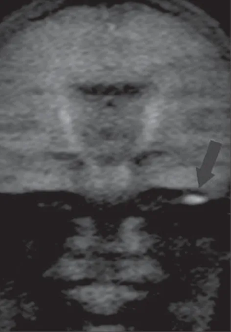
Figure 2C. MRI, coronal plane, HASTE diffusion-weighted (non echo-planar) sequence. Note diffu-sion restriction in congenital cholesteatoma at right (arrow).
assessment of cerebral ischemia. It is based on the demonstration of the movement of free water molecules and possible restric-tion to such a movement in pathological cases. Fitzek et al. were the first ones to demonstrate that cholesteatoma was visu-alized with high signal intensity at diffu-sion EPI sequences, more specifically at b1000 images. However, diffusion EPI pre-sents low spatial resolution, relatively thick sections and higher susceptibility to
arti-facts in regions of air-bone interface, rep-resenting disadvantages which many times hinder the diagnosis of cholesteatomas, particularly in cases of lesions measuring < 5 mm in diameter(8). Such a limitation can
be clearly observed on Figure 2D, where the artifact hinders the visualization of the focus of diffusion restriction, while at the image on Figure 2C acquired with non echo-planar sequence, the diffusion restric-tion was clearly demonstrated. Toyama et al.,
Figure 2B. MRI, coronal plane, spin echo, T1-weighted sequence with fat suppression, 45 min-utes intravenous paramagnetic contrast injection. Note the subtle peripheral contrast-enhancement, a typical finding of cholesteatoma (arrow). Figure 2A. CT, coronal plane. Congenital
choleste-atoma involving the region of the right geniculate ganglion (arrow).
Figure 1D. The same patient, one year later. MRI, coronal plane, spin echo T1-weighted sequence with fat suppression, 45 minutes after intravenous paramagnetic contrast injection. Note the intense contrast-enhancement in the whole lesion, corre-sponding to inflammatory granulomatous tissue (arrow).
in a study with 17 patients, have found 91.5% sensitivity, 60% specificity, 84.6% positive predictive value, and 75% negative predictive value, concluding that diffusion EPI combined with contrast-enhanced im-aging is useful in the differential diagno-sis of recurrent cholesteatoma and granu-lation tissue(9).
Jeunen et al. have found sensitivity, specificity, PPV and NPV of 54%, 90%, 92% and 47%, respectively in a study in-volving 31 patients and utilizing diffusion EPI. In the case of residual or recurrent cho-lesteatomas, sensitivity, specificity, PPV and NPV were 83%, 82%, 83% and 82%, respectively. In such study, residual cho-lesteatomas were correctly identified in 15 out of 18 patients, and small-sized lesions (2–5 mm) were missed in three patients(10).
Recent studies have demonstrated the value of non-EPI diffusion weighted imag-ing in the diagnosis of primary cholesteato-mas (Figures 2A to 2D)and also in post-operative recurrence. Single shot turbo spin echo (SSTSE) diffusion-weighted or multi-shot fast spin echo (FSE) imaging present lower susceptibility to artifacts and can be acquired with thinner sections, with higher spatial resolution (Figures 3A and 3B), al-lowing the detection of small-sized lesions, such as the one shown on Figure 4, with 3 mm in diameter. Dubrulle et al. have evalu-ated TSE diffusion-weighted imaging for detecting recurrent cholesteatomas, but the
Figure 3A. MRI, coronal plane, HASTE (non eplanar) sequence. Note diffusion restriction in cho-lesteatoma at right (arrow).
Figure 3B. ADC map of the same case confirming diffusion restriction (arrow).
Figure 4. MRI, axial plane, PROPELER diffusion-weighted (non echo-planar) sequence. Presence of a focus of diffusion restriction in a small (3 mm) cholesteatoma at left (arrow).
threshold for detection of small-sized cho-lesteatomas in this study was 5 mm, equal to the threshold for EPI sequences. Other studies indicate that SSTSE presents high sensitivity and specificity, detecting cho-lesteatomas with up to 2 mm in diameter(11).
De Foer et al. have found 90% sensitivity, 100% specificity, 100% PPV, and 96% NPV for non-EPI sequences in the study of patients with residual cholesteatoma. In this study, cholesteatomas measuring 2 to 6 mm in diameter were diagnosed(6).
Non-EPI diffusion-weighted imaging acquisition time is longer than for echo-pla-nar imaging and presents two significant limitations. Motion artifacts may blur hyperintense b1000 images, provoking sig-nal iso-sigsig-nal intensity and conditioning a false-negative result. Spontaneously evacu-ated cholesteatoma is also a possible cause o false-negative result, once the contents of desquamated and degraded keratin respon-sible for the diffusion restriction shall not be present in cases of automastoidectomy. In such cases, the cholesteatoma content is evacuated into the external auditory me-atus, and can displace the matrix far away from its original positioning in the middle ear and in the mastoid antrum. Thus, it is important to highlight that both diffusion weighted EPI and non-EPI imaging may fail in the detection of cholesteatoma due to the absence of keratin (responsible for the hyper signal intensity).
Figures 5A and 5B exemplify such method limitation, demonstrating a case of bilateral acquired cholesteatoma with automastoidectomy at left. In such an ex-ample, non-echo-planar HASTE imaging demonstrated diffusion restriction at right and absence of restriction at left, despite the matrix permanence after auto evacuation of the keratin content.
Images acquisition with MRI T1-weighted sequences with fat saturation, 30–45 minutes after gadolinium injection has been originally described by Williams and collaborators based on the fact that cholesteatoma is a non-enhancing avascu-lar tissue whereas inflammatory, granulo-matous and cicatricial tissues are poorly vascularized and do present slow contrast enhancement. Such T1-weighted sequence could demonstrate the exact location of the cholesteatoma in the middle ear and mas-toid process, as well as the surrounding in-flammatory reaction(2,4).
The combination of post-contrast MRI with non-EPI diffusion-weighted se-quences presents higher sensitivity and specificity than CT for detecting residual cholesteatomas, conditioning the reduction of the number of tympanic cavity revision surgeries. However, the utilization of non-EPI diffusion-weighted sequences without necessity of delayed contrast-enhanced imaging has been defended by some au-thors, since the combined utilization of such sequences adds little diagnostic sen-sitivity and specificity to the study. Also, diffusion-weighted imaging alone presents further advantages, namely, reduction of the total images acquisition time and costs reduction(13).
REFERENCES
1. Semaan MT, Megerian CA. The pathophysiology of cholesteatoma. Otolaryngol Clin North Am. 2006;39:1143–59.
2. Lemmerling M, De Foer B. Imaging of cholestea-tomatous and non-cholesteacholestea-tomatous middle ear
Figure 5A. CT, coronal plane. Bilateral acquired cholesteatoma with automastoidectomy at left (arrow). Figure 5B. MRI, coronal plane, HASTE diffusion-weighted (non echo-planar) sequence. Note diffu-sion restriction in cholesteatoma at right (arrow head). There is no diffusion restriction at left, due to the method limitation to demonstrate the re-sidual matrix in auto-evacuated cholesteatoma (arrow).
disease. In: Lemmerling M, Kollias S, editors. Radiology of the petrous bone. Berlin: Springer; 2004. p. 31–47.
3. De Foer B, Vercruysse JP, Bernaerts A, et al. De-tection of postoperative residual cholesteatoma with non-echo-planar diffusion-weighted mag-netic resonance imaging. Otol Neurotol. 2008;29: 513–7.
4. Aquino JEAP, Cruz Filho NA, Aquino JNP. Cho-lesteatoma surgery in children and adolescents. Analysis in 200 patients. Intl Arch Otorhinolaryn-gol. 2006;10:55–61.
5. Dornelles C, Costa SS, Meurer L, et al. Some con-siderations about acquired adult and pediatric cholesteatomas. Rev Bras Otorrinolaringol. 2005; 71:536–46.
6. De Foer B, Vercruysse JP, Bernaerts A, et al. The value of single-shot turbo spin-echo diffusion-weighted MR imaging in the detection of middle ear cholesteatoma. Neuroradiology. 2007;49:841– 8.
7. Lemmerling MM, De Foer B, Verbist BM, et al. Imaging of inflammatory and infectious diseases in the temporal bone. Neuroimaging Clin N Am. 2009;19:321–37.
8. Fitzek C, Mewes T, Fitzek S, et al. Diffusion-weighted MRI of cholesteatomas of the petrous bone. J Magn Reson Imaging. 2002;15:636–41. 9. Toyama C, Leite CC, Baraúna Filho IS, et al. The
role of magnetic resonance imaging in the post-operative management of cholesteatomas. Rev Bras Otorrinolaringol. 2008;74:693–6. 10. Jeunen G, Desloovere C, Hermans R, et al. The
value of magnetic resonance imaging in the di-agnosis of residual or recurrent acquired choleste-atoma after canal wall-up tympanoplasty. Otol Neurotol. 2008;29:16–8.
11. Dubrulle F, Souillard R, Chechin D, et al. Diffu-sion-weighted MR imaging sequence in the de-tection of postoperative recurrent cholesteatoma. Radiology. 2006;238:604–10.
12. Ayache D, Williams MT, Lejeune D, et al. Use-fulness of delayed postcontrast magnetic reso-nance imaging in the detection of residual cho-lesteatoma after canal wall-up tympanoplasty. Laryngoscope. 2005;115:607–10.