Estimates of DNA damage by the comet assay in the direct-developing frog
Eleutherodactylus johnstonei
(Anura, Eleutherodactylidae)
Laura Carolina Valencia
1, Adriana García
1, Martha Patricia Ramírez-Pinilla
2and Jorge Luis Fuentes
11
Laboratorio de Microbiología y Mutagénesis Ambiental, Escuela de Biología,
Universidad Industrial de Santander, Bucaramanga, Colombia.
2
Laboratorio de Biología Reproductiva de Vertebrados, Escuela de Biología,
Universidad Industrial de Santander, Bucaramanga, Colombia.
Abstract
The aim of this study was to use the Comet assay to assess genetic damage in the direct-developing frog Eleutherodactylus johnstonei. A DNA diffusion assay was used to evaluate the effectiveness of alkaline, enzymatic and alkaline/enzymatic treatments for lysingE. johnstonei blood cells and to determine the amount of DNA strand breakage associated with apoptosis and necrosis. Cell sensitivity to the mutagens bleomycin (BLM) and 4-nitro-quinoline-1-oxide (4NQO) was also assessed using the Comet assay, as was the assay reproducibility. Alkaline treatment did not lyse the cytoplasmic and nuclear membranes ofE. johnstonei blood cells, whereas enzymatic di-gestion with proteinase K (40mg/mL) yielded naked nuclei. The contribution of apoptosis and necrosis (assessed by the DNA diffusion assay) to DNA damage was estimated to range from 0% to 8%. BLM and 4NQO induced DNA damage inE. johnstonei blood cells at different concentrations and exposure times. Dose-effect curves with both mutagens were highly reproducible and showed consistently low coefficients of variation (CV£10%). The results are discussed with regard to the potential use of the modified Comet assay for assessing the exposure ofE. johnstonei to herbicides in ecotoxicological studies.
Key words:bleomycin, Comet assay, DNA diffusion assay,Eleutherodactylus johnstonei, 4-nitroquinoline-1-oxide.
Received: April 29, 2011; Accepted: July 13, 2011.
Introduction
Amphibians are useful biological indicators because their sensitive skins and occurrence in aquatic and terres-trial habitats makes them vulnerable to environmental change (Blaustein and Wake, 1990; Lips, 1998). Studies using frogs as models to measure the environmental impact of xenobiotics have used mainly larval phases of biphasic frog species such as Anaxyrus americanus, Hyla versicolor, Lithobates catesbeianus,Lithobates clamitans and Lithobates pipiens (Berrill et al., 1994; Relyea, 2004a,b). Most Neotropical frogs have direct development and reproduce on land (Hedgeset al., 2008). However, few studies have assessed the usefulness of these species as en-vironmental bioindicators.
Eleutherodactylus johnstonei(Anura: Eleutherodac-tylidae) is a direct-developing frog (Hedgeset al., 2008) from the Lesser Antilles with recently established popula-tions in several Caribbean islands (Kaiser, 1997), as well as in Central and South America (Ortegaet al., 2001; Kaiseret
al., 2002). As a consequence of its widespread distribution and certain life history features (reproductive flexibility and high environmental adaptability) (Ortegaet al., 2005), E. jonhstonei has been considered a successful invasive species (Rödder, 2009). These attributes also suggest that this species could be a useful model for evaluating the genotoxicological impact of environmental xenobiotics such as pesticides.
DNA damage by environmental xenobiotics is fre-quently assessed by single cell gel electrophoresis (SCGE) or the Comet assay (Singhet al., 1988), which detects DNA strand breakage and alkali-labile sites by measuring the mi-gration of DNA from immobilized individual cell nuclei. In this assay, the cells are embedded in agarose gel on micro-scopic slides, lysed and then electrophoresed under alkaline condition. Cells with damaged DNA show increased mi-gration of DNA fragments from the nucleus and the length of the migration indicates the amount of DNA strand break-age; the latter can be estimated by manual and computer-ized image scoring procedures (Kumaravel et al., 2009). The technique is highly sensitive for detecting DNA dam-age in any eukaryotic cell type and requires only a few cells. The minimal technical requirements for doing this
as-Send correspondence to Jorge Luis Fuentes. Laboratorio de Micro-biología y Mutagénesis Ambiental, Escuela de Biología, Univer-sidad Industrial de Santander, A.A. 678, Bucaramanga, Colombia. E-mail: [email protected]; [email protected].
sayin vitroandin vivoare well established (Cotelle and Férard, 1999; Ticeet al., 2000; Hartmannet al., 2003). The Comet assay is sufficiently sensitive for detecting DNA damage in frogs (Dhawanet al., 2009).
The main aim of this work was to assess the useful-ness of the Comet assay for detecting DNA damage inE. johnstonei. The efficiency of alkaline, enzymatic and alka-line/enzymatic treatments for lysing E. johnstonei blood cells was also assessed using a DNA diffusion assay. Since positive Comet results do not necessarily reflect genotoxi-city because DNA strand breakage may be associated with cellular apoptosis and necrosis, we used the DNA diffusion assay (Singh, 2000a) to determine the percentage of DNA strand breakage associated with apoptosis and necrosis (%NAp/N) and thereby estimate the proportion of DNA strand breakage that was unrelated to apoptosis and necro-sis. Cell sensitivity to the mutagens bleomycin (BLM) and 4-nitroquinoline-1-oxide (4NQO) was examined based on DNA strand breakage detected with the Comet assay (Bao-honget al., 2005; Kumaravel and Jha, 2006); the repro-ducibility of the assay in this species was also addressed.
Materials and Methods
Chemicals
Bleomycin (BML), dimethyl sulfoxide (DMSO), phosphotungstic acid, molecular grade and low gel temper-ature (LGT) agaroses, 4-nitroquinoline-1-oxide (4-NQO), silver nitrate and sodium N-lauryl sarcosine were obtained from Sigma-Aldrich Co. (St. Louis, MO, USA). RNase-and DNase-free proteinase K RNase-and PK buffer were obtained from Promega Co. (Madison, WI, USA). Glycerol was pur-chased from Merck (Darmstadt, Germany). The other re-agents and solvents were obtained from J.T. Baker (Phil-lipsburg, NJ, USA).
Capture and maintenance of frogs
Eleutherodactylus johnstonei specimens were cap-tured at several sites in the Bucaramanga metropolitan area (Santander, Colombia). Research and collecting permission was given by the Corporación Regional para la Defensa de la Meseta de Bucaramanga (File PC-0014-2008, Resolution 001368). Specimen sex was determined based on species sexual dimorphism and male calling. The frogs were main-tained in glass terrariums at 24±2 °C on a 12 h light/dark photoperiod, in conditions (vegetation, air circulation, hu-midity, illumination, etc.) that simulated the wild habitat. The frogs were fed flies, crickets, spiders, ants and mosqui-toes that were captured on the campus of the Universidad In-dustrial de Santander (Bucaramanga, Colombia).
Blood sampling, cell counts and exposure to mutagens
Blood obtained by cardiac puncture of cold-anes-thetized frogs was collected in heparinized Eppendorf tubes
and placed on ice until assayed. Blood cells were counted in a Neubauer counting chamber and then diluted in 0.9% (w/v) NaCl solution to a cell density of 3.55 x 10⁶cells/mL. Aliquots of blood cells were treated with BLM (0.6-152.0mg/mL) or 4NQO (1.9-60.0mM) for 2, 4, 6, 10 and 12 h (selected on the basis of preliminary experiments). Treatments were done at 6±2 °C to minimize basal DNA strand breakage. A negative control (0.9% NaCl solution) was always included in each assay. The experiments were done at least three times.
Estimation of DNA strand breakage inE. johnstonei blood cells
DNA strand breakage inE. johnstoneiblood cells was initially assayed by using the alkaline Comet assay, as de-scribed by Singhet al.(1988) but with silver staining. Sub-sequently, DNA strand breakage was detected by the Co-met assay as follows: E. johnstonei blood cells were centrifuged (10,000 rpm) and the pellet suspended in pro-teinase K solution (20mL) prepared in PK buffer (50 mM Tris-HCl, 10 mM CaCl2, pH 8) at 40mg/mL (concentration determined empirically). Aliquots (20mL) of the cell sus-pension were mixed with 75mL of 1% low melting point agarose and the mixture spread on slides containing a layer of 1.3% molecular grade agarose. The slides were covered with coverslips and incubated at 6±2 °C for the agarose to solidify. After enzymatic lysis and agarose polymerization, the coverslips were removed and the slides were placed in a Comet assay tank (Cleaver Scientific Ltd, UK) containing cold alkaline electrophoresis buffer (0.3 N NaOH, 1 mM EDTA, pH 13) for 25 min. Electrophoresis was done at 25 V and current adjusted to 300 mA. The slides were rou-tinely exposed to this current in the dark at 6±2 °C for 30 min. After electrophoresis, the slides were placed in a staining tray and covered with a neutralizing buffer (0.4 M Tris-HCl, pH 7.5) in the dark for 5 min. Silver staining was done as indicated by Díazet al.(2009).
DNA damage was expressed as arbitrary units based on the classification of comets into five categories (0-4) proposed by Collinset al.(1997). The total amount of DNA strand breakage was expressed in total arbitrary units (AUT) defined as: AUT= N0x 0 + N1x 1 + N2x 2 + N3x 3 + N4x 4, where Niis the number of nuclei scored in each
cate-gory (Collins, 2002). One hundred cells per slide and two slides per blood sample were analyzed using a Nikon Eclip-se E200 microscope and the results from at least three inde-pendent experiments were averaged to obtain the AUTfor each treatment.
apoptotic/ne-crotic nuclei in each field were counted (minimum of 100 fields per slide) and the latter then expressed as a percent-age of the former. As in the Comet assay, two slides per blood sample were analyzed and the results of at least three independent experiments were averaged to obtain the per-centage of apoptotic/necrotic nuclei (%NAp/N) for each ex-periment.
Based on the AUTand %NAp/Nestimates, the propor-tion of remaining DNA strand breakages was calculated (in arbitrary units) as:
AU AU %N AU
100
R T
AP / N T
= - ´
where AUR corresponds to non-apoptotic/necrotic DNA strand breakages.
Statistical analysis
In all cases, the data passed the Kolmogorov-Smirnov and F-maximum tests for normality and variance homoge-neity, respectively, so that parametric tests were used in subsequent data analyses. When a significant F-value was obtained in one-way analysis of variance (ANOVA) the groups were subsequently compared with Studentst-test. Product-moment (Pearson) correlation analysis was used to examine the relationship between mutagen doses and the average estimates of DNA damage. A value of p < 0.05 in-dicated significance. All statistical analyses were done with STATISTICA V.6 software (StatSoft Inc).
Results
Cellular lysis assays
In the present work, we used the Comet assay de-scribed by Singhet al.(1988) to detect DNA strand break-age inE. johnstoneiblood cells. These cells were not lysed by alkaline conditions and naked nuclei were not obtained. Although cells varied in their sensitivity to silver staining (Figure 1), neither the pH (between 10 and 13) of the incu-bation solution nor the length of incuincu-bation (2 h and 24 h) had any effect on the susceptibility to lysis (data not shown).
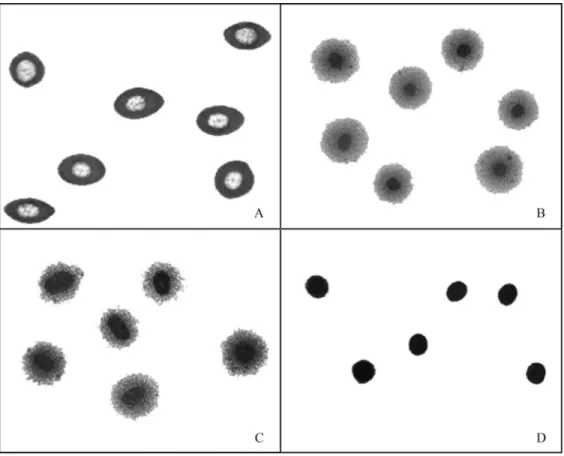
Since naked nuclei could not be obtained fromE. johnstoneiblood cells by the standard Comet procedure (Singhet al., 1988), we compared the ability of alkaline, enzymatic and alkaline/enzymatic treatments to produce these nuclei, as assessed by the DNA diffusion assay (Singh, 2000a). Treatments that included enzymatic lysis (Figure 2B,C) were effective in producing naked nuclei from blood cells, in contrast to lysis by alkaline treatment (Figure 2A). Combined alkaline/enzymatic treatment (Fig-ure 2B) was more aggressive to nuclear stability than enzy-matic treatment (Figure 2C), as shown by the nuclear diam-eter. The Comet assay showed that alkaline/enzymatic treatment produced DNA damage after a very short expo-sure to alkaline lysis (AU5 min = 295 ± 11,
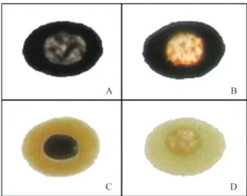
AU10 min= 323±7, AU15 min= 326±9, AU30 min= 330±11 and AU45 min= 361±5). In this assay, the negative control had a mean arbitrary unit value (AU0 min) of 38±5. In con-trast to the enzymatic treatment at 37 °C overnight (Figure 2C), the nuclei were still intact after treatment at 6±2 °C during agarose solidification (Figure 2D). Hence, subse-quent experiments involving cell lysis were done using only enzymatic treatment at 6±2 °C. Figure 3 shows im-ages of the Comet categories established forE. johnstonei blood cells.
Estimation of DNA strand breakage inE. johnstonei blood cells
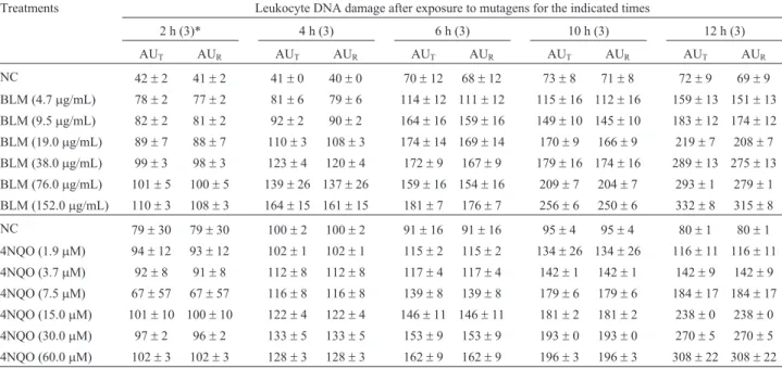
Table 1 shows the DNA strand breakage induced by BLM and 4NQO inE. johnstoneiblood cells at different doses and incubation times. A marked dose-response rela-tionship was observed for both doses and incubation times. Dose-response curves to BLM and 4NQO after a 12 h expo-sure (Table 2) showed marked correlations (R = 0.83 and 0.90, p £ 0.05; respectively). DNA strand breakage in-creased significantly (p£ 0.05) from a concentration of 4.7mg/mL of BLM and 1.9mM of 4NQO. The %NAp/NinE. johnstoneiranged from 0% to 8% (mean: 2.8%). The assay reproducibility under the conditions in this model was con-sistently high, with coefficients of variation£10%.
Discussion
In this work, we used a modified Comet assay to de-tect DNA strand breakage in the direct-developing frogE. johnstonei. Previous studies of DNA damage in frogs used alkaline treatment to lyse the cells prior to analysis by the Comet assay (Table 3). Alkaline conditions are generally sufficient to cause cellular lysis in all frog species.
Eleutherodactylus johnstoneiblood cells were resistant to tolerant to alkaline treatment in the standard procedure (Singh et al., 1988). This finding suggests that E. johnstonei contains alkali-resistant but proteinase K-sensitive proteins that stabilize and/or protect the nu-clei. The Comet assay with enzymatic (proteinase K) lysis has been used to assess DNA intactness in mammalian
sperm cells (Baumgartneret al., 2009), which have highly compact nuclear DNA (Ward and Coffey, 1991). Proteinase K digests proteins associated with nuclei and eliminates DNA-protein bonds generated by some xenobiotics, action that facilitates the electrophoretic mi-gration of damaged DNA (Merk et al., 2000; Singh, 2000b).
Figure 2- DNA diffusion assay images fromE. johnstoneiblood cells after: (A) alkaline lysis, (B) alkaline/enzymatic (40mg/mL proteinase K) lysis at 37 °C overnight, (C) enzymatic (40mg/mL proteinase K) lysis at 37 °C overnight, and (D) enzymatic (40mg/mL proteinase K) lysis at 6±2 °C during agarose solidification (10 min).
Table 1- Estimates of DNA damage inE. johnstoneiblood cells exposed to BLM and 4NQO for different times.
Treatments Leukocyte DNA damage after exposure to mutagens for the indicated times
2 h (3)* 4 h (3) 6 h (3) 10 h (3) 12 h (3)
AUT AUR AUT AUR AUT AUR AUT AUR AUT AUR
NC 42±2 41±2 41±0 40±0 70±12 68±12 73±8 71±8 72±9 69±9
BLM (4.7mg/mL) 78±2 77±2 81±6 79±6 114±12 111±12 115±16 112±16 159±13 151±13
BLM (9.5mg/mL) 82±2 81±2 92±2 90±2 164±16 159±16 149±10 145±10 183±12 174±12
BLM (19.0mg/mL) 89±7 88±7 110±3 108±3 174±14 169±14 170±9 166±9 219±7 208±7
BLM (38.0mg/mL) 99±3 98±3 123±4 120±4 172±9 167±9 179±16 174±16 289±13 275±13
BLM (76.0mg/mL) 101±5 100±5 139±26 137±26 159±16 154±16 209±7 204±7 293±1 279±1
BLM (152.0mg/mL) 110±3 108±3 164±15 161±15 181±7 176±7 256±6 250±6 332±8 315±8
NC 79±30 79±30 100±2 100±2 91±16 91±16 95±4 95±4 80±1 80±1
4NQO (1.9mM) 94±12 93±12 102±1 102±1 115±2 115±2 134±26 134±26 116±11 116±11
4NQO (3.7mM) 92±8 91±8 112±8 112±8 117±4 117±4 142±1 142±1 142±9 142±9
4NQO (7.5mM) 67±57 67±57 116±8 116±8 139±8 139±8 179±6 179±6 184±17 184±17
4NQO (15.0mM) 101±10 100±10 122±4 122±4 146±11 146±11 181±2 181±2 238±0 238±0
4NQO (30.0mM) 97±2 96±2 133±5 133±5 153±9 153±9 193±0 193±0 270±5 270±5
4NQO (60.0mM) 102±3 102±3 128±3 128±3 162±9 162±9 196±3 196±3 308±22 308±22
The values are the mean±SEM from at least three independent experiments with two replicate slides in each. *The total number of male frogs used per experiment with each mutagen. AU - arbitrary units, AUT- total DNA damage measured with the Comet assay, AUR- the remaining
non-apoptotic/ne-crotic DNA damage, BLM - bleomycin, 4NQO - 4-nitroquinoline-1-oxide and NC - negative control (0.9% NaCl solution). The %NAp/N(see Materials
and Methods) was the percentage of apoptotic/necrotic nuclei counted in 100 slide fields, and ranged from 0% to 8%.
Table 2- Dose-response relationships between BLM and 4NQO concentrations and the estimated DNA damage. An exposure time of 12 h was used in all experiments.
Treatment Leukocyte DNA damage after exposure to mutagen
Exp. 1 (3) Exp. 2 (3) Exp. 3 (3) Mean±SEM CV (%)
AUT AUR AUT AUR AUT AUR AUT AUR
NC 72±9 69±9 87±8 80±8 65±6 62±6 75±9 70±9 10
BLM (4.7mg/mL) 159±13 * 151±13 * 142±16 * 131±16 * 145±10 * 139±10 * 149±7 140±7 6
BLM (9.5mg/mL) 183±12 * 174±12 * 171±16 * 157±16 * 183±10 * 176±10 * 179±6 169±6 4
BLM (19.0mg/mL) 219±7 * 208±7 * 220±4 * 202±4 * 240±13 * 231±13 * 226±9 213±9 5
BLM (38.0mg/mL) 289±13 * 275±13 * 265±10 * 244±10 * 276±14 * 265±14 * 277±10 261±10 4
BLM (76.0mg/mL) 293±1 * 279±1 * 299±3 * 275±3 * 296±19 * 285±19 * 296±2 279±2 1
BLM (152.0mg/mL) 332±8 * 315±8 * 338±9 * 311±9 * 343±3 * 330±3 * 338±4 318±4 2
r = 0.83 (p£0.05)
NC 78±7 78±7 80±1 80±1 93±6 93±6 84±7 84±7 10
4NQO (1.9mM) 133±6 * 133±6 * 116±11 * 116±11 * 129±3 * 129±3 * 126±7 126±7 7
4NQO (3.7mM) 147±2 * 147±2 * 142±9 * 142±9 * 155±6 * 155±6 * 148±5 148±5 4
4NQO (7.5mM) 176±16 * 176±16 * 184±17 * 184±17 * 190±3 * 190±3 * 183±6 183±6 4
4NQO (15.0mM) 227±9 * 227±9 * 238±0 * 238±0 * 222±11 * 222±11 * 229±7 229±7 4
4NQO (30.0mM) 261±2 * 261±2 * 270±5 * 270±5 * 257±5 * 257±5 * 263±5 263±5 3
4NQO (60.0mM) 316±8 * 316±8 * 308±22 * 308±22 * 299±21 * 299±21 * 308±7 308±7 3
r = 0.90 (p£0.05)
The values are the mean±SEM from three independent experiments with two replicate slides in each. The total number of male frogs used per experiment with each mutagen is shown in parentheses. The average values from the three experiments are shown. AU - arbitrary units, AUT- total DNA damage
measured with the Comet assay, AUR the remaining nonapoptotic/necrotic DNA damage, BLM bleomycin, CV coefficient of variation (%), 4NQO
-4-nitroquinoline-1-oxide, NC - negative control (0.9% NaCl solution) and r - Pearson correlation coefficient. The %NAp/N(see Materials and Methods)
The intactness of sperm DNA is regularly analyzed with the Comet assay after alkaline and enzymatic treat-ments (Speit et al., 2009). However, alkaline/enzymatic treatment was particularly aggressive to nuclear stability in E. johnstoneiblood cells. For this reason, we used a neutral (pH 8) and single enzymatic digestion with proteinase Kin situin agarose gels; this procedure considerably reduced the assay costs and time. The temperature during cellular lysis is another critical variable that affects basal DNA damage in the Comet assay, as indicated in previous reports (Speitet al., 1999; Banáthet al., 2001). In E. johnstonei erythrocytes, enzymatic lysis at low temperature (6±2 °C) was ideal for obtaining naked nuclei with low levels of basal DNA damage.
The results described here show that the Comet assay can provide a good estimation of DNA damage in E. johnstonei. The assay was reproducible and sensitive enough to detect DNA strand breakage in E. johnstonei blood cells. The basal DNA damage estimated for the spe-cies agreed with previously reported values (Collinset al., 1997). In addition, the DNA damage observed here was poorly associated to apoptosis and necrosis, in contrast to
the situation in humans (Ticeet al., 2000), sea lions (El-Zeinet al., 2006) and dolphins (Díazet al., 2009).
This study has shown the usefulness of amphibians as bio-indicators. A simultaneous study (Meza-Joyaet al., in preparation) in our laboratory examined the toxic and geno-toxic effects of a glyphosate-based herbicide (Roundup®SL - Cosmoflux®411F) on E. johnstonei. The study again showed that the Comet assay was highly sensitive for de-tecting DNA damage induced by this herbicide. This find-ing suggests that the Comet assay is an accurate method for detecting DNA damage inE. johnstoneiafter exposure to environmental xenobiotics.
In conclusion, the alkaline Comet assay (Singhet al., 1988) was inappropriate for measuring DNA strand break-age inE. johnstonei. Alkaline lysis can be replaced by en-zymatic lysis (proteinase K), with good results. In contrast, combined alkaline/enzymatic treatment or long incubations (overnight) at 37 °C with proteinase K generate unstable nuclei and result in consistently elevated basal DNA dam-age. The contribution of apoptosis and necrosis to the over-all DNA damage in E. johnstonei was negligible, as assessed by the Comet assay. The Comet assay is a
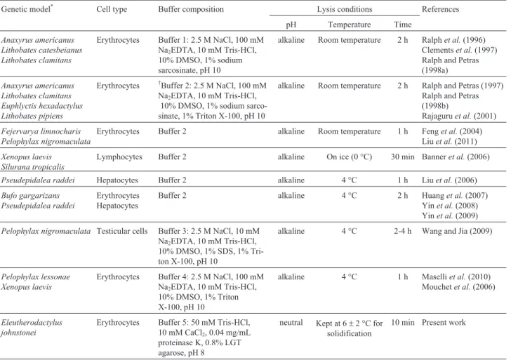
repro-Table 3- Cellular lysis conditions used in different studies to detect DNA damage by the Comet assay in frogs.
Genetic model* Cell type Buffer composition Lysis conditions References
pH Temperature Time
Anaxyrus americanus Lithobates catesbeianus Lithobates clamitans
Erythrocytes Buffer 1: 2.5 M NaCl, 100 mM Na2EDTA, 10 mM Tris-HCl,
10% DMSO, 1% sodium sarcosinate, pH 10
alkaline Room temperature 2 h Ralphet al.(1996) Clementset al.(1997) Ralph and Petras (1998a)
Anaxyrus americanus Lithobates clamitans Euphlyctis hexadactylus Lithobates pipiens
Erythrocytes †Buffer 2: 2.5 M NaCl, 100 mM
Na2EDTA, 10 mM Tris-HCl,
10% DMSO, 1% sodium sarco-sinate, 1% Triton X-100, pH 10
alkaline Room temperature 2 h Ralph and Petras (1997) Ralph and Petras (1998b)
Rajaguruet al.(2001)
Fejervarya limnocharis Pelophylax nigromaculata
Erythrocytes Buffer 2 alkaline Room temperature 1 h Fenget al.(2004) Liuet al.(2011)
Xenopus laevis Silurana tropicalis
Lymphocytes Buffer 2 alkaline On ice (0 °C) 30 min Banneret al.(2006)
Pseudepidalea raddei Hepatocytes Buffer 2 alkaline 4 °C 1 h Liuet al.(2006)
Bufo gargarizans Pseudepidalea raddei
Erythrocytes Hepatocytes
Buffer 2 alkaline 4 °C 2 h Huanget al.(2007) Yinet al.(2008) Yinet al.(2009)
Pelophylax nigromaculata Testicular cells Buffer 3: 2.5 M NaCl, 10 mM Na2EDTA, 10 mM Tris-HCl,
10% DMSO, 1% SDS, 1% Tri-ton X-100, pH 10
alkaline 4 °C 2-4 h Wang and Jia (2009)
Pelophylax lessonae Xenopus laevis
Erythrocytes Buffer 4: 2.5 M NaCl, 100 mM Na2EDTA, 10 mM Tris-HCl,
10% DMSO, 1% Triton X-100, pH 10
alkaline 4 °C 1 h Maselliet al.(2010) Mouchetet al.(2006)
Eleutherodactylus johnstonei
Erythrocytes Buffer 5: 50 mM Tris-HCl, 10 mM CaCl2, 0.04 mg/mL
proteinase K, 0.8% LGT agarose, pH 8
neutral Kept at 6±2 °C for solidification
10 min Present work
ducible, sensitive method for detecting DNA strand break-age inE. johnstonei.
Acknowledgments
The Corporación Regional para la Defensa de la Me-seta de Bucaramanga (CDMB) provided the research and collecting permits. This work was supported by the Vicer-rectoría de Investigaciones y Extensión, Universidad In-dustrial de Santander, Colombia (Grant No. 5163).
References
Banáth JP, Kim A and Olive PL (2001) Overnight lysis improves the efficiency of detection of DNA damage in the alkaline Comet assay. Radiat Res 155:564-571.
Banner SH, Ruben LN and Johnson RO (2006) Bleomycin-in-duced DNA damage and repair in Xenopus laevis and Xenopus tropicalis.J Exp Zool 307A:84-90.
Baohong W, Jiliang H, Lifen J, Deqiang L, Wei Z, Jianlin L and Hongpin D (2005) Studying the synergistic damage effects induced by 1.8 GHz radiofrequency field radiation (RFR) with four chemical mutagens on lymphocyte DNA using comet assayin vitro. Mutat Res 578:149-157.
Baumgartner A, Cemeli E and Anderson D (2009) The Comet as-say in male reproductive toxicology. Cell Biol Toxicol 25:81-98.
Berrill M, Bertram S, McGillivray L, Kolohon M and Paull B (1994) Effects of low concentrations of forest-use pesticides on frog embryos and tadpole. Environ Toxicol Chem 13:657-664.
Blaustein AR and Wake DB (1990) Declining amphibian popula-tions: a global phenomenon. Trends Ecol Evol 5:203-204. Clements C, Ralph S and Petras M (1997) Genotoxicity of select
herbicides inRana catesbeianatadpoles using the alkaline single-cell gel DNA electrophoresis (Comet) assay. Environ Mol Mutagen 29:277-288.
Collins AR (2002) The comet assay, principles, applications and limitations. In: Didenko VV (ed.)In SituDetection of DNA Damage. Methods and Protocols, v. 203. Humana Press Inc., Totowa, pp 163-177.
Collins AR, Dusinska A, Franklin M, Somorovska M, Petrovska H, Duthie S, Fillion L, Panayoitidis M, Raslova K and Vaughan N (1997) Comet assay in human biomonitoring studies: reliability, validation and applications. Environ Mol Mutagen 30:139-146.
Cotelle S and Férard JF (1999) Comet assay in genetic ecotoxi-cology: a review. Environ Mol Mutagen 34:246-255. Dhawan A, Bajpayee M and Parmar D (2009) Comet assay: a
reli-able tool for the assessment of DNA damage in different models. Cell Biol Toxicol 25:5-32.
Díaz A, Carro S, Santiago L, Estévez J, Guevara C, Blanco M, Sánchez L, Sánchez L, López N, Cruz D,et al.(2009) Esti-mates of DNA strand breakage in bottlenose dolphin (Tursiops truncatus) leukocytes measured with the Comet and DNA diffusion assays. Genet Mol Biol 32:367-372. El-Zein RA, Hastings-Smith DA, Ammenheuser MM,
Trinen-Moslen M, Gulland FM and Ward JB (2006) Evaluation of two different biomarkers for use in the assessment of toxic chemical exposure in California sea lions (Zalophus californianus). Marine Pollut Bull 52:104-120.
Feng S, Kong Z, Wang X, Zhao L and Peng P (2004) Acute toxic-ity and genotoxictoxic-ity of two novel pesticides on amphibian, Rana n. hallowell. Chemosphere 56:457-463.
Hartmann A, Agurell E, Beevers C, Brendler-Schwaab S, Burlin-son B, Clay P, Collins A, Smith, A, Speit G, Thybaud V,et al.(2003) Recommendations for conducting thein vivo al-kaline Comet assay. Mutagenesis 18:45-51.
Hedges SB, Duellman WE and Heinicke MP (2008) New World direct-developing frogs (Anura, Terrarana): Molecular phy-logeny, classification, biogeography, and conservation. Zo-otaxa 1737:1-182.
Huang D, Zhang Y, Wang Y, Xie Z and Ji W (2007) Assessment of the genotoxicity in toadBufo raddeiexposed to petro-chemical contaminants in Lanzhou region, China. Mutat Res 629:81-88.
Kaiser H (1997) Origins and introductions of the Caribbean frog, Eleutherodactylus johnstonei (Leptodactylidae): Manage-ment and conservation concerns. Biodiv Conserv 6:1391-1407.
Kaiser H, Barrio-Amorós CL, Trujillo JD and Lynch JD (2002) Expansion of Eleutherodactylus johnstonei in northern South America: Rapid dispersal through human interac-tions. Herpetol Rev 33:290-294.
Kumaravel TS and Jha AN (2006) Reliable comet assay measure-ments for detecting DNA damage induced by ionizing radia-tion and chemicals. Mutat Res 605:7-16.
Kumaravel TS, Vilhar B, Faux SP and Jha AN (2009) Comet as-say measurements: A perspective. Cell Biol Toxicol 25:53-64.
Lips KR (1998) Decline of a tropical montane amphibian fauna. Conserv Biol 12:106-112.
Liu WY, Wang CY, Wang TS, Feller GM, Lai BC and Kam YC (2011) Impacts of the herbicide butachlor on the larvae of a paddy field breeding frog (Fejervarya limnocharis) in sub-tropical Taiwan. Ecotoxicology 20:377-384.
Liu Y, Zhang Y and Huang D (2006) The role of reactive oxygen species in the herbicide acetochlor-induced DNA damage on Bufo raddeitadpole liver. Aquat Toxicol 78:21-26. Maselli V, Polese G, Rippa D, Ligrone R, Rastogi RK and
Ful-gione D (2010) Frogs, sentinels of DNA damage induced by pollution in Naples and the neighbouring provinces. Eco-toxicol Environ Saf 73:1525-1529.
Merk O, Reiser K and Speit G (2000) Analysis of chromate-induced DNA-protein crosslinks with the comet assay. Mu-tat Res 471:71-80.
Mouchet F, Gauthier L, Mailhes C, Jourdain MJ, Ferrier V, Triffault G and Devaux A (2006) Biomonitoring of the genotoxic potential of aqueous extracts of soils and bottom ash resulting from municipal solid waste incineration, using the Comet and micronucleus tests on amphibian (Xenopus laevis) larvae and bacterial assays (Mutatox and Ames tests). Sci Total Environ 355:232-246.
Nigro M, Frenzilli G, Scarcelli V, Gorbi S and Regoli F (2002) In-duction of DNA strands breakage and apoptosis in the eel Anguilla anguilla. Marine Environ Res 54:517-520. Ortega JE, Jerez A and Ramírez-Pinilla MP (2001)
Eleutherodactylus johnstonei.Herpetol Rev 32:269. Ortega JE, Serrano VH and Ramírez-Pinilla MP (2005)
Rajaguru P, Kalpana R, Hema A, Suba S, Baskarasethupathi B, Kumar PA and Kalaiselvi K (2001) Genotoxicity of some sulfur dyes on tadpoles (Rana hexadactyla) measured using the Comet assay. Environ Mol Mutag 38:316-322. Ralph S and Petras M (1997) Genotoxicity monitoring of small
bodies of water using two species of tadpoles and the alka-line single cell gel (Comet) assay. Environ Mol Mutag 29:418-430.
Ralph S and Petras M (1998a) Comparison of sensitivity to methyl methanesulphonate among tadpole developmental stages using the alkaline single cell gel DNA electrophoresis (Comet) assay. Environ Mol Mutag 31:374-382.
Ralph S and Petras M (1998b) Caged amphibian tadpoles and in situ genotoxicity monitoring of aquatic environments with the alkaline single cell gel electrophoresis (Comet) assay. Mutat Res 413:235-250.
Ralph S, Petras M, Pandrangi R and Vrzoc M (1996) Alkaline sin-gle-cell gel (Comet) assay and genotoxicity monitoring us-ing two species of tadpoles. Environ Mol Mutag 28:112-120.
Relyea RA (2004a) Synergistic impacts of Malathion and preda-tory stress on six species of North American tadpoles. Envi-ron Toxicol Chem 23:1080-1084.
Relyea RA (2004b) Grown and survival of five amphibian species exposed to combinations of pesticides. Environ Toxicol Chem 23:1735-1742.
Rödder D (2009) Human footprint, facilitated jump dispersal, and the potential distribution of the invasiveEleutherodactylus johnstonei Barbour 1914 (Anura, Eleutherodactylidae). Trop Zool 22:205-217.
Singh NP (2000a) A simple method for accurate estimation of apoptotic cells. Exp Cell Res 256:328-337.
Singh NP (2000b) Microgels for estimation of DNA strand breaks, DNA protein crosslinks and apoptosis. Mutat Res 455:111-127.
Singh NP, McCoy MT, Tice RR and Schneider EL (1988) A sim-ple technique for quantitation of low levels of DNA damage in individual cells. Exp Cell Res 175:184-191.
Speit G, Trenz K, Schütz P, Rothfuss A and Merk O (1999) The influence of temperature during alkaline treatment and elec-trophoresis on results obtained with the Comet assay. To-xicol Lett 110:73-78.
Speit G, Vasquez M and Hartman A (2009) The Comet assay as an indicator test for germ cell genotoxicity. Mutat Res 681:3-12.
Tice RR, Agurell E, Anderson D, Burlinson B, Hartmann A, Kobayashi H, Miyamae Y, Rojas E, Ryu JC and Sasaki YF (2000) Single cell gel/Comet assay: guidelines forin vitro
and in vivo genetic toxicology testing. Environ Mol
Mutagen 35:206-221.
Wang MZ and Jia XY (2009) Low levels of lead exposure induce oxidative damage and DNA damage in the testes of the frog Rana nigromaculata. Ecotoxicology 18:94-99.
Ward WS and Coffey DS (1991) DNA packaging and organiza-tion in mammalian spermatozoa: Comparison with somatic cells. Biol Reprod 44:569-574.
Yin XH, Li SN, Zhang L, Zhu GN and Zhuang HS (2008) Evalua-tion of DNA damage in Chinese toad (Bufo bufo gargarizans) afterin vivoexposure to sublethal concentra-tion of four herbicides using the comet assay. Ecotoxicology 17:280-286.
Yin XH, Zhu GN, Li XB and Liu SY (2009) Genotoxicity evalua-tion of chlopyrifos to amphibian Chinese toad (Amphibian, Anura) by Comet assay and micronucleus test. Mutat Res 680:2-6.
Internet Resources
Frost DR (2011) Amphibian Species of the World: An Online Reference, v. 5.5. Electronic Database accessible at http://research.amnh.org/vz/herpetology/amphibia/. Ameri-can Museum of Natural History, New York, USA (Accessed on February 24, 2011).
Associate Editor: Daisy Maria F. Salvadori