The role of complement in the
modulation by fluid-phase IgG of the
production of reactive oxygen species
by polymorphonuclear leukocytes
stimulated with IgG immune complexes
Departamento de Bioquímica e Imunologia, Faculdade de Medicina de Ribeirão Preto,
Universidade de São Paulo, Ribeirão Preto, SP, Brasil S. Chedraoui-Silva
and B. Mantovani
Abstract
The production of reactive oxygen species (ROS) by polymorpho-nuclear leukocytes (PMN) can be induced by immune complexes and is an important component of phagocytosis in the killing of microor-ganisms, but can also be involved in inflammatory reactions when immune complexes are deposited in tissues. We have observed that fluid-phase IgG can inhibit the generation of ROS by rabbit PMN stimulated with precipitated immune complexes of IgG (ICIgG) in a dose-dependent manner, acting as a modulatory factor in the range of physiological IgG concentrations. This inhibitory effect is compatible with the known affinity (Kd) of monomeric IgG for the receptors involved (FcRII and FcRIII). The presence of complement compo-nents in the immune complexes results in a higher stimulation of ROS production. In this case, however, there is no inhibition by fluid-phase IgG. The effect of complement is strongly dependent on the presence of divalent cations (Ca2+ or Mg2+) in the medium, whereas the stimu-lation of ICIgG (without complement) does not depend on these cations. We have obtained some evidence indicating that iC3b should be the component involved in the effect of complement through interaction with the CR3 receptor. The absence of the inhibitory effect of fluid-phase IgG in ROS production when complement is present in the immune complex shows that complement may be important in vivo
not only in the production of chemotactic factors for PMN, but also in the next phase of the process, i.e., the generation of ROS.
Correspondence B. Mantovani
Departamento de Bioquímica e Imunologia, FMRP, USP 14049-900 Ribeirão Preto, SP Brasil
Fax: +55-16-633-6840 E-mail: [email protected]
Research supported by FAPESP and FAEPA.
Received December 13, 2002 Accepted July 31, 2003
Key words
•Polymorphonuclear
leukocytes
•Reactive oxygen species •Immune complex •IgG
•Complement
Introduction
It is well known that polymorphonuclear leukocytes (PMN) can undergo a rapid meta-bolic change, known as the respiratory burst, when exposed to a variety of stimuli (1); this includes an increased rate of oxygen
con-sumption, a marked activation of the pen-tose-phosphate pathway and the generation of reactive oxygen species (ROS; O2-, H2O2,
im-plicated in the mechanisms of defense. How-ever, in some instances it may be involved in inflammatory reactions, since these com-pounds may be released from the cells and contribute to tissue damage (2).
It is well established that the interaction of particles or surfaces with leukocytes me-diated by Fcγ receptors (immune complexes
of IgG - ICIgG) can trigger the respiratory burst. However, divergent findings have been reported as to the ability of complement (C3b and iC3b) to induce the generation of ROS upon interaction with the cells. Some experiments have shown that C3b or iC3b is not able to induce the generation of ROS by human monocytes and PMN (3); other ob-servations, however, have indicated an ac-tive role of these complement components in triggering the respiratory burst (4,5). Also, some evidence has been presented indicat-ing that the production of ROS by PMN is dependent on a synergic mechanism involv-ing Fcγ and complement receptors (6).
Regardless of the importance of deter-mining which kind of receptors are able to trigger this mechanism by themselves, an-other relevant issue is to know the behavior of these cells in the production of these compounds under physiological conditions, when the phagocytes interact with immune complexes which contain IgG or IgG and complement components, since in vivo there is a high concentration of fluid-phase IgG which possibly could compete for the Fcγ
receptors. We analyze this question by in vitro experiments with rabbit PMN using as a stimulus two types of immune complexes in the form of precipitates: ICIgG and oval-bumin and the same immune complexes with complement components incorporated by previous incubation with whole serum (ICIgG-C).
Material and Methods
The preparation of the antigen (chicken ovalbumin), the purification of rabbit IgG
anti-ovalbumin antibodies, the preparation of ICIgG with ovalbumin in the equivalence zone, as well as the isolation of rabbit blood PMN were performed as described (7).
Media and solutions
Phosphate-buffered saline (PBS) contain-ing 0.9% NaCl and 8 mM sodium phosphate buffer, pH 7.2, was used. Hanks’ medium was prepared as described (8). Luminol (Sigma, St. Louis, MO, USA) was dissolved in dimethylsulfoxide (DMSO) at a concen-tration of 2 mM; for the experiments this stock solution was diluted with Hanks’ me-dium (the final concentration of DMSO was 0.45%, v/v).
Animals
New Zealand rabbits were used as blood donors for the isolation of PMN as well as of normal or immune serum.
Preparation of antigen-antibody-complement complexes
increased by 44%. To test for the comple-ment-fixing capacity of the ICIgG, the re-sidual CH50 (50% hemolytic unit of
comple-ment) was determined (10) in the superna-tant after incubation of the immune com-plexes with serum. It was found that 182 µg of ICIgG consumed 29.8 CH50 units.
Treatment of rabbit serum to inactivate some complement components
Rabbit serum used as a source of comple-ment was subjected to three types of treat-ment in order to selectively inactivate comple-ment components: a) heat inactivation at 56ºC for 30 min to block the activity of C1, C2 and factor B; b) depletion of C3 by treatment of serum with zymosan using 15 mg of zymosan per ml of serum at 37ºC for 1 h (10); c) inactivation of C3 and C4 by treatment of serum with 50 mM hydrazine, incubated with serum at 37ºC for 2 h, fol-lowed by dialysis with PBS containing Ca2+
and Mg2+ at the concentrations of Hanks’
medium (11).
Measurement of the production of ROS by chemiluminescence
The production of ROS by PMN was assayed by the generation of chemilumines-cence in the presence of luminol (12). This is an indirect measurement of the amount of superoxide produced: the stimulation of the cells leads to the activation of NADPH oxi-dase, resulting in the formation of O2
which, upon the action of superoxide dismutase (SOD), undergoes a dismutation to H2O2
which, in the presence of Cl- and myeloper-oxidase, forms OCl-. Some of these com-pounds react with luminol bringing the mol-ecule to an excited state whose decay results in light emission (13). The kinetics of chemi-luminescence was recorded with a BioOrbit luminometer, model 1251 (Bio-Orbit Oy, Turku, Finland). PMN suspensions (106 cells)
in Hanks’ medium containing 15 mM
HEPES, pH 7.2, 0.1% gelatin and 10 µM luminol were added to the cuvettes and pre-incubated for 15 min at 37ºC. The cuvettes were then inserted into the luminometer, and the stimulus (immune complex) was added to start the experiment; the final volume was 1.1 ml. The cuvettes were maintained at 37ºC and the rate of photon emission (re-ported in mV) was recorded every 51 s. The first measurement was made after 25 s.
Competition experiments with fluid-phase IgG
PMN suspensions were preincubated with fluid-phase IgG at 37ºC for 15 min in Hanks’ medium with the desired concentrations of IgG before stimulation with the immune com-plexes, also in the presence of IgG. The IgG solution used (prepared by DEAE cellulose chromatography) was tested for the presence of aggregated molecules by polyacrylamide gel electrophoresis using 3.5% acrylamide gels with 0.5% agarose as described previ-ously (14), and no aggregation was found. In addition we demonstrated that centrifuga-tion of IgG solucentrifuga-tion at 100,000 g for 30 min just before these experiments had no
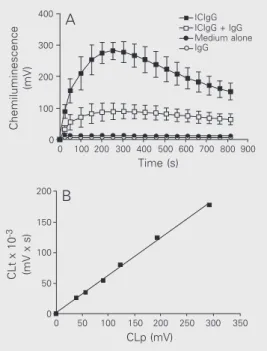
appre-Figure 1. Kinetics of chemilumi-nescence, a measure of the rate of reactive oxygen species pro-duction, by rabbit polymorpho-nuclear leukocytes stimulated with IgG immune complexes (ICIgG) in the absence and in the presence of fluid-phase IgG.
A, Cells (106) were preincubated
in Hanks’ medium alone or con-taining 10 mg/ml IgG for 15 min at 37ºC and then stimulated with 300 µg ICIgG. Chemilumines-cence was recorded in a final volume of 1.1 ml. Two controls are shown: only cells (medium alone) and cells plus 10 mg/ml IgG (IgG). Data are reported as means ± SD for N = 4 (cells from different animals). B, Cor-relation between chemilumines-cence in the peak (CLp) and to-tal chemiluminescence (CLt) for 13.6 min (calculated by integra-tion of the peak). The six points correspond to six different con-centrations (9.4 to 300 µg/ml) of ICIgG. Each point corresponds to the mean value of 4 inde-pendent experiments (different animals).
Chemiluminescence
(mV)
400 300 200 100 0
100 200 300 400 500 600 700 800 900 ICIgG ICIgG + IgG IgG Medium alone
Time (s)
0
A
CLt x 10
-3
(mV x s)
100 150 200
50 0
100
0 50 150 200 250 300 350
ciable effect on the inhibitory action of fluid-phase IgG on ROS production by PMN stim-ulated with ICIgG. In view of this result, this previous centrifugation was performed in some, but not all, experiments.
Results
The kinetics of chemiluminescence pro-duction by PMN induced by ICIgG is pre-sented in Figure 1A. The presence of fluid-phase IgG at a concentration representative of that existing in plasma (10 mg/ml) greatly reduced the stimulatory effect of the im-mune complex. The chemiluminescence with PMN alone as well as with PMN plus 10 mg/ ml fluid-phase IgG was very low and negli-gible compared with that induced by the immune complex. Figure 1B shows the cor-relation between the values of chemilumi-nescence in the peak and the total chemilu-minescence after 13.6 min (calculated by integration over this period of time) using six
different concentrations of ICIgG (from 9.4 to 300 µg/ml). Since there was a close corre-lation between the two measurements, we used only the chemiluminescence in the peak, that corresponds to the maximum rate of ROS production, for quantification in the subsequent experiments.
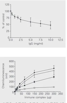
Figure 2 shows the inhibitory effect of increasing concentrations of fluid-phase IgG on the stimulation of ROS production by PMN induced by ICIgG (300 µg/ml). The inhibition was progressive with increasing IgG concentration and started to be demon-strable at around 0.9 mg/ml IgG; with 10 mg/ ml IgG the effect reported as percent of control (without fluid-phase IgG) was in the range of 34 to 64% in four independent experiments (47 ± 14%, mean ± SD).
In another set of experiments we ana-lyzed the possible effect of complement in-corporated into the immune complex of IgG on its stimulatory effect on ROS production, as well as the susceptibility to inhibition by the soluble immunoglobulin, using several immune complex concentrations (Figure 3). In the range of immune complex concentra-tions from 9.4 to 300 µg/ml, the presence of IgG greatly reduced ROS production by ICIgG; however, when complement was pres-ent in the immune complex, no inhibition was observed. The stimulatory effect of ICIgG-C was higher than the effect of ICIgG, albeit it was not affected by the presence of soluble IgG (the two curves were practically coincident in the entire range of immune complex concentrations).
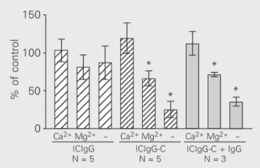
The experiments in Figure 4 show the influence of extracellular divalent cations (Ca2+ and Mg2+) on ROS production induced
by ICIgG and ICIgG-C. For each type of immune complex the control was the value of chemiluminescence in the peak obtained in complete Hanks’ medium (1.26 mM Ca2+
and 0.9 mM Mg2+).
Stimulation with ICIgG was practically independent of the presence of these cations in the medium. The effect of ICIgG-C,
how-Figure 2. Inhibition of reactive oxygen species production by in-creasing concentrations of fluid-phase IgG. Cells (106) were
pre-incubated with fluid-phase IgG for 15 min at 37ºC and then stim-ulated with 300 µg ICIgG. The control cells were preincubated in the medium without IgG. Re-sults are reported as percent chemiluminescence in the peak in relation to control values (mean ± SD, N = 4 with cells from different animals).
Figure 3. Effect of complement components incorporated into immune complexes of IgG (ICIgG-C) on the stimulation of reactive oxygen species produc-tion by polymorphonuclear leu-kocytes and the influence of the presence of fluid-phase IgG. Cells (106) were preincubated in
Hanks’ medium with or without 10 mg/ml fluid-phase IgG for 15 min at 37ºC before stimulation with different amounts of im-mune complexes. For ICIgG-C the mass of immune complexes indicated on the abscissa corre-sponds to its mass before the addition of complement. ICIgG-C was prepared using 182 µg ICIgG per ml of serum. Data are reported as means ± SEM for N = 4 (different animals).
% of control
125
100
75
50
25
0
0.0 2.5 5.0 7.5 10.0 12.5
IgG (mg/ml)
Chemiluminescence
(mV)
600
350 500
400
300
200
100
00 50 100 150 200 250 300
Immune complex (µg)
ever, was strongly dependent on them, with Ca2+ alone being sufficient for the full effect,
whereas Mg2+ produced a lower stimulation
(around 60% of control). Moreover, when fluid-phase IgG was present (10 mg/ml) the same dependence on Ca2+ and Mg2+ was
obtained for the effect of ICIgG-C.
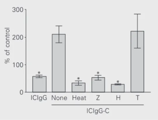
In order to have some indication of which components of complement incorporated into the immune complex were responsible for the effect of ICIgG-C, we prepared this im-mune complex using as a source of comple-ment rabbit serum previously treated by three different procedures: heat inactivation at 56ºC for 30 min blocking the activity of C1, C2 and factor B, depletion of C3 by treatment with zymosan, and inactivation of C3 and C4 by treatment with hydrazine. The experi-ments in Figure 5 were done in the presence of 10 mg/ml fluid-phase IgG.
All three treatments abolished the effect of complement (in these experiments the control was the effect of ICIgG without com-petition with soluble IgG). These results in-dicate that iC3b must be involved in the effect of complement incorporated into im-mune complexes.
Discussion
Fcγ receptors, which recognize the Fc
domain of IgG, are involved in a number of cellular functions such as phagocytosis, su-peroxide production and cytokine release. Three types of Fcγ receptors have been de-scribed in human and mouse PMN: FcRI, FcRII and FcRIII (15). FcRI is a high affinity receptor for monomeric IgG with a dissocia-tion constant ranging from 10 to 0.1 nM, and is expressed in INF-γ-treated neutrophils.
FcRII and FcRIII are normally present in non-activated PMN and have a lower af-finity for IgG, with a dissociation constant ranging from 10 to 0.1 µM (16-18). The same types of receptors may be present in rabbit PMN. Thus, in our experiments the low affinity receptors FcRII and FcRIII might
mediate the interaction between ICIgG and the cells. In competition experiments using 10 mg/ml fluid-phase IgG we had an IgG concentration of 67 µM, a value in the range of the dissociation constant for FcRII and FcRIII (10 to 0.1 µM). It is thus reasonable to assume that fluid-phase IgG could compete with the immune complexes for the binding to the receptors, resulting in partial inhibi-tion of the stimulainhibi-tion of ROS producinhibi-tion, as observed here.
We have shown that the presence of complement components in the immune com-plexes of IgG appreciably increases the ca-pacity of ROS production by PMN. Also, when complement is present in the immune complexes, fluid-phase IgG has no effect on the induction of ROS production. Comple-ment receptors would not be affected by the occupation of Fcγ receptors by fluid-phase
IgG, so that the ICIgG-C can by-pass the competitive inhibition that is observed with ICIgG. In this case, the interaction mediated by complement receptors may increase the binding of immune complexes to the cells or may by itself also stimulate ROS generation. One possibility we should consider is that the immune complex could be partially solubilized upon the addition of serum for the preparation of ICIgG-C, as previously reported (19,20). In our experiments this was improbable since the relation between serum volume and the ICIgG mass used by us was much smaller than that needed to solubilize the immune complexes. Compared to the volume used by the cited investigators with IgG antibody and BSA, we used a
se-Figure 4. Effect of extracellular Ca2+ and Mg2+ on reactive
oxy-gen species production induced by ICIgG and ICIgG-C. Polymor-phonuclear leukocytes (106
cells) were suspended in Hanks’ medium without Ca2+ and Mg2+
(-), or in medium containing ei-ther one of these cations at the concentrations used in Hanks’ medium (1.26 mM Ca2+ and 0.9
mM Mg2+). The
chemilumines-cence in the peak with 300 µg ICIgG was measured and the re-sults are indicated as % of con-trol; for each of the three groups the control value was that ob-tained with complete Hanks’ medium. ICIgG-C + IgG indi-cates the experiments in the presence of 10 mg/ml fluid-phase IgG. The values obtained with only Ca2+ in the medium
were not statistically different from the corresponding controls (with Ca2+ and Mg2+). Data are
reported as means ± SEM. *P < 0.02 for comparison of the con-ditions with Mg2+ or (-) with the
corresponding values with Ca2+
in each of the three groups (t -test).
% of control
150 12 12 12 12 12 12 12 12 12 12 12 12 12 12 12 12 12 12 12 12 12 12 12 12 12 12 12 12 12 12 123 123 123 123 123 123 123 123 123 123 123 123 123 123 123 123 123 123 123 1234 1234 1234 1234 1234 1234 1234 1234 1234 1234 1234 123 123 123 123 * * * * 100 50 0
N = 5 Ca2+Mg2+
-ICIgG
N = 3 Ca2+Mg2+
-ICIgG-C + IgG N = 5
rum volume about 18 to 20 times smaller than that required for solubilization. But, even in the case of a small amount of im-mune precipitate being solubilized (despite the increase in mass of the immune complex by incorporation of complement compo-nents), this does not interfere with the inter-pretation of the results since ICIgG-C in-duces a higher ROS production than ICIgG (obviously it would interfere if the reverse were obtained).
It is known that, upon activation of the complement system by immune complexes, several activated proteins (derived from C1, C4, C2, and C3) become tightly bound to the complexes (9,21,22). PMN have been shown to have receptors for some of these compo-nents which may be involved in various biological functions (23). Thus, interaction through C3b/iC3b can promote the binding of immune complexes; it was shown, how-ever, that the interaction through C3b and iC3b cannot induce phagocytosis (24). C1q bound to latex beads stimulates the respira-tory burst and the hexose phosphate pathway in these cells (25). As to the capacity of C3b/ iC3b to trigger the generation of ROS, some studies have led to conflicting results (3-6). Possibly the differences in physical form of the stimulus could explain these differences. None of these studies on the function of complement receptors used precipitated im-mune complexes as done in our experiments, a fact that might be important in inflamma-tory reactions involving PMN. Our finding
that complement can reverse the inhibition by fluid-phase IgG suggests that its compo-nents incorporated into the immune com-plexes could have a stimulatory effect by themselves. However, one cannot exclude the possibility that Fc receptors could be involved because, if complement promotes the binding of the immune complex to the cell membrane, the IgG molecules present in the immune precipitate would be forced to interact with its receptors and the Fc-medi-ated triggering of stimulation would be fea-sible.
We have observed that the effects of the presence of complement in the immune com-plexes on ROS generation are strongly de-pendent on divalent cations (Ca2+ or Mg2+),
whereas the stimulation of ROS production by ICIgG practically does not require these cations in the medium. This is evidence for the involvement of other kinds of receptors (CR3 and CR4 which bind to iC3b or CR1 which binds to C3b). This is consistent with the observation that neutrophil activation by IgG immune complexes was less dependent on extracellular Ca2+ than that induced by
C5a (26).
It is interesting to note that in the experi-ments with Ca2+ and Mg2+ (Figure 4) the
effect of ICIgG-C in the absence of these cations was very low even without IgG in the fluid phase. At first this would not have been expected since IgG is present in the immune complex and could interact with Fcγ recep-tors, and this interaction, as shown in the same experiment, does not depend on these ions. One possible explanation would be that the incorporation of complement into ICIgG blocks to some extent the binding site for the receptor in the Fc domains. This is plausible since it was found that FcRII and FcRIII bind to IgG in the lower hinge region (27), which is also close to the site of fixation of C1q to the immunoglobulin (28).
The experiments illustrated in Figure 5, in which the serum used as a source of complement was subjected to some
treat-Figure 5. Effect of three kinds of treatment of rabbit serum used as a source of complement in the preparation of ICIgG-C on the experiments of competition with fluid-phase IgG. Polymor-phonuclear leukocytes (106
cells) were stimulated with the immune complexes (300 µg) and the chemiluminescence in the peak was recorded (general con-ditions were the same as de-scribed in previous experi-ments). The values presented were obtained in the presence of 10 mg/ml fluid-phase IgG, and are reported as percent of con-trol (the chemiluminescence in the peak obtained with ICIgG without fluid-phase IgG). Serum treatments: H = treatment with hydrazine, Heat = incubation at 56ºC for 30 min, T = the same conditions for treatment with hy-drazine but without this reagent, Z = absorption with zymosan. Data are reported as means ± SEM for N = 4-6 with different cell preparations (with the ex-ception of H and T in which 2 cell preparations were used (2 determinations for each one). *P < 0.004 compared to ICIgG-C (t-test).
% of control
300
200
100
0 *
* * *
ICIgG None Heat Z H T
ments in order to selectively inhibit comple-ment components, indicate that the compo-nents involved in the interaction of ICIgG-C with PMN should be C3b (involving CR1) or iC3b (involving CR3 and CR4). The direct participation of C1q, for which there is a specific receptor in PMN (25), may be ex-cluded since treatment with hydrazine or absorption of serum with zymosan does not interfere with this component. It is most likely that iC3b may be responsible for the binding to the cells, since C3b (which binds to CR1) has a half-life of about 90 s (29) and, in the preparation of ICIgG-C, ICIgG was incubated with serum for 30 min. So, the receptor involved should be CR3. Further evidence for excluding CR1 in the interac-tion is that, although this receptor can also recognize iC3b, it does not require divalent cations for binding (15). It is also interesting to note that CR3 has a long extracellular domain containing three calmodulin-like consensus Ca2+-binding sequences (30), in
agreement with the effect of this cation in our experiments.
It is known that PMN can migrate to diverse sites of the organism where variable concentrations of IgG might exist. Thus we may say that the fluid-phase IgG present at these sites can exert an important modula-tion of the process of ROS producmodula-tion by PMN induced by IgG immune precipitates.
PMN have been implicated in many dis-eases as possible mediators of tissue dam-age. The production of ROS by these cells upon stimulation may be one of the mechan-isms involved in this effect (2). Since ICIgG is a powerful stimulant of ROS generation, it
is possible that the formation of these com-pounds by PMN could be involved in several pathological states dependent on deposition of ICIgG on tissues. Complement has been implicated in several conditions of tissue injury induced by immune complexes (31-33), although it may not be necessary, de-pending on the animal species and tissue site (34,35). It has been well established that activation of complement results initially in the production of the potent chemotactic molecule (C5a) which promotes PMN re-cruitment to sites of inflammation (36). Our results indicate another important role for complement mediated by the incorporation of its components into ICIgG in the next phase of the process, i.e., the stimulation of ROS production. Although the presence of complement in the immune complex may not be an absolute requirement for this func-tion (because the competifunc-tion by physiologi-cal concentrations of IgG in tissue fluids cannot be complete), it is quantitatively most relevant. This result is also consistent with previous observations (37) showing the same important role of complement in the induc-tion of lysosomal enzyme release by PMN, which might be another factor involved in inflammatory reactions.
Acknowledgments
The authors thank Dr. Victor Diaz Galban for valuable help with graphic computation, Maria Thereza Rodrigues for expert secre-tarial assistance, and José Antonio da Silva for technical assistance.
References
1. Seifert R & Schultz G (1991). The superoxide-forming NADPH oxidase of phagocytes. An enzyme system regulated by multiple mechanisms. Reviews of Physiology, Biochemistry and Pharma-cology, 117: 5-17.
2. Halliwell B & Gutteridge JMC (1995). Free Radicals in Biology and Medicine. 2nd edn. Oxford Clarendon Press, Oxford, UK, 416-508.
3. Wright SD & Silverstein SC (1983). Receptors for C3b and C3bi promote phagocytosis but not the release of toxic oxygen from human phagocytes. Journal of Experimental Medicine, 158: 2016-2023.
phagocytosis by a new inflammatory mediator. Journal of Immunol-ogy, 134: 3339-3345.
5. Hoogerwerf M, Weening RS, Hack CE & Ross D (1990). Comple-ment fragComple-ments C3b and iC3b coupled to latex induce a respiratory burst in human neutrophils. Molecular Immunology, 27: 159-167. 6. Zhou M & Brown EJ (1994). CR3 (Mac-1, αmß2, CD11b/CD18) and
Fcγ-RIII cooperate in generation of a neutrophil respiratory burst: requirement for Fcγ-RII and tyrosine phosphorylation. Journal of Cell Biology, 125: 1407-1416.
7. Lucisano YM & Mantovani B (1984). Lysosomal enzyme release from polymorphonuclear leukocytes induced by immune complexes of IgM and of IgG. Journal of Immunology, 132: 2015-2020. 8. Paul J (1970). Cell and Tissue Culture. 4th edn. E. & S. Livingstone,
Edinburgh and London, 86-119.
9. Gadd KJ & Reid KB (1981). The binding of complement component C3 to antibody-antigen aggregates after activation of the alternative pathway in human serum. Biochemical Journal, 195: 471-480. 10. Kabat EA & Mayer MM (1971). Experimental Immunochemistry.
2nd edn. Charles C. Thomas, Springfield, IL, USA, 133-240. 11. Harrison RA & Lachmann PJ (1986). Complement technology. In:
Weir DM (Editor), Handbook of Experimental Immunology. 4th edn. Vol. 1. Blackwell Scientific Publications, Oxford, UK, 39.1-39.49. 12. Williams AJ & Cole PJ (1981). The onset of polymorphonuclear
leukocyte membrane-stimulated metabolic activity. Immunology, 43: 733-739.
13. De Chatelet LR, Long GD, Shirley PS, Bass DA, Thomas MJ, Hen-derson FW & Cohen MS (1982). Mechanism of the luminol-depend-ent chemiluminescence of human neutrophils. Journal of Immunol-ogy, 129: 1589-1593.
14. Kurlander RJ & Batker J (1982). The binding of human immunoglo-bulin G1 monomer and small, covalently cross-linked polymers of immunoglobulin G1 to human peripheral blood monocytes and poly-morphonuclear leukocytes. Journal of Clinical Investigation, 69: 1-8. 15. Greenberg S & Silverstein SC (1993). Phagocytosis. In: Paul WE (Editor), Fundamental Immunology. 3rd edn. Raven Press, New York, 941-964.
16. Hulett MD & Hogarth PM (1994). Molecular basis of Fc receptor function. Advances in Immunology, 57: 1-127.
17. Galon J, Robertson MW, Galinha A, Mazieres N, Spagnoli R, Fridman WH & Sautes C (1997). Affinity of the interaction between Fc gamma receptor type III (FcγRIII) and monomeric human IgG
sub-classes. Role of FcγRIII glycosylation. European Journal of
Immu-nology, 27: 1928-1932.
18. Powell MS, Barton PA, Emmanouilidis D, Wines BD, Newmann GM, Peitersz GA, Maxwell KF, Garrett TP & Hogart PM (1999). Biochemi-cal analysis and crystallisation of Fc gamma RIIa, the low affinity receptor for IgG. Immunology Letters, 68: 17-23.
19. Czop J & Nussenzweig V (1976). Studies on the mechanism of solubilization of immune precipitates by serum. Journal of Experi-mental Medicine, 143: 615-630.
20. Takahashi M, Tack BF & Nussenzweig V (1977). Requirements for the solubilization of immune aggregates by complement. Journal of Experimental Medicine, 145: 86-100.
21. Goers JW & Porter RR (1978). The assembly of early components of complement on antibody antigen aggregates and on antibody-coated erythrocytes. Biochemical Journal, 175: 675-684.
22. Campbell RD, Dodds AW & Porter RR (1980). The binding of human
complement component C4 to antibody-antigen aggregates. Bio-chemical Journal, 189: 67-80.
23. Carrol MC (1998). The role of complement and complement recep-tors in induction and regulation of immunity. Annual Review of Immunology, 16: 545-568.
24. Mantovani B (1975). Different roles of IgG and complement recep-tors in phagocytosis by polymorphonuclear leukocytes. Journal of Immunology, 115: 15-17.
25. Tenner AJ & Cooper NR (1982). Stimulation of a human polymor-phonuclear leukocyte oxidative response by the Clq subunit of the first complement component. Journal of Immunology, 128: 2547-2552.
26. Shirato M, Takahashi K, Nagasawa S & Koyama J (1988). Different sensitivities of the responses of human neutrophils stimulated with immune complexes and C5a anaphylatoxin to pertussis toxin. FEBS Letters, 234: 231-234.
27. Radaev S & Sun PD (2001). Recognition of IgG by Fcγ receptor. The role of Fc glycosylation and the binding of peptide inhibitors. Journal of Biological Chemistry, 276: 16478-16483.
28. Yasmeen D, Ellerson JR, Dorrington KJ & Painter RH (1976). The structure and function of immunoglobulin domains. IV. The distribu-tion of some effector funcdistribu-tions among the C gamma2 and C gamma3 homology regions of human immunoglobulin G1. Journal of Immunology, 116: 518-526.
29. Ross GP, Walport MJ & Hogg N (1989). Receptors for IgG Fc and fixed C3. In: Zembala M & Asherson GL (Editors), Human Mono-cytes. Academic Press, London, UK, 123-139.
30. Arnaout MA, Gupta SK, Pierce MW & Tenen DG (1988). Amino acid sequence of the alpha subunit of human leukocyte adhesion recep-tor Mo1 (complement receprecep-tor type 3). Journal of Cell Biology, 106: 2153-2158.
31. Baumann U, Köhl J, Tschernig T, Schwerter-Strumpf K, Verbeek JS, Schmidt RE & Gessner JE (2000). A codominant role of FcγRI/III and C5aR in the reverse Arthur reaction. Journal of Immunology, 164: 1065-1070.
32. Ward PA (1996). Role of complement, chemokines, and regulatory cytokines in acute lung injury. Annals of the New York Academy of Sciences, 796: 104-112.
33. Heller T, Gessner JE, Schmidt RE, Klos A, Bautsch W & Köhl J (1999). Cutting edge: Fc receptor type I for IgG on macrophages and complement mediate the inflammatory response in immune com-plex peritonitis. Journal of Immunology, 162: 5657-5661.
34. Sylvestre D, Clynes R, Ma M, Warren H, Carroll MC & Ravetch JV (1996). Immunoglobulin G-mediated inflammatory responses de-velop normally in complement-deficient mice. Journal of Experi-mental Medicine, 184: 2385-2392.
35. Ravetch JV & Clynes RA (1998). Divergent roles for Fc receptors and complement in vivo. Annual Review of Immunology, 16: 421-432.
36. Gerard C & Gerard NP (1994). C5a anaphylatoxin and its seven transmembrane-segment receptors. Annual Review of Immunol-ogy, 12: 775-808.
37. Lucisano YM & Mantovani B (1988). The role of complement in the stimulation of lysosomal enzyme release by polymorphonuclear leucocytes induced by immune complexes of IgG and of IgM.