Genotoxicity Induced by Foetal and Infant
Exposure to Magnetic Fields and Modulation
of Ionising Radiation Effects
Ion Udroiu1*, Antonio Antoccia1, Caterina Tanzarella1, Livio Giuliani2, Francesca Pacchierotti3, Eugenia Cordelli3, Patrizia Eleuteri3, Paola Villani3, Antonella Sgura1
1Dept. of Science, University of Rome“Roma Tre”, Rome, Italy,2Research Center of Monteporzio Catone, INAIL, Rome, Italy,3Technical Unit for Radiation Biology and Human Health, ENEA, Rome, Italy
Abstract
Background
Few studies have investigated the toxicity and genotoxicity of extremely low frequency mag-netic fields (ELF-MF) during prenatal and neonatal development. These phases of life are characterized by cell proliferation and differentiation, which might make them sensitive to environmental stressors. Althoughin vitroevidences suggest that ELF-MF may modify the effects of ionizing radiation, no research has been conducted so farin vivoon the genotoxic effects of ELF-MF combined with X-rays.
Aim and methods
Aim of this study was to investigate in somatic and germ cells the effects of chronic ELF-MF exposure from mid gestation until weaning, and any possible modulation produced by ELF-MF exposure on ionizing radiation-induced damage. Mice were exposed to 50 Hz, 65μT
magnetic field, 24 hours/day, for a total of 30 days, starting from 12 days post-conception. Another group was irradiated with 1 Gy X-rays immediately before ELF-MF exposure, other groups were only X-irradiated or sham-exposed. Micronucleus test on blood erythrocytes was performed at multiple times from 1 to 140 days after birth. Additionally, 42 days after birth, genotoxic and cytotoxic effects on male germ cells were assessed by comet assay and flow cytometric analysis.
Results
ELF-MF exposure had no teratogenic effect and did not affect survival, growth and develop-ment. The micronucleus test indicated that ELF-MF induced a slight genotoxic damage only after the maximum exposure time and that this effect faded away in the months following the end of exposure. ELF-MF had no effects on ionizing radiation (IR)-induced genotoxicity in erythrocytes. Differently, ELF–MF appeared to modulate the response of male germ cells to X-rays with an impact on proliferation/differentiation processes. These results point to the
OPEN ACCESS
Citation:Udroiu I, Antoccia A, Tanzarella C, Giuliani L, Pacchierotti F, Cordelli E, et al. (2015) Genotoxicity Induced by Foetal and Infant Exposure to Magnetic Fields and Modulation of Ionising Radiation Effects. PLoS ONE 10(11): e0142259. doi:10.1371/journal. pone.0142259
Editor:Maria Rosaria Scarfi, National Research Council, ITALY
Received:June 4, 2015
Accepted:October 20, 2015
Published:November 11, 2015
Copyright:© 2015 Udroiu et al. This is an open access article distributed under the terms of the Creative Commons Attribution License, which permits unrestricted use, distribution, and reproduction in any medium, provided the original author and source are credited.
Data Availability Statement:All relevant data are within the paper and its Supporting Information files.
Funding:This work was supported by "Istituto nazionale per l'assicurazione contro gli infortuni sul lavoro", grant number B23/DIPIA/2009 (www.inail.it). LG (INAIL).
importance of tissue specificity and development on the impact of ELF-MF on the early stages of life and indicate the need of further research on the molecular mechanisms under-lying ELF-MF biological effects.
Introduction
The possible increased risk of cancer–especially childhood leukaemia–related with extremely low frequency magnetic fields (ELF-MF) is cause of concern [1,2]. Many epidemiological stud-ies have been published, but a clear association between exposure to ELF-MF and cancer has not been unequivocally demonstrated [3]. Although IARC has classified this physical agent as
“possibly carcinogenic to humans (group 2B)”[4], the over 1,000 mechanistic studies
con-ducted so far have not yet revealed the possible biologic mechanism by which ELF-MF can cause any health effect [5].
Since DNA damage is considered to be the primary cause of cancer, many studies investi-gated the ability of ELF-MF to harm the genome. These comprise a large number of investiga-tions, bothin vitroandin vivo, but the results are still contradictory (as reviewed in [6]). Among genotoxicity assays, the micronucleus test is one of the most used, because of its sim-plicity, sensitivity and reliability. This test plays an important role due to its good predictivity for carcinogenic hazards [7]. So far, there are few reports onin vivogenotoxic effects of ELF-MF using micronucleus assays. Svedenstal and Johanson [8] detected no differences in micronucleated erythrocytes between adult mice exposed for 90 days to a 14μT magnetic field
and those unexposed; the same result was observed by Abramsson-Zetterberg and Grawé [9], using an equal field, both in adult and newborn mice. Conversely, positive results were found analyzing erythrocytes of newborn mice prenatally exposed to 650μT [10,11], adult rats
exposed to 1 mT for 45 days [12], adult mice exposed to 5μT for 40 days [13] and adult mice
exposed to 200μT for 7 days [14]. Since one of the major causes of concern regarding
non-ion-izing radiation is their possible association with childhood cancer, it would be meaningful to study ELF-MF effects during infancy. Moreover, studying the effects induced by ELF-MF in foetal and neonatal life stages may be useful to disclose their genotoxic properties, because infant cells [15–18] and even more fetal cells [19] showed a greater sensitivity to genotoxic insults than adult cells. To our knowledge, only two works investigated genotoxic effects of ELF-MF in rodents exposedin utero, giving opposite results [9,10]. The importance to investi-gatein uteroexposure for assessing potential carcinogenicity of ELF-MF has also been pointed out [20].
Although there is some evidence that ELF-MF may interfere with the DNA damage response elicited by IRin vitro[30–32], no study has been conductedin vivoon the possible genotoxic effects of a combined exposure to ELF-MF and X-rays.
Hence, in this work we aimed at studying the effects of low-level, chronic ELF-MF exposure in mouse during a very sensitive period such as the foetal and neonatal life and any possible modulation that ELF-MF exposure might exert on damage induced by IR. Furthermore, since it has long been demonstrated [33] that IR can produce delayed effects (de novoeffects in the unirradiated descendants or neighbours of irradiated cells), but there are no data on the influ-ence that combined exposures to multiple physical agents may have on delayed effects, we also investigated the outcomes of combined exposures on genomic instability.
Materials and Methods
Animals and exposure system
Pregnant CD-1 Swiss (outbred) mice (Charles River, Italy) were divided into four groups, com-prising two dams each. One group was unexposed and served as control (C, 27 pups); another group (E, 20 pups) was exposed to ELF-MF from day 11.5 post conception (p.c.) until weaning, for a total of 30 days; another group (X, 25 pups) was X-irradiated [1 Gy) on day 11.5 p.c.; the last group (XE, 31 pups) was X-irradiated (1 Gy) on day 11.5 p.c. and immediately exposed to ELF magnetic fields until weaning (30 days in total).
For this experiment, we employed a magnetic field of 65μT. Since mice require a 12-26-fold
greater MF exposure than that required by humans to induce similar current density within the body [34], the field we used is (in term of biological effects) comparable to a 2–5μT MF for
humans. These values are usually present in most households.
The 50 Hz, 65μT magnetic field was generated by a solenoid working 24 h per day. The
solenoid was 0.8 m in length and 0.13 m in radius, with 552 turns of 2.5 mm2copper wire, wound in two layers in continuous forward-backward fashion around a cylinder of PVC. It was supplied by 50 Hz main power through a transformer. A voltage of 6.5 V (rms) was applied to obtain a flux density of 65μT (rms) at the centre of the solenoid. The field was uniform
between ±5% in the volume where the mice were exposed. The solenoid was not shielded for the electric field, as the induced electric field was negligible due to the low voltage used. Expo-sure to 1 Gy X-rays was performed in a Gilardoni apparatus (Gilardoni, Italy) at a dose rate of 0.5 Gy/min.
Animals were housed in polycarbonate cages put inside an operating solenoid (groups E and XE) or a switched off solenoid (groups C and X). The temperature and the relative humid-ity of the animal room were 22°C and 40%, respectively. Artificial lighting was from 8 am to 8 pm and commercial pellets and tap water were available ad libitum throughout the experimen-tal period. The temperature inside the coils was the same as in the room.
Litter size was determined at birth. Pups were weighed at delivery and at day 11, 21, 42 and 140 after birth. Survival rate was assessed at weaning (day 21). Pups were daily monitored for appearance of physiological landmarks of development.
Forty-three male mice were sacrificed at 42 days after birth for the analyses on the reproduc-tive system. The remaining animals were sacrificed at day 140 after birth. Sacrifice was per-formed by cervical dislocation in anesthetized animals.
Micronucleus test on erythrocytes
Blood smears were obtained puncturing the tail vein of the mice at birth and on day 11 (infancy), 21 (weaning), 42 (sexual maturity) and 140 (adulthood) after birth. The slides were fixed in absolute methanol and maintained at -20°C until staining. Smears were stained with acridine orange (Sigma-Aldrich, Italy, 20μg/mL in pH 6.8 Sørensen buffer). The samples were
coded and scored blindly by the same analyst. Micronuclei were scored at 1000× magnification using a Zeiss Axiophot (Zeiss, Germany) fluorescence microscope (494 nm excitation filter, 523 nm barrier filter). For each animal, 2,000 erythrocytes were analyzed [10].
Comet assay on epididymal sperm
After sacrifice, both epididymal caudae were surgically removed, placed in a Petri dish contain-ing TNE buffer (0.15 M NaCl, 0.01 M Tris-HCl, 0.001 M Na2EDTA, pH 7.4) and minced with
curved scissors. Sperm suspensions were filtered through a 0.2 mm nylon mesh, centrifuged (5 min, 1800 g), resuspended in TNE buffer plus 10% glycerol, aliquoted, and frozen at -80°C. Alkaline (pH 12.1) and neutral (pH 8.0) protocols of Comet assay were performed to evaluate DNA damage in spermatozoa. The assay was performed essentially according to Cordelli et al. [35]. Briefly the slides were immersed in a lysing solution (2.5 M NaCl, 100 mM Na2EDTA,
10 mM Tris, pH 10.0) containing 10% DMSO (Carlo Erba, Italy) and 1% Triton X-100 (Sigma-Aldrich), overnight at 4°C. At the end of lysis slides were immersed for 30 minutes in 10 mM dithiothreitol (Sigma-Aldrich) in lysis solution to decondense the extremely compacted sperm chromatin and allow the migration of DNA. Slides were then placed in a horizontal gel electro-phoresis tank where electroelectro-phoresis was performed under the following conditions: 10 minutes at 4°C in alkaline electrophoresis buffer (300 mM NaOH, 1 mM Na2EDTA; HCl was added to
reach pH 12.1), followed by 7 minutes of 27 V (0.8 V/cm), 300 mA electrophoresis, at 4°C, for the alkaline assay; 20 minutes in TBE buffer (2 mM Na2EDTA, 90 mM Tris, 90 mM boric acid;
pH 8.0) at 4°C, followed by 7 minutes of 27 V (0.8 V/cm), 10 mA electrophoresis, at 4°C, for the neutral assay. After electrophoresis, the slides were fixed for 5 minutes in Tris 0.4 M pH 7.5, and for 5 minutes in absolute ethanol and were air-dried at room temperature. Immedi-ately before scoring, slides were stained with 12μg/mL ethidium bromide (Sigma-Aldrich) and
examined at 200× magnification with an Olympus fluorescence microscope. Slides were ana-lyzed blindly with a computerized image analysis system (Delta Sistemi, Italy). To evaluate the amount of DNA damage, 100 cells were analysed from two different slides, and computer gen-erated percentage of tail DNA values (tail intensity) were used.
Flow cytometric analysis of testicular cell subpopulations distribution
After removal of the tunica albuginea, the testes were minced with surgical scissors and treated with 0.1% pepsin solution (1.5 milliAnson units/mg; Serva, Germany) for 10 minutes at room temperature under magnetic stirring to maximise the release of germ cells from the seminifer-ous tubules. The cell suspension was fixed in 70% cold ethanol. Samples were then stored at -20°C prior to analysis. Fixed cells were treated with 0.5% pepsin solution for 10 minutes at room temperature under magnetic stirring and were stained with a solution containing 50μg/
ml propidium iodide (Sigma-Aldrich), and 40μg/ml RNase (Sigma-Aldrich), in PBS. The
spermatids. Diploid G1-phase spermatogonia, and secondary spermatocytes are recorded in the second peak (2C), together with terminally differentiated testis somatic cells. The third peak (4C) includes some G2/M spermatogonia but, mostly, primary spermatocytes. Actively DNA-synthesizing S-phase cells are located in the region between the second and the third peaks.
A total of 10,000 events were recorded for each histogram. The relative frequencies of testic-ular cell types were calculated using the Cell Quest software.
Statistical analysis
Survival differences between the different groups were analysed through Mantel’s procedure of log-rank test. Multiple linear regressions were performed to assess if weight and micronucleus frequencies were dependent upon sex, litter size and treatment. Since no differences (using the Student’s t-test) were detected between the two litters of the same group, a mean value relative to all the pups treated in the same way was calculated and used for inter-groups comparison. Welch’s t-test was used to compare the micronucleus frequencies of the different treatment groups. One-way ANOVA and Duncan’s test was used for post-hoc comparison of male germ cell parameters and comet assay data among the different treatment groups. The level of signif-icance was established at p<0.05. All tests were performed using Statistica 10 (Statsoft., Inc.,
OK, USA).
Results
Birth and development
Table 1shows litter size, weights at birth and at weaning, and survival at weaning of the four experimental groups. Neither magnetic fields nor X-rays seemed to affect litter size. Survival was not significantly different between the groups. Multiple regression analysis showed that the effects of sex and litter size on weight were greater than the effect of treatment.
Micronucleus test on erythrocytes
A multiple regression analysis showed that micronucleus frequencies were not significantly associated either with sex or litter size. No significant differences were detected between the mean micronucleus frequencies of the two litters of the same treatment group.
Fig 1shows the mean frequencies of micronucleated erythrocytes in blood sampled at differ-ent times in the four groups. At birth, the differences of micronucleus (MN) frequencies were statistically significant between groups C and X and between groups C and XE (p<0.001,
both). Similarly, at day 11, the differences of MN frequencies between groups C and X and between groups C and XE were also statistically significant (p= 0.002 andp= 0.0057, respec-tively). At day 21, the mean frequencies of MN in C, E, X, and XE groups did not differ signifi-cantly. At day 42, the mean frequencies of MN in E, X and XE were significantly different compared to C (respectivelyp= 0.008,p= 0.02 andp<0.001). At day 140 the group XE showed
Effects on the male reproductive system
For the analyses of the effects on the male reproductive system, all the animals belonging to the 4 experimental groups were sacrificed 42 days after birth. The impact of the exposure(s) was evaluated by the capacity of the system to produce terminally differentiated germ cells and the level of damage in their DNA. Prenatal irradiation with a single X-rays dose at the time of early
Table 1.
Number of pups (two litters) Sex ratio (M:F) Weight at birth (means±SD) Weight at weaning (means±SD) Survival at weaning
C 27 0.93 1.52±0.09 g 9.99±0.81 g 88.9%
E 20 1.22 1.53±0.20 g 9.20±1.48 g 95%
X 25 1.50 1.22±0.10 g 8.15±1.45 g 92%
XE 31 0.82 1.34±0.10 g 7.60±1.25 g 80.6%
In agreement with literature data [36], appearance of physiological landmarks (pinna detachment, eye opening, fur development, and testes descent) was delayed in the X-rays treated compared to the control group (data not shown). No differences were recorded between groups C and E and between groups X and XE.
doi:10.1371/journal.pone.0142259.t001
Fig 1. Micronucleus frequencies in peripheral blood erythrocytes.The number of animals in each group is reported inside the histogram columns. Bars represent standard error. Significance compared to C:*p<0.05;**p<0.01;***p<0.001.
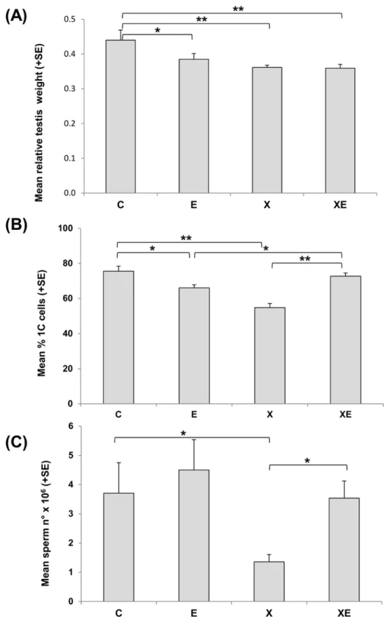
gonad differentiation induced a significant decrease of testis weight suggestive of a toxic effect (Fig 2A). ELF-MF exposure from 11.5 days post-conception to 21 days after birth induced a slight decrease of relative testis weight mainly due to a small increase of body weight. After combined exposure to ionizing radiation and ELF-MF, the level of testis weight reduction was comparable to that induced by X-rays exposure.
By flow cytometric analyses of testicular cells, the relative percentages of 1C, 2C, S-phase and 4C cells were calculated. A significant 28% reduction of postmeiotic 1C cells was observed after X-rays exposure, and a small but significant 13% reduction was detected also after ELF-MF exposure (Fig 2B). Surprisingly, after the combined exposure, the percentage of 1C cells was significantly higher than after the single treatments and equal to that of controls. The evidence of X-rays induced toxicity was magnified by the analysis of terminally differentiated cells in cauda epididymis where a 73% sperm count reduction was detected (Fig 2C). ELF-MF exposure did not induce a significant variation of sperm count with respect to controls. Consis-tently with the data on the relative percentages of testicular 1C cells, epididymal sperm count after combined exposure was significantly higher than after X-rays alone and not different from unexposed controls. Data on epididymis weight were consistent with those on sperm count (data not shown).
The effects of single and combined exposures to ELF-MF and X-rays were further evaluated by comet analysis in epididymal sperm. No effect of single treatment with ELF-MF or X-rays was shown under both neutral and alkaline conditions (Fig 3A). Also the combined exposure did not induce significant variation with respect to controls, but when the data were compared with those collected after X-rays alone, a statistically significant reduction of the mean tail intensity was detected. Under neutral conditions, an arbitrary tail intensity cut off point was established, above which sperm were considered clearly damaged. Consistently with the data on the mean tail intensity, the percentages of clearly damaged sperm were not significantly dif-ferent from controls under any treatment conditions, but the percentage of clearly damaged sperm induced by the combined exposure was significantly less than that induced by X-rays alone (Fig 3B).
Discussion
The main aim of this study was to employ a sensitive model to detect the genotoxic properties of ELF-MF, since–after decades of studies–proofs are still contradictory. Hence, we investigated the genotoxic effects of ELF-MFin vivoon somatic and germ cells after exposure during prena-tal and neonaprena-tal developmenprena-tal stages, known to be sensitive to environmenprena-tal stress. In addi-tion, genetic changes arising during this period of life might represent a co-factor at the origin of childhood cancer [37,38]. We also aimed at investigatingin vivoa possible influence of ELF-MF on the cell response to ionizing radiation. Chronic exposure to ELF-MF from day 12 p.c. until weaning had no teratogenic effect and did not affect survival, growth and develop-ment. X-irradiation at day 12 p.c. resulted in stunted pups and retarded development, in agree-ment with literature data [36]. No synergistic effects of ELF-MF and X-rays exposure on these parameters were observed.
Beside these developmental endpoints, we studied the genotoxic effects on the erythroid and male germ lines, using techniques validated in the respective tissues.
pregnancy, stopped it at birth and sampled blood from the prenatally exposed mice at the age of 35 days, finding no increase in MN frequencies. In fact, the differences in the duration and developmental windows of exposure might explain the different results.
The observed weak genotoxic effect of ELF-MF, only observed 42 days after birth, could be due to a perturbation of the cellular defence capacity against endogenous sources of DNA lesions.For the effect to be detectable both the exposure and expression times would be critical: a continuous exposure from mid-gestation until weaning, followed by a“latency”period, with a further haematopoietic system development seemed to be necessary. After 4 months the sys-tem appeared to have recovered its homeostatic capacity. Specific experiments should be
Fig 3. Comet assay in epididymal sperm.(A) Mean tail intensity values obtained with alkaline and neutral comet assay. (B) Percentage of sperm carrying clearly damaged DNA (with tail intensity values higher than 10%) after neutral assay. Columns represent the mean values of 4 controls (C), 5 ELF-MF exposed (E), 8 X-rays exposed (X), or 6 animals exposed to combined treatments (XE).
conducted to identify the molecular mechanisms leading to genotoxic damage. Disturbance of the oxidative balance might be a candidate because many authors suggested that this mecha-nism is implicated in ELF-MF-induced biological effects [39,40].
Differently, exposure to X-rays seems to produce two distinct periods of detectable geno-toxic effects. An elevated frequency of MN was observed at birth as a direct effect of X-irradia-tion. At the time when this happened (day 12 p.c.), the foetal liver was beginning
erythropoiesis thanks to recently arrived hematopoietic stem cells [41] and micronucleated erythrocytes deriving from those progenitors/precursors were detected in circulating blood at birth. Consistently with the kinetics of prenatal erythropoiesis, the frequency of micronu-cleated erythrocytes observed in blood was one order of magnitude less than that detected in the foetal haematopoietic liver at the time of maximum expression (16–18 h) after a compara-ble radiation treatment [42]. As more time passed by, these micronucleated cells were further diluted in the bloodstream by erythrocytes formed from unirradiated precursors. In fact, MN frequency of the X group at day 11 is lower than at birth and reaches the control value at day 21. Beside this acute, early outcome, what it seems to be a delayed effect became detectable at day 42 and persisted until day 140. Finally, the data show that ELF-MF exposure did not mod-ify the effect of X-rays at any tested time.
The effects on the male reproductive system were evaluated 42 days after birth when all germ cell sub-populations are present in the testis, the spermiogenesis has started and the first wave of spermatozoa is present in the epididymus. However, at this age the mouse germinal tis-sue is still in development as shown by a few comparative data: the testis weight was slightly lower than that of adults (0.11 vs 0.14 g respectively) and the number of spermatozoa was less than 15% of that found in adult mice.
Prenatal X-rays exposure at day 11.5 p.c. had an impact on testicular development as shown by the decrease of testis weight, of the relative frequency of post-meiotic cells (1C) and of the number of epididymal spermatozoa at 42 days after birth. These results are in agreement with the known radiosensitivity of germ and somatic cells in the foetal gonads [43].
ELF-MF exposure from 11.5 days post-conception to 21 days after birth induced a slight rel-ative decrease of post-meiotic cells, which was not reflected in a decrease of spermatozoa. Stud-ies on the effect ofin uteroexposure to ELF-MF on male reproductive system are few and results are controversial. A disorganization of spermatogenic epithelium was observed in adult rats as a result of oxidative stress induced byin uteroexposure to ELF-MF, 3 mT, 4 h/day [28]. Testicular alteration was observed also in rats exposed from 13 days p.c. to 21 days after birth to ELF-MF 1 mT, 3 applications of 30 minutes per day [29]. An increase of weight in epididy-mus, prostate and seminal vesicles without differences in epididymal sperm count, was observed in adult rats exposedin uteroto 15 Hz pulsed electromagnetic field from day 15 to day 20 of gestation [27]. On the other hand,in uteroand neonatal exposure of rats to ELF-MF at field strengths up to 500μT did not produce any alteration in the offspring spermatogenesis
and fertility [26]. No reproductive or developmental toxic effects were shown in a multigenera-tion study in rats exposed 18.5 hours/day to ELF-MF at field strengths up to 1 mT [25].
apoptosis; in addition, an impact of ELF-MF could be envisaged on proliferation of cells surviv-ing the X-rays exposure, which could also influence the onset of puberty.
In our study we also aimed at assessing possible delayed effects on DNA of terminally differ-entiated spermatozoa. The induction of delayed effects in male germ cells has been supported by the observation of DNA damage in spermatozoa deriving from X-rays exposed adult sper-matogonia [35,45–47]. In the present study a small, not significant increase of DNA damage was observed afterin uteroexposure to 1 Gy X-rays. It should be noted that the level of DNA damage in spermatozoa of untreated 42 days old mice was higher and more heterogeneous than in adult mice (laboratory historical data not shown). This result is in agreement with the characteristics of sperm populations produced by the first wave of spermatogenesis, which, in comparison with adult sperm show abnormalities in sperm head morphology and abnormal chromatin structure [48]. No effects were detected after continuous exposure to 50 Hz, 65μT
ELF-MF. Surprisingly, an evident reduction of DNA damage, homogeneous in all animals of the group, was observed when X-rays were followed by ELF-MF exposure. This effect, together with the observed increase of sperm number, suggests that ELF-MF is able to modulate male germ cell response to X-rays. This finding could be due to an influence of ELF-MF on early DNA damage response leading to a reduction of X-rays cell killing. Moreover, ELF-MF could influence post-natal germ cell proliferation/differentiation pathways, possibly anticipating the starting of spermatogenesis at puberty; this would result in a dilution of the first wave of natu-rally occurring abnormal spermatozoa with unaltered sperm deriving from a second wave of spermatogenesis. Although such an influence of ELF-MF on experimentally altered spermato-genesis has never been studied, the interaction of ELF-MF with damaged specific biological sys-tems can modulate the proliferation/differentiation process such as the promotion of healing of bone fractures [4,49].
The intensity of the magnetic field we used was quite high; nonetheless, it is in the order of magnitude of some domestic exposure, for example in the case of holding a laptop computer on the womb. In the germinal tissue, we were able to detect a biological effect of ELF-MF par-ticularly as modulation of the cell response to the toxic insult induced by a single X-rays dose. This result confirms the importance to investigate the effects of combined exposure to ELF-MF and other stressors because the modulation of a stress response may be as critical for the cell fate as the stress itself. Our data supported the hypothesis that the plasticity of developing tis-sues make them vulnerable to external stressors. Moreover, if ELF-MF may indeed interfere with proliferation/differentiation processes during early phases of life, it is important to investi-gate different tissues/organs because each one has its own specific differentiation pattern in a specific time window of development. This in turn points to the need of a deeper comprehen-sion of the molecular mechanisms underlying ELF-MF biological effects.
Supporting Information
S1 Data. Individual values for the micronucleus test (Dataset1) and for the male reproduc-tive system (Dataset2).
(PDF)
Acknowledgments
Author Contributions
Conceived and designed the experiments: CT AA AS IU. Performed the experiments: EC PV PE IU. Analyzed the data: FP EC PV PE IU AS. Contributed reagents/materials/analysis tools: FP EC PV PE AS LG. Wrote the paper: FP EC PV IU AS.
References
1. Wertheimer N, Leeper E. Electrical wiring configurations and childhood cancer. Am J Epidemiol. 1979; 109(3): 273–284. PMID:453167
2. Teepen JC, van Dijck JA. Impact of high electromagnetic field levels on childhood leukemia incidence. Int J Cancer. 2012; 131(4): 769–778. doi:10.1002/ijc.27542PMID:22437882
3. Repacholi M. Concern that "EMF" magnetic fields from power lines cause cancer. Sci Total Environ. 2012; 426:454–458. doi:10.1016/j.scitotenv.2012.03.030PMID:22534362
4. IARC Working Group on the Evaluation of Carcinogenic Risks to Humans. Non-ionizing radiation, Part 1: static and extremely low-frequency (ELF) electric and magnetic fields. IARC Monogr Eval Carcinog Risks Hum. 2002; 80: 1–395. PMID:12071196
5. World Health Organization. Environmental health criteria 238, Extremely Low Frequency Fields; 2007.
6. Udroiu I, Giuliani L, Ieradi LA. Genotoxic properties of extremely low frequency electromagnetic fields. In: Giuliani L, Soffritti M, editors. Non-thermal effects and mechanisms of interaction between electro-magnetic fields and living matter. Fidenza (Italy): Mattioli 1885; 2010. p.123–134.
7. Morita T, Asano N, Awogi T, Sasaki YF, Sato S, Shimada H, et al. Evaluation of the rodent micronu-cleus assay in the screening of IARC carcinogens (groups 1, 2A and 2B) the summary report of the 6th collaborative study by CSGMT/JEMS MMS. Collaborative Study of the Micronucleus Group Test. Mam-malian Mutagenicity Study Group. Mutat Res. 1997; 389(1): 3–122. PMID:9062586
8. Svedenstal BM, Johanson KJ. Leukocytes and micronucleated erythrocytes in peripheral blood from mice exposed to 50-Hz or 20-kHz magnetic fields. Electro- Magnetobiol. 1998; 17: 127–143.
9. Abramsson-Zetterberg L, Grawé J. Extended exposure of adult and fetal mice to 50 Hz magnetic field does not increase the incidence of micronuclei in erythrocytes. Bioelectromagnetics. 2001; 22(5): 351– 357. PMID:11424159
10. Udroiu I, Cristaldi M, Ieradi LA, Bedini A, Giuliani L, Tanzarella C. Clastogenicity and aneuploidy in new-born and adult mice exposed to 50 Hz magnetic fields. Int J Radiat Biol. 2006; 82(8): 561–567. PMID: 16966183
11. Udroiu I, Cristaldi M, Ieradi LA, Bedini A, Giuliani L. Genotoxic and hematotoxic damage induced by ELF magnetic fields. Eur J Oncol. 2008; 13: 239–244.
12. Erdal N, Gürgül S, Celik A. Cytogenetic effects of extremely low frequency magnetic field on Wistar rat bone marrow. Mutat Res. 2007; 630(1–2): 69–77. PMID:17452120
13. Okudan N, Celik I, Salbacak A, Cicekcibasi AE, Buyukmumcu M, Gökbel H. Effects of long-term 50 Hz magnetic field exposure on the micronucleated polychromatic erythrocyte and blood lymphocyte fre-quency and argyrophilic nucleolar organizer regions in lymphocytes of mice. Neurol Endocrinol Lett. 2010; 31: 208–214.
14. Alcaraz M, Olmos E, Alcaraz-Saura M, Achel DG, Castillo J. Effect of long-term 50 Hz magnetic field exposure on the micronucleated polychromatic erythrocytes of mice. Electromagn Biol Med. 2014; 33 (1): 51–57. doi:10.3109/15368378.2013.783851PMID:23781994
15. Sanderson BJ, Clark AM. Micronuclei in adult and foetal mice exposed in vivo to heliotrine, urethane, monocrotaline and benzidine. Mutat Res. 1993; 285(1): 27–33. PMID:7678129
16. Udroiu I, Ieradi LA, Cristaldi M, Tanzarella C. Detection of clastogenic and aneugenic damage in new-born rats. Environ Mol Mutagen. 2006; 47(5): 320–324. PMID:16538686
17. Zúñiga-González G, Ramírez-Muñoz MP, Torres-Bugarín O, Pérez-Jiménez J, Ramos-Mora A, Zamora-Pérez A, Gallegos-Arreola MP, Sánchez-Corona J. Induction of micronuclei in the domestic cat (Felis domesticus) peripheral blood by colchicine and cytosine-arabinoside. Mutat Res. 1998; 413 (2): 187–189. PMID:9639702
18. Neri M, Ceppi M, Knudsen LE, Merlo DF, Barale R, Puntoni R, Bonassi S. Baseline micronuclei fre-quency in children: estimates from meta- and pooled analyses. Environ Health Perspect. 2005; 113(9): 1226–1229. PMID:16140632
20. Li P, McLaughlin J, Infante-Rivard C. Maternal occupational exposure to extremely low frequency mag-netic fields and the risk of brain cancer in the offspring. Cancer Causes Control. 2009; 20(6): 945–955. doi:10.1007/s10552-009-9311-5PMID:19224378
21. Vergouwen RP, Jacobs SG, Huiskamp R, Davids JA, de Rooij DG. Proliferative activity of gonocytes, Sertoli cells and interstitial cells during testicular development in mice. J Reprod Fertil. 1991; 93(1): 233–243. PMID:1920294
22. Chernoff N, Rogers JM, Kavet R. A review of the literature on potential reproductive and developmental toxicity of electric and magnetic fields. Toxicology. 1992; 74(2–3): 91–126. PMID:1519247
23. Lee SK, Park S, Gimm YM, Kim YW. Extremely low frequency magnetic fields induce spermatogenic germ cell apoptosis: possible mechanism. Biomed Res Int. 2014; 2014:567183. doi:10.1155/2014/ 567183PMID:25025060
24. Duan W, Liu C, Wu H, Chen C, Zhang T, Gao P, Luo X, Yu Z, Zhou Z. Effects of exposure to extremely low frequency magnetic fields on spermatogenesis in adult rats. Bioelectromagnetics. 2014; 35(1): 58– 69. doi:10.1002/bem.21816PMID:24122970
25. Ryan BM, Symanski RR, Pomeranz LE, Johnson TR, Gauger JR, McCormick DL. Multigeneration reproductive toxicity assessment of 60-Hz magnetic fields using a continuous breeding protocol in rats. Teratology. 1999; 59(3): 156–162. PMID:10194806
26. Chung MK, Lee SJ, Kim YB, Park SC, Shin DH, Kim SH, Kim JC. Evaluation of spermatogenesis and fertility in F1 male rats after in utero and neonatal exposure to extremely low frequency electromagnetic fields. Asian J Androl. 2005; 7(2): 189–194. PMID:15897976
27. McGivern RF, Sokol RZ, Adey WR. Prenatal exposure to a low-frequency electromagnetic field demas-culinizes adult scent marking behavior and increases accessory sex organ weights in rats. Teratology. 1990; 41(1): 1–8. PMID:2106174
28. Gharamaleki H, Parivar K, Soleimani Rad J, Roshangar L, Shariati M. Effects of electromagnetic field exposure during the prenatal period on biomarkers of oxidative stress and pathology of testis and tes-tosterone level of adult rats in F1 generation. Acta Endo. (Buc.) 2014; 10: 577–587.
29. Tenorio BM, Jimenez GC, Morais RN, Torres SM, Albuquerque Nogueira R, Silva Junior VA. Testicular development evaluation in rats exposed to 60 Hz and 1 mT electromagnetic field. J Appl Toxicol. 2011; 31(3): 223–230. doi:10.1002/jat.1584PMID:20936650
30. Hintenlang DE. Synergistic effects of ionizing radiation and 60 Hz magnetic fields. Bioelectromagnetics. 1993; 14(6): 545–551. PMID:8297398
31. Miyakoshi J, Yoshida M, Shibuya K, Hiraoka M. Exposure to strong magnetic fields at power frequency potentiates X-ray-induced DNA strand breaks. J Radiat Res. 2000; 41(3): 293–302. PMID:11210830
32. Yoon HE, Lee JS, Myung SH, Lee YS. Increasedγ-H2AX by exposure to a 60-Hz magnetic fields
com-bined with ionizing radiation, but not hydrogen peroxide, in non-tumorigenic human cell lines. Int J Radiat Biol. 2014; 90(4): 291–298. doi:10.3109/09553002.2014.887866PMID:24467330
33. Karotki AV, Baverstock K. What mechanisms/processes underlie radiation-induced genomic instabil-ity? Cell Mol Life Sci. 2012; 69(20):3351–60. doi:10.1007/s00018-012-1148-5PMID:22955377
34. Xi W, Stuchly MA, Gandji OP. Induced electric currents in models of man and rodents from 60 Hz mag-netic fields. IEEE TransBiomed Eng. 1994; 41: 1018–1023.
35. Cordelli E, Fresegna AM, Leter G, Eleuteri P, SpanòM, Villani P. Evaluation of DNA damage in different stages of mouse spermatogenesis after testicular X irradiation. Radiat Res. 2003; 160(4): 443–451. PMID:12968930
36. Brent RL. Radiation teratogenesis. Teratology. 1980; 21(3): 281–298. PMID:7006137
37. Marshall GM, Carter DR, Cheung BB, Liu T, Mateos MK, Meyerowitz JG, Weiss WA. The prenatal ori-gins of cancer. Nat Rev Cancer. 2014; 14(4): 277–289. doi:10.1038/nrc3679PMID:24599217
38. Kim AS, Eastmond DA, Preston RJ. Childhood acute lymphocytic leukemia and perspectives on risk assessment of early-life stage exposures. Mutat Res. 2006; 613(2–3): 138–160. PMID:17049456
39. Luukkonen J, Liimatainen A, Juutilainen J, Naarala J. Induction of genomic instability, oxidative pro-cesses, and mitochondrial activity by 50Hz magnetic fields in human SH-SY5Y neuroblastoma cells. Mutat Res. 2014; 760:33–41. doi:10.1016/j.mrfmmm.2013.12.002PMID:24374227
40. Mattsson MO, Simkó M. Grouping of experimental conditions as an approach to evaluate effects of extremely low-frequency magnetic fields on oxidative response in in vitro studies. Front Public Health. 2014; 2:132. doi:10.3389/fpubh.2014.00132PMID:25229055
42. Cole RJ, Taylor N, Cole J, Arlett CF. Short-term tests for transplacentally active carcinogens. I. Micro-nucleus formation in fetal and maternal mouse erythroblasts. Mutat Res. 1981; 80(1): 141–157. PMID: 7207479
43. Vergouwen RP, Huiskamp R, Bas RJ, Roepers-Gajadien HL, Davids JA, de Rooij DG. Radiosensitivity of testicular cells in the fetal mouse. Radiat Res. 1995; 141(1): 66–73. PMID:7997516
44. Pafkova H, Jerabek J. Interaction of MF 50 Hz, 10 mT with high dose of X-rays: evaluation of embryo-toxicity in chick embryos. Rev Environ Health. 1994; 10(3–4): 235–241. PMID:7724884
45. Haines GA, Hendry JH, Daniel CP, Morris ID. Increased levels of comet-detected spermatozoa DNA damage following in vivo isotopic- or X-irradiation of spermatogonia. Mutat Res. 2001; 495(1–2): 21– 32. PMID:11448639
46. Haines GA, Hendry JH, Daniel CP, Morris ID. Germ cell and dose-dependent DNA damage measured by the comet assay in murine spermatozoaa after testicular X-irradiation. Biol Reprod. 2002; 67(3): 854–861. PMID:12193394
47. Cordelli E, Eleuteri P, Grollino MG, Benassi B, Blandino G, Bartoleschi C, Pardini MC, Di Caprio EV, SpanòM, Pacchierotti F, Villani P. Direct and delayed X-ray-induced DNA damage in male mouse germ cells. Environ Mol Mutagen. 2012; 53(6): 429–439. doi:10.1002/em.21703PMID:22730201
48. Janca FC, Jost LK, Evenson DP. Mouse testicular and sperm cell development characterized from birth to adulthood by dual parameter flow cytometry. Biol Reprod. 1986; 34(4): 613–623. PMID: 3708046