Insight into magnetite nanoparticle phase
evolution in solvothermal synthesis through a
simple method based on iron chloride and metallic
iron
F. B. F. Silva,aE. C. Paris,bG. M. da Costacand C. Ribeiro*b
This paper presents the development and optimization of the synthesis of magnetic nanoparticles composed of iron oxides by a solvothermal method with benzyl alcohol using iron(III) chloride hexahydrate as a metallic precursor in the substitution of iron(III) acetylacetonate. The synthesis parameters were studied and varied to obtain a high yield of magnetite. Metallic iron was evaluated as a reducing agent and iron source. Furthermore, the use of urea as a precipitation agent was crucial to increasing the yield of magnetite, at once reducing the acidity of the medium and inducing the precipitation of Fe2+ ions. Longer treatment times and no stirring led to an increase in the yield of magnetite, whose formation process followed the Ostwald step rule. The particles were characterized by X-ray diffraction, M¨ossbauer spectroscopy, FTIR, TEM, SEM-FEG and VSM (Vibrating Sample Magnetometry). The phases were quantified by the Rietveld method and M¨ossbauer spectroscopy, and a 97% maximum yield of magnetite was achieved. The polymerization of benzyl alcohol during the solvothermal treatment was shown to play an important role in the nanoparticle capping. The polymerized resin was characterized by FTIR and NMR-1H, and a mechanism for the polymerization reaction was proposed to elucidate the synthetic route and its relationship to particle formation and stability.
Introduction
Recent advances in colloidal synthesis have allowed for the exploration and manipulation of the magnetic properties of solid materials at the nanometer scale and enabled a range of novel applications of great interest to researchers from several disciplines, including magneticuids, catalysis, biotechnology/ biomedicine, magnetic resonance imaging, data storage and environmental remediation.1–3 Magnetic nanoparticles, when
functionalized, can be very promising for applications in which, being so small and magnetically separable, the particles may act as quasi-homogeneous systems, combining high reactivity, high dispersion and easy separation. These features are espe-cially interesting in catalysis, and many publications have described the application of such magnetic nanoparticles in facilitating the effective separation and recycling of expensive catalysts.4Certain technological enzymatic processes can also
benet by the functionalization of magnetic nanoparticles with
enzymes, at once enabling the recovery and the reuse of soluble enzymes.5–7
Iron oxide nanoparticles are most likely the best-studied magnetic nanomaterial due to their nontoxicity and biocom-patibility. Nevertheless, the formation process of these magnetic nanoparticles is still not clear for many reaction systems.8–10 New strategies for synthesizing these particles by
controlling their shape and size (and surface) and for protecting them against oxidation are fundamental to the use of these materials in a wide range of specic applications, especially on a large scale. These limitations can be overcome by nonaqueous approaches, which provide a more effective control of size distribution due mainly to the moderate reactivity of the oxygen–carbon bond, leading to slower reaction rates, and to the surfactant role play by certain organic solvents.10–12 For
example, the thermal decomposition of organometallic or metal organic iron precursors such as iron(III) cupferronate (FeCup3),
iron(III) acetylacetonate (Fe(acac)3) and pentacarbonyl iron
(Fe(CO)5) in high-boiling organic solvents containing stabilizing
surfactants leads to a very narrow size distribution of small magnetite or maghemite nanoparticles.13–19Pinnaet al.(2005)
described a surfactant-free route to synthesizing magnetite nanoparticles by using benzyl alcohol as a solvent and a ligand at the same time to overcome the drawbacks of
surfactant-aDepartamento de Qu´ımica
–UFSCar, Rod. Washington Luiz, km235, CEP 13565-905 S˜ao Carlos, SP, Brazil
bEmbrapa Instrumentaç˜ao, Rua XV de Novembro, 1452, CEP 13560-970, S˜ao Carlos,
SP, Brazil. E-mail: [email protected]
cDepartamento de Qu´ımica
–Universidade Federal de Ouro Preto, CEP 35400-000, Ouro Preto, MG, Brazil
Cite this:RSC Adv., 2014,4, 53265
Received 3rd July 2014 Accepted 7th October 2014
DOI: 10.1039/c4ra06620k
www.rsc.org/advances
PAPER
Published on 07 October 2014. Downloaded by Universidade Federal de Ouro Preto on 26/02/2015 21:40:42.
controlled approaches. The direct reaction between benzyl alcohol and iron(III) acetylacetonate under solvothermal
condi-tions has been observed to produce high-purity magnetite nanocrystals with sizes ranging from 12 to 25 nm.20The
“benzyl alcohol route”has proven to be a suitable strategy for synthe-sizing several binary metal oxide nanoparticles, perovskites and hybrid materials.21–23This simple and robust method leads to
low amounts of organic impurities in the products, therefore making it suitable for studying nanoparticle formation.11
The“benzyl alcohol route”has been applied to synthesize iron oxide nanoparticles using mainly iron(III) acetylacetonate
as a precursor; however, common precursors employed in sol–
gel synthesis, such as Fe(III) and Fe(II) chlorides, nitrates,
sulfates or even metallic iron, have not been reported in the literature as possible precursors for this method.8 This fact
suggests that the phase evolution in the aforementioned system is still poorly understood, despite several other oxides having been synthesized using metal halides.24,25
In this work, we report the synthesis of magnetite nano-particles following the benzyl alcohol route by using iron(III)
chloride hexahydrate and metallic iron as iron precursors to investigate the mechanisms involved in Fe3O4phase evolution
under solvothermal conditions. Other processes involved in the synthesis, such as the polymerization of benzyl alcohol, were also investigated to understand their role in the phase forma-tion and stability of the products.
Experimental
All materials were prepared under solvothermal conditions with different proportions of iron chloride hexahydrate (FeCl3$6H2O)
and metallic iron (Fe0) while varying the synthetic parameters
(temperature, time, stirring and addition of urea). In a typical synthesis procedure, carried out in a glovebox (H2O < 0.1 ppm),
the iron precursors (totaling 4.5 mmol of iron) were added to 30 mL of benzyl alcohol, containing 1.0 g of powdered urea (16.6 mmol). A similar experiment was performed without the addi-tion of urea. The mixtures were homogenized in a borosilicate glass cup with an inner volume of 60 mL, under magnetic stirring, and then transferred to a stainless steel solvothermal reactor that was carefully sealed. The apparatus was removed from the glovebox and heated to 250C for 24, 48 and 96 hours.
The materials obtained aer the solvothermal treatment were cooled to room temperature, centrifuged and thoroughly washed with acetone aer sonication in an ultrasonic bath for 3 minutes. This procedure was performed to remove the organic residues on the particles' surface. A second washing step with ethanol was necessary when urea was added to the reaction mixture to remove the ammonium chloride byproduct. Aer the washing steps, the precipitates were dried in air at 70C.
The particles were structurally characterized in an X-ray diffractometer (Shimadzu XRD 6100) with CuKa radiation,
using aq–2qconguration, at 30 kV and 30 mA. The 2qrange
used in the measurement was 10–110, with a step size of 0.02
and a step time of 4.0 s. Quantitative analysis by Rietveld renement was performed with GSAS soware.26 M¨ossbauer
spectra (MS) were obtained at room temperature with a
spectrometer using a constant-acceleration drive with a trian-gular reference signal and 1024 channels and in the velocity range of 11 to 11 mm s 1(increment of0.046 mm s 1). The
velocity was calibrated from the MS of a standard alpha-Fe foil at room temperature. The spectra were computer-tted either with discrete Lorentzian sextets or doublets.
The sample size and morphologies were examined byeld emission gun-scanning electron microscopy (FEG-SEM) in a JEOL JSM6701F microscope operating at 5.0 kV and by trans-mission electron microscopy (TEM) in a Philips CM-120 microscope (operating at 120 kV) through bright eld (BF), darkeld (DF) images and electron diffraction (ED) of selected particles. The TEM samples were dispersed in ethanol, and a few drops of the samples were deposited on a cooper-carbon grid. Hysteresis loops of the samples were obtained by vibrating sample magnetometry, performed at room tempera-ture with a vibrating sample measurement (EG&G Princeton Applied Research equipment) in a home-made setup.
Fourier-transform infrared spectroscopy (FTIR) was per-formed in a Bruker/Equinox 55 instrument, from 4000 to 400 cm 1, by conducting an average of 100 scans and using trans-mission and diffuse-reectance modes. To analyze the eventual polymerization of the solvent (benzyl alcohol), high-resolution
1H nuclear magnetic resonance spectroscopy (Bruker 600 MHz
equipment, operating under 14.1 T) was performed. The chemical shivalues were calibrated using tetramethylsilane as a reference. To identify the shis in a proposed molecular structure, a simulation was performed using the NMR-Predictor program (available at http://www.nmrdb.org/).
Results and discussion
Because in magnetite FeIII and FeII species are present, the syntheses were initially carried out using different proportions of FeCl2 and FeCl3, under similar solvothermal conditions.
However, in all cases, the formation of hematite as a major phase was observed, and only small amounts of magnetite were obtained. These results indicated that a redox pair would be necessary to achieve the proper phase formation. Thus, we opted to introduce metallic iron (Fe0) as a reductant, aiming to
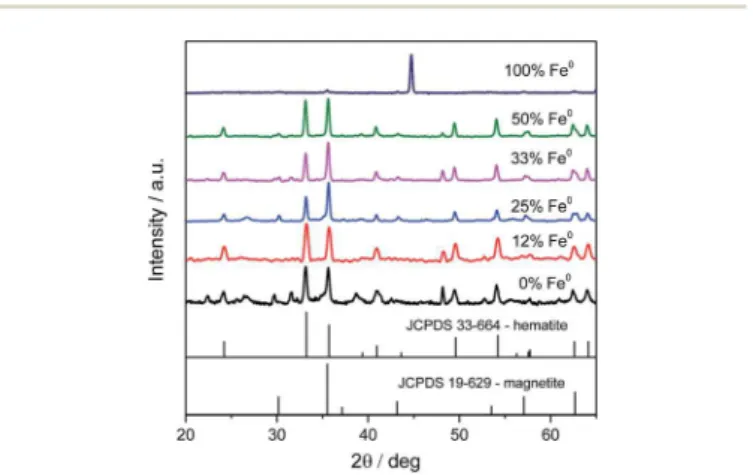
Fig. 1 XRD patterns of the particles synthesized with different amounts of metallic iron.
reduce Fe3+ions during the solvothermal process because Fe0
can act as an iron precursor and reducing agent.27Fig. 1 shows
the XRD patterns obtained for different Fe0/Fe3+molar ratios, at 250C/24 h under stirring. All diffractograms show a mixture of
hematite and magnetite, except the one correspondent to 100% metallic iron, in which the only observed phase is the metallic iron. These results indicate that the solvent by itself, even at high temperature and pressure, does not play an oxidative role during the synthesis procedures. It also shows that Fe0does in fact interact with Fe3+during the redox process. It was possible to observe the dependence of the magnetite yield on the amount of metallic iron. This relationship could be easily observed by comparing the relative intensity between the peaks at 33.2, the
most intense peak of hematite, and 35.6, the most intense peak
of magnetite. The best result was reached with 25% of metallic iron, indicating an optimum range for phase formation. In an attempt to improve the magnetite yield, urea (CO(NH2)2) was
added to provide a basic medium, adequate for the formation of magnetite phase because Fe(OH)2, the most probable precursor
of FeII, is less soluble under this condition. FeCl3$6H2O is a
fairly strong Lewis acid, and the stability of Fe(OH)2depends on
a source of OH–, which can be obtained by the decomposition of urea.27–29
As expected, magnetite production was signicantly improved (Fig. 2a). Furthermore, Tamaura suggested that Cl retards magnetite formation by hindering the condensation of neighboring OH groups and consequent formation of Fe–O–Fe linkages. Thus, this contaminant can be easily separated because NH4+ions formed during the decomposition of urea
are able to capture Cl ions by forming NH4Cl, which is
insol-uble in the reaction medium.30
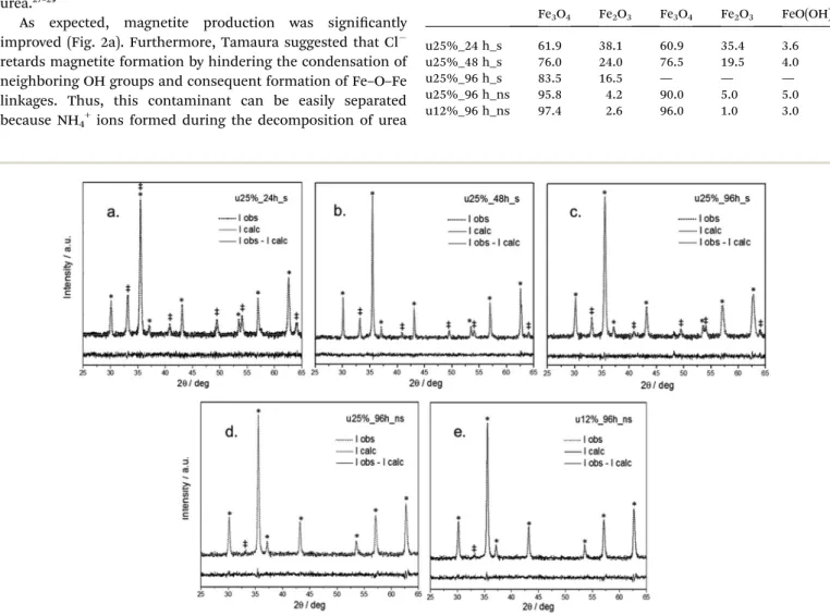
Having established the optimum metallic iron fraction to be 25% and adding an excess of urea, the reaction time was extended from 24 (Fig. 2a) to 48 (Fig. 2b) to 96 h (Fig. 2c). These
gures clearly show the increase in the yield of magnetite, indicating the benecial effect of longer reaction times. It can also be observed that the calculated XRD lineshapes resulting from the Rietveld renement closely match the experimental data. Considering the absence of amorphous phases and the existence of only hematite and magnetite, a quantitative anal-yses was performed, the results of which are presented in Table 1. It is noteworthy that for longer periods, the magnetite yield was higher than 80%, showing that the formation of hematite most likely preceded that of magnetite. Additionally, the role of Fe0 as a reductant species was also con
rmed. However,
Fig. 2 Rietveld refinement plots of the samples synthesized with urea under various conditions: (a) 25% Fe0for 24 hours; (b) 25% Fe0for 48 hours; (c) 25% Fe0for 96 hours; (d) 25% Fe0for 96 hours and no stirring; (e) 12% Fe0for 96 hours and no stirring. Di
ffraction peaks assigned to hematite phase are marked with double dagger symbols (‡), and magnetite phase are marked with an asterisk (*).
Table 1 Phase quantification (weight%) obtained by Rietveld refi ne-ments and M¨ossbauer spectroscopy
Rietveld M¨ossbauer
Fe3O4 Fe2O3 Fe3O4 Fe2O3 FeO(OH)
u25%_24 h_s 61.9 38.1 60.9 35.4 3.6
u25%_48 h_s 76.0 24.0 76.5 19.5 4.0
u25%_96 h_s 83.5 16.5 — — —
u25%_96 h_ns 95.8 4.2 90.0 5.0 5.0
u12%_96 h_ns 97.4 2.6 96.0 1.0 3.0
because stirring may interfere with the phase formation, this parameter was also evaluated. The magnetite yield for the sample prepared with 25% Fe0 for 96 hours and no stirring (Fig. 2d) was improved to 96%, as shown in Table 1.
This improvement can be explained by considering that stirring can induce the agglomeration of as-formed by magnetic interactions, reducing the total effective area for reaction and leading to possible segregation. Additionally, the residual hematite content may be related to an excess of Fe0 in the reaction. In fact, when performing the same experiment but using 12% Fe0instead of 25% Fe0, (Fig. 2e) we observed a slight
increase in the magnetite yield to 97.4%. Thus, the optimum Fe0content appears to be between 12% and 25%.
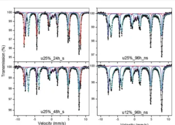
Magnetite and hematite can be satisfactorily distinguished by their XRD patterns. On the other hand, magnetite and maghemite have very similar patterns and cannot be distin-guished by this technique. Therefore, M¨ossbauer spectroscopy performed at room temperature was used to elucidate the true composition of these magnetic particles. Thetted spectra are shown in Fig. 3, whereas the calculated amounts of magnetite and hematite are presented in Table 1. The spectrum of all samples showed magnetite contents ranging from 60.9 to 96.0%, values that are in a good agreement with the results of the Rietveld method (see Table 1). However, the relative area ratios of the two magnetite sextets deviate substantially from the ideal value of 1.88 for pure magnetite. Thisnding can be attributed either to the presence of non-stoichiometric magnetite or to the presence of some maghemite. The pres-ence of a low-intensity doublet may be correlated to a small amount of goethite.
Fig. 4a to c show BF-TEM images of selected samples, which indicate particle sizes ranging from 50 to 100 nm. Also, from a representative zoom out FEG-SEM image (Fig. 4d) it was possible to analyze the particle size by direct image analysis of more than 200 particles. The results are shown in a histogram (Fig. 4e), conrming the average diameter of 92.8 nm. At these sizes, it is expected that the magnetite phase would be more stable, in accord with the results of other characterizations. The sample synthesized over a period of 96 hours showed higher agglomeration and size dispersion, which is expected since longer treatment times would lead to grain growth. Further-more, under this condition, higher particle sizes may alter the magnetic behavior from superparamagnetism to ferrimagne-tism, leading to permanent magnetization.
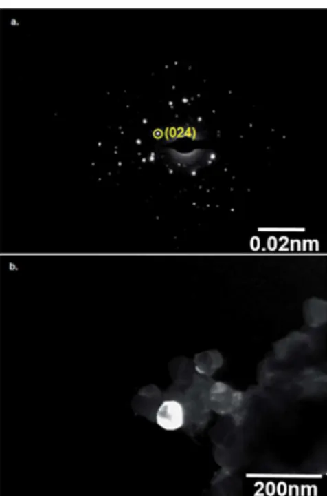
Fig. 5 shows a DF-TEM image and ED for the sample synthesized in 96 hours, where the diffraction pattern is
Fig. 3 M¨ossbauer spectra and their curve-fitting results for the samples u25%_24 h_s, u25%_48 h_s, u25%_96 h_ns and u12%_96 h_ns.
Fig. 4 TEM images of samples: (a) u25%_24 h_s; (b) u25%_48 h_s; (c) u25%_96 h_s. Image (d) is a zoom-out FEG-SEM image and (e) is a histogram showing the particle size distribution.
characteristic of a few agglomerated single crystals. The DF image shows a single crystal identied as hematite by the (024) plane in ED, which conrms the individual formation of sepa-rate nanoparticles of hematite and magnetite.
From the results presented, we can speculate that the synthesis process is governed by the Ostwald step rule. The small amount of goethite detected by M¨ossbauer spectroscopy is most likely the rst step in crystallization from solution, followed by the conversion to hematite, whose formation can be
observed aer a few hours of solvothermal treatment. Magnetite formation is the last step in phase conversion because magne-tite has a lower free energy of formation under standard conditions31and is even more stable under alkaline reducing
conditions. The yields of magnetite increased signicantly with the solvothermal treatment time at temperatures above 200C;
however, Pinnaet al.noted that temperature higher than 175C
favored the formation of hematite.20This contradiction is due to
the differences in the mechanisms governing the formation of magnetite in each approach. The formation of magnetite from the reaction between iron(III) acetylacetonate and benzyl alcohol
is only possible due to the partial reduction of the metallic center to iron(II), made viable by the oxidation of
4-phenyl-2butanone, which produced during the synthesis.
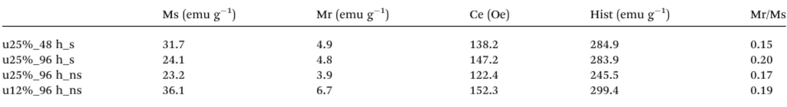
In order to analyze the magnetic properties of these mate-rials, at room temperature (300 K), vibrating sample magne-tometry (VSM) was applied to selected samples, as shown in Fig. 6. Despite the effective amounts of Fe3O4and the deviation
in particle sizes, as observed in Fig. 4, the magnetic prole was not strongly affected between the samples. The saturation magnetization was around 23 to 37 emu g 1to all samples, as showed in Table 2, which is comparable to observed by Morel
et al.32and Xuet al.33for particles around the same size
distri-bution. Also, the remanence magnetization, coercitivity and hysteresis indicate a permanent magnetization behavior, but close to a superparamagnetic prole. These results show the applicability of these materials for magnetic purposes.
To investigate the role of the solvent as a capping material, we performed specic experiments to analyze the organic products formed over the particles. Fig. 7 displays the FTIR spectra of benzyl alcohol (BA), used as the solvent; benzyl alcohol aer the solvothermal process, obtained as a byproduct (separated by centrifugation in the Fe3O4synthesis done in 96 Fig. 5 (a) Electron diffraction pattern and (b) DF-TEM image of u25%
_96 h_s.
Fig. 6 Hysteresis loops obtained by VSM at room temperature for selected samples: (a) u25%_48 h_s; (b) u25%_96 h_s; (c) u25%_96 h_ns and (d) u12%_96 h_ns.
hours); and the nanoparticles synthesized over periods of 24, 48 and 96 hours. The FTIR spectra of the benzyl alcohol before and aer the solvothermal process show similar peaks assigned to the C–H stretching of the aromatic ring from 3100 to 3000 cm 1, out-of-plane aromatic C–H bending at 726, C–C ring stretching at 1494 and 1452 cm 1and ring C–C bending at 698 cm 1. The peaks assigned to O–H bending at 1208 cm 1 and to C–O
stretching at 1020 cm 1nearly disappeared in the spectrum of
benzyl alcohol aer the solvothermal process, indicating a reduction in the number of O–H groups as the primary alcohol. This result is an indicative of possible benzyl alcohol polymer-ization. The characteristic intermolecular O–H stretching band centered at 3456 cm 1remained in the spectrum but was less intense and shied to higher wavelengths due the decrease in
the concentration of alcoholic hydroxyl groups. Methylene C–H stretching peaks appeared at 2916 and 2846 cm 1, shied to
shorter wavelengths relative to the same peaks in the benzyl alcohol spectrum.
The spectra of particles synthesized in 24, 48 and 96 hours, obtained by the diffuse reectance mode, also showed the peaks of organic residues on the particles' surface forming an over-lapping band of the peaks observed between 1770 and 1380 cm 1in the spectrum of the polymerized benzyl alcohol, related
to C–C ring stretching, whose shis were due to the interaction with the particles' surface. At 1415 cm 1, a peak can be observed only in the spectra of the particles synthesized in 48 and 96 hours. The presence of this peak, assigned to C–C ring stretching, suggests increasingly effective bonding of the organic residues to the particles as the time of solvothermal treatment and the polymerization degree increased. Addition-ally, the spectra exhibit the characteristic magnetite band of Fe–
O centered at approximately 590 cm 1. It can be observed that the band is displaced to shorter wavelengths according to the
Table 2 The saturation magnetization (Ms), remanence (Mr), coercivity (Ce) and hysteresis (Hist) for selected samples obtained by VSM
Ms (emu g 1) Mr (emu g 1) Ce (Oe) Hist (emu g 1) Mr/Ms
u25%_48 h_s 31.7 4.9 138.2 284.9 0.15
u25%_96 h_s 24.1 4.8 147.2 283.9 0.20
u25%_96 h_ns 23.2 3.9 122.4 245.5 0.17
u12%_96 h_ns 36.1 6.7 152.3 299.4 0.19
Fig. 7 FTIR spectra of benzyl alcohol (BA), benzyl alcohol after the solvothermal process (BA#) and the particles synthesized in 24 hours (u25%_24 h_s), 48 hours (u25%_48 h_s) and 96 hours (u25%_96 h_s).
Scheme 1 Reaction mechanism for the polymerization of benzyl alcohol during the solvothermal treatment.
hematite concentration in the phase mixture because the main characteristic Fe–O band for this phase is centered at 550 cm 1. As cited, during the solvothermal treatment, a possible polymerization of benzyl alcohol occurred, leading to the formation of a dark and sticky supernatant with water release. This process can be associated with the Friedel–Cras
alkyl-ation reaction because FeCl3$6H2O is a catalyst for this reaction
and benzyl alcohol is susceptible to the reaction.34A possible
mechanism for the reaction is shown in Scheme 1. Therst step is the reaction of alkyl halide elimination, leading to the formation of benzyl chloride and Fe–OH species, which repre-sents the starting point of nanoparticle formation. We consid-ered the direct elimination of benzyl chloride instead of iron alkoxo-halide formation because benzyl groups are able to stabilize a positive charge as a stable carbocation for the SN1 reaction.11,35,36
A polymerization reaction follows the formation of benzyl chloride, which reacts with other benzyl chloride or benzyl alcohol molecules, following the Friedel–Cras mechanism.
Benzyl chloride reacts with the catalyst by the attachment of the chlorine atom of the former, creating a more electrophilic carbon on the methylene group. The p electrons on the
aromatic rings of a benzyl chloride or a benzyl alcohol molecule act as a nucleophile, attacking the electrophilic carbon. Proton removal from sp3carbon reforms the aromatic ring and leads to the formation of HCl and catalyst regeneration. The mechanism drawn shows the second step as an SN2 reaction, although it could be an SN1 reaction because the benzylic cation is stable. Friedel and Cras were therst to observe the polymerization of benzyl chloride, followed by Jacobson,37Dermer and Hooper,38
who reported the formation of a soluble, dark-brown resin with the empirical formula C7H6as the main reaction product of iron
chloride and benzyl chloride. The polymer formed during the synthesis and the mechanism proposed are in agreement with the observations of the aforementioned researchers and with the corresponding FTIR spectrum, which maintains the bands assigned to the aromatic ring but not the bands related to the hydroxyl groups. Furthermore, the polymerization of benzyl alcohol during the synthesis of metal oxide nanoparticles was previously reported by Olliges-Stadleret al.,39who obtained a
hard and brittle, dark-green-blue monolith with the same empirical formula, but through a different mechanism, while synthesizing tungsten oxide nanoparticles. Differences in the viscosity and color of this polymer are related to the nature of the metallic catalyst, which can lead to different degrees of polymerization.38
The polymerization of benzyl alcohol was also veried by nuclear magnetic resonance (NMR) spectroscopy (Fig. 8) using the pure benzyl alcohol (BA) as reference and a sample of the polymerized solvent separated by centrifugation (BApol).
In the BA spectrum, characteristic peaks due to the three different chemical environments are observed: aromatic ring (7.2–7.4 ppm), methylene group (4.6 ppm) and alcohol group (1.8 ppm). The peak assigned to the hydrogen atom of the OH group (1.8 ppm) in the BA1H-NMR spectrum is suppressed in the spectrum of BApol, which conrms the condensation reaction with water release. The spectrum of BApol displays
broad and partly split peaks in three main regions correspon-dent to the protons of the aromatic rings (6.9–7.6 ppm); to the protons of the aliphatic methylene groups (3.8–4.1 ppm), dis-placed and outspread from the original positions due to the multiplicity of chemical environments aer polymerization;
and to the primary hydrogen atoms at terminations (2.1–2.4 ppm).
The polymerization reaction was noticed to be a parallel process to the phase evolution of iron oxides. It occurred independently of the phases obtained and its extent was func-tion of the amount of FeCl3$6H2O, the Friedel–Cras catalyst,
and solvothermal treatment period. Even though it is not related to the phase evolution, the polymerization of benzyl alcohol has a synergic effect in the global process by capping the as-synthesized magnetite particles and preventing their oxida-tion for long term stability. Experiments performed in the same samples, stored in dry form during 1 year showed the same results observed for fresh materials, indicating that good stability.
Conclusions
Based on the results presented herein, we can conclude that magnetite formation under solvothermal conditions follows the Ostwald step rule (from the formation of metastable to that of more stable phases). Goethite formation is most likely therst step in the synthesis process, followed by hematite formation. Metallic iron most likely interacts with the surface of the particles formed, reducing part of the FeIIIin the crystal struc-ture of the particles to FeII, yielding the Fe3O4stoichiometry.
This hypothesis is consistent with the ndings of Matthews, who studied, in detail, magnetite formation under hydro-thermal conditions from previously prepared hematite particles through reduction by metallic iron and observed the occurrence
Fig. 8 1H-NMR spectra of benzyl alcohol (BA) and polymerized benzyl alcohol (BApol).
of two simultaneous processes: oxidation of metallic iron and reduction of hematite to magnetite.40
Acknowledgements
The authors acknowledge thenancial support from the Bra-zilian Agencies CNPq, CAPES, FAPESP, FINEP and Embrapa (Agronano Network). The authors also acknowledge the tech-nical support from LCE-DEMa/UFSCar (for TEM facility), GSM-DF/UFSCar (VSM facility) and LIEC/UFSCar (FTIR facility).
Notes and references
1 U. Jeong, X. W. Teng, Y. Wang, H. Yang and Y. N. Xia,Adv. Mater., 2007,19, 33–60.
2 S. Laurent, D. Forge, M. Port, A. Roch, C. Robic, L. V. Elst and R. N. Muller,Chem. Rev., 2010,110, 2574.
3 A.-H. Lu, E. L. Salabas and F. Sch¨uth,Angew. Chem., Int. Ed., 2007,46, 1222–1244.
4 A. Hu, G. T. Yee and W. Lin,J. Am. Chem. Soc., 2005,127, 12486–12487.
5 J. Jordan, C. S. S. R. Kumar and C. Theegala,J. Mol. Catal. B: Enzym., 2011,68, 139–146.
6 D.-H. Chen and M.-H. Liao,J. Mol. Catal. B: Enzym., 2002,16, 283–291.
7 A. Garcia, S. Oh and C. R. Engler,Biotechnol. Bioeng., 1989,
33, 321–326.
8 I. M. Grabs, C. Bradtmoller, D. Menzel and G. Garnweitner,
Cryst. Growth Des., 2012,12, 1469–1475.
9 P. Hu, L. J. Yu, A. H. Zuo, C. Y. Guo and F. L. Yuan,J. Phys. Chem. C, 2009,113, 900–906.
10 M. Niederberger, G. Garnweitner, J. Buha, J. Polleux, J. H. Ba and N. Pinna,J. Sol–Gel Sci. Technol., 2006,40, 259–266.
11 M. Niederberger and N. Pinna,Metal Oxide Nanoparticles in Organic Solvents: Synthesis, Formation, Assembly and Application, Springer, London, 2009.
12 G. Garnweitner and M. Niederberger, J. Am. Ceram. Soc., 2006,89, 1801–1808.
13 W. Cai and J. Q. Wan,J. Colloid Interface Sci., 2007,305, 366–
370.
14 T. Hyeon, S. S. Lee, J. Park, Y. Chung and H. Bin Na,J. Am. Chem. Soc., 2001,123, 12798–12801.
15 D. Maity, S. N. Kale, R. Kaul-Ghanekar, J. M. Xue and J. Ding,
J. Magn. Magn. Mater., 2009,321, 3093–3098.
16 J. Park, K. J. An, Y. S. Hwang, J. G. Park, H. J. Noh, J. Y. Kim, J. H. Park, N. M. Hwang and T. Hyeon,Nat. Mater., 2004,3, 891–895.
17 S. Si, C. Li, X. Wang, D. Yu, Q. Peng and Y. Li,Cryst. Growth Des., 2005,5, 391–393.
18 D. B. Yu, X. Q. Sun, J. W. Zou, Z. R. Wang, F. Wang and K. Tang,J. Phys. Chem. B, 2006,110, 21667–21671.
19 G. Z. Li, W. C. Peng, X. Y. Li, X. B. Fan, X. J. Li, G. L. Zhang and F. B. Zhang,Appl. Surf. Sci., 2008,254, 4970–4979. 20 N. Pinna, S. Grancharov, P. Beato, P. Bonville, M. Antonietti
and M. Niederberger,Chem. Mater., 2005,17, 3044–3049. 21 N. Pinna,J. Mater. Chem., 2007,17, 2769–2774.
22 N. Pinna, M. Karmaoui and M. G. Willinger,J. Sol–Gel Sci.
Technol., 2011,57, 323–329.
23 Y. Masubuchi, S. Yamashita, T. Motohashi, S. Kikkawa and M. Niederberger,J. Eur. Ceram. Soc., 2011,31, 2471–2474. 24 M. Niederberger, M. H. Bartl and G. D. Stucky,J. Am. Chem.
Soc., 2002,124, 13642–13643.
25 M. Niederberger, M. H. Bartl and G. D. Stucky,Chem. Mater., 2002,14, 4364–4370.
26 B. H. Toby,J. Appl. Crystallogr., 2001,34, 210–213.
27 S. Chaleawlert-umpon and N. Pimpha,Mater. Chem. Phys., 2012,135, 1–5.
28 S. Y. Lian, E. Wang, Z. H. Kang, Y. P. Bai, L. Gao, M. Jiang, C. W. Hu and L. Xu,Solid State Commun., 2004,129, 485–490. 29 I. J. Bruce, J. Taylor, M. Todd, M. J. Davies, E. Borioni, C. Sangregorio and T. Sen, J. Magn. Magn. Mater., 2004,
284, 145–160.
30 Y. Tamaura, P. V. Buduan and T. Katsura, J. Chem. Soc., Dalton Trans., 1981, 1807–1811.
31 A. Navrotsky,Proc. Natl. Acad. Sci. U. S. A., 2004,101, 12096–
12101.
32 M. Morel, F. Martinez and E. Mosquera, J. Magn. Magn. Mater., 2013,343, 76–81.
33 J. Xu, H. B. Yang, W. Y. Fu, K. Du, Y. M. Sui, J. J. Chen, Y. Zeng, M. H. Li and G. Zou,J. Magn. Magn. Mater., 2007,
309, 307–311.
34 I. Iovel, K. Mertins, J. Kischel, A. Zapf and M. Beller,Angew. Chem., Int. Ed., 2005,44, 3913–3917.
35 M. Niederberger and G. Garnweitner,Chem.–Eur. J., 2006,12,
7282–7302.
36 A. Vioux,Chem. Mater., 1997,9, 2292–2299.
37 R. A. Jacobson,J. Am. Chem. Soc., 1932,54, 1513–1518. 38 O. C. Dermer and E. Hooper,J. Am. Chem. Soc., 1941,63,
3525–3526.
39 I. Olliges-Stadler, M. D. Rossell and M. Niederberger,Small, 2010,6, 960–966.
40 A. Matthews,Am. Mineral., 1976,61, 927–932.