Pathway Operates under Oxic Conditions
Po-Hsiang Wang1., Tzong-Huei Lee2., Wael Ismail3
, Ching-Yen Tsai1, Ching-Wen Lin1, Yu-Wen Tsai1, Yin-Ru Chiang1*
1Biodiversity Research Center, Academia Sinica, Taipei, Taiwan,2Graduate Institute of Pharmacognosy, Taipei Medical University, Taipei, Taiwan,3Biotechnology Program, College of Graduate Studies, Arabian Gulf University, Manama, Kingdom of Bahrain
Abstract
Cholesterol is one of the most ubiquitous compounds in nature. The 9,10-seco-pathway for the aerobic degradation of cholesterol was established thirty years ago. This pathway is characterized by the extensive use of oxygen and oxygenases for substrate activation and ring fission. The classical pathway was the only catabolic pathway adopted by all studies on cholesterol-degrading bacteria.Sterolibacterium denitrificanscan degrade cholesterol regardless of the presence of oxygen. Here, we aerobically grew the model organism with13C-labeled cholesterol, and substrate consumption and intermediate production were monitored over time. Based on the detected13C-labeled intermediates, this study proposes an alternative cholesterol catabolic pathway. This alternative pathway differs from the classical 9,10-seco-pathway in numerous important aspects. First, substrate activation proceeds through anaerobic C-25 hydroxylation and subsequent isomerization to form 26-hydroxycholest-4-en-3-one. Second, after the side chain degradation, the resulting androgen intermediate is activated by adding water to the C-1/C-2 double bond. Third, the cleavage of the core ring structure starts at the A-ring via a hydrolytic mechanism. The18O-incorporation experiments confirmed that water is the sole oxygen donor in this catabolic pathway.
Citation:Wang P-H, Lee T-H, Ismail W, Tsai C-Y, Lin C-W, et al. (2013) An Oxygenase-Independent Cholesterol Catabolic Pathway Operates under Oxic Conditions. PLoS ONE 8(6): e66675. doi:10.1371/journal.pone.0066675
Editor:Ivo G. Boneca, Institut Pasteur Paris, France
ReceivedFebruary 7, 2013;AcceptedMay 9, 2013;PublishedJune 24, 2013
Copyright:ß2013 Wang et al. This is an open-access article distributed under the terms of the Creative Commons Attribution License, which permits unrestricted use, distribution, and reproduction in any medium, provided the original author and source are credited.
Funding:This study was funded by the National Science Council (NSC 100-2311-B-182-005-MY3) and Chang-Gung Memorial Hospital (CMRPD1A0072) of Taiwan. The funders had no role in study design, data collection and analysis, decision to publish, or preparation of the manuscript.
Competing Interests:The authors have declared that no competing interests exist.
* E-mail: yinru915@gate.sinica.edu.tw
.These authors contributed equally to this work.
Introduction
Steroids are ubiquitous and structurally diverse in nature. Cholesterol is an essential structural component of animal cell membranes where it acts as a regulator of membrane fluidity and permeability. In addition, cholesterol serves as a crucial precursor for the biosynthesis of steroid hormones, bile acids, and vitamin D. Plants [1,2] and fungi [3,4] also synthesize small quantities of cholesterol. Although eukaryotes are the main producers of steroids, they lack degradation pathways for recycling the carbon content of these compounds. Hence, the degradation of steroids is dominated by bacteria [5]. Because steroids have limited functional groups, they are usually attacked by bacterial oxygen-ases using molecular oxygen as a co-substrate [6,7].
The ubiquity and abundance of cholesterol renders the biodegradation of the C27sterol a crucial issue in biogeochemistry.
In previous years, the microbial transformation of steroids has attracted considerable attention because of its potential effects on biotechnological, pharmaceutical, and clinical applications [8,9]. The investigation of cholesterol-degrading microorganisms began 70 years ago. In 1942, Tak observed that several Mycobacterium
species could use cholesterol as their sole carbon and energy source [10]. Subsequent studies detected cholesterol-derived intermediates by growing various positive and Gram-negative bacteria with cholesterol [11]. The use of metabolic inhibitors such asa,a9, -dipyridyl (a,a9-D) enabled the significant
accumulation of cholesterol-derived intermediates including an-drost-4-en-3,17-dione (AD) and androsta-1,4-diene-3,17-dione (ADD) [12–15].
In the pioneering studies conducted by Sih et al. [16,17], the side-chain degradation of cholesterol by microbial activities was described. Sih et al. [18–20] also established the mechanisms of oxygenolytic cleavage of steroidal rings. Kieslich then proposed a complete, oxygenase-dependent catabolic pathway for cholesterol in 1985 [6]. This pathway is characterized by the cleavage of the steroidal core ring between C-9 and C-10 (Figure 1A) and is called the 9,10-seco-pathway [21]. Following degradation of the aliphatic side-chain, several oxygenases cleave and degrade the core ring system of C19steroid substrates. Introducing a hydroxyl group into
ADD results in an extremely unstable intermediate, 9a -hydroxy-androsta-1,4-diene-3,17-dione. This compound thus undergoes simultaneous aromatization of the A-ring and cleavage of the B-ring (via a non-enzymatic reaction) to form 3-hydroxy-9,10-seco -androsta-1,3,5(10)-triene-9,17-dione. Further cleavage of the ring system proceeds through a hydroxylation at C-4. The aromatic A-ring then splits through the well-knownmeta-cleavage (Figure 1A). The aerobic testosterone catabolism of Comamonas testosteroni
exhibits similar oxygenolytic ring cleavage mechanisms [7,22]. The 9,10-seco-pathway is the only catabolic pathway for the microbial degradation of steroids described to date.
Mycobacterium tuberculosisin the lungs of chronically infected animals [23,24]. Other studies have reported the purification and characterization of the key enzymes involved in the 9,10-seco -pathway [steroid C26-hydroxylase (CYP125) for substrate activa-tion and 3-ketosteroid 9a-hydroxylase (KSH) for oxygenolytic core ring cleavage] from M. tuberculosis and its closely related strains [25–28].
A few reports have suggested the possibility of alternative catabolic pathways for the aerobic degradation of cholesterol [29,30]. For example, the draft genome sequence ofSterolibacterium denitrificans DSMZ 13999 contains no steroid-transforming oxy-genases [30]. This indirect evidence prompted us to study the aerobic cholesterol catabolism by the b-proteobacterium S. denitrificans, which is capable of growing aerobically and anaero-bically with cholesterol using oxygen and nitrate as the terminal electron acceptors, respectively [31]. In a previous study, the initial steps of anaerobic cholesterol catabolism by S. denitrificans were investigated, and 25-hydroxycholest-4-en-3-one was the last detected intermediate [32] (for its structure, see Figure 1B). Very similar steps for substrate activation were suggested to occur in aerobic cholesterol catabolism by the same organism [33]. Recently, the molybdoenzyme of S. denitrificans that catalyzes catalyzing the anaerobic hydroxylation of the tertiary carbon (C-25) of C27steroid substrates was purified and characterized [30].
Here, we adopted a13C-metabolomic approach to detect the
13
C-labeled intermediates involved in the aerobic cholesterol catabolism of S. denitrificans. Many detected intermediates are different from those of the classical 9,10-seco-pathway. Based on the13C-metabolomics data and the time course data of cholesterol consumption and intermediates production, this study proposes an alternative cholesterol catabolic pathway, that does not require oxygenases for substrate activation and steroidal core ring cleavage (Figure 1B). The18O-incorporation experiments conducted in this study confirm the O2-independent mechanisms.
Results
Cholesterol Catabolism byS. denitrificansis not Inhibited bya,a9-D
To investigate the effect ofa,a9-D on the cholesterol metabo-lism ofS. denitrificans, we addeda,a9-D (5 mM) to the culture after 1 mM cholesterol was consumed.Gordonia cholesterolivoransDSMZ 45229 was also tested for comparison. The addition ofa,a9-D to theG.cholesterolivoransculture resulted in the accumulation of AD and ADD, indicating an interruption in the cholesterol catabolic pathway (Figure 2AII). The cholesterol-derived intermediates detected in the G. cholesterolivorans cultures were summarized in Table 1. HPLC analysis showed that two intermediates exhibited the characteristic maximal UV absorption at approximately 280 nm, indicating the presence of a phenolic A-ring (data not shown). These data indicated that G. cholesterolivorans uses the classical 9,10-seco-pathway to degrade cholesterol. On the contrary, a,a9-D did not inhibit the cholesterol degradation by
S. denitrificans(Figure 2AIV).
Steroid C26-hydroxylase activity was detected in aerobically cholesterol-grownG. cholesterolivoranscells, but not inS. denitrificans
cells (Table S1). These results suggested that S. denitrificansmay
adopt an alternative pathway to degrade cholesterol. This alternative pathway does not require monooxygenase-catalyzed hydroxylations at C-9 and C-26 of steroid substrates.
In vivoTransformation of [4C-13C]Cholesterol byS. denitrificansCells
The S. denitrificans cells were grown with 1 mM [4C-13 C]cho-lesterol. The time course of substrate consumption and interme-diate production is shown in Figure 3A. The strong negative slope for cholest-4-en-3-one indicates that it is the first accumulated intermediate, which drastically decreased after 2 h of incubation. The strong positive slope for 1,17-dioxo-2,3-seco-androstan-3-oic acid (DSAO) indicates that it is the end product. The ADD and androstan-1,3,17-trione behaved like intermediates between cholest-4-en-3-one and DSAO. The 13C-labeled intermediates present in the ethyl acetate extracts were detected using ultra-performance liquid chromatography - high-resolution mass spectrometry (UPLC-HRMS), and their mass spectra are given in Figures 3B and S1. We detected a series of C27,C22 acidic
metabolites and C19androgens. The acidic intermediates are the
same as those shown in Table 1. These results indicate that inS. denitrificanscells, after the oxidation of the A-ring of cholesterol to form a 4-en-3-one structure, a series ofb-oxidation and retro-aldol reactions degrade the aliphatic side-chain of cholesterol, as in the 9,10-seco-pathway. We also detected certain13C-labeled interme-diates that do not occur in the 9,10-seco-pathway (Figures 3BIV, 3BV, and S1A). One of them, identified as 1,17-dioxo-2,3-seco -androstan-3-oic acid by mass and NMR analyses, has an open A-ring structure.
In a previous in vitro study [32], 25-hydroxysteroids was transformed from cholest-4-en-3-one through an anaerobic hydroxylation mechanism. Here, we detected a tiny amount of
13
C-labeled 25-hydroxycholest-4-en-3-one in the aerobically [4C-13C]cholesterol-grown S. denitrificans cells (Figure S1A). We then demonstrated that the presence of a competitive inhibitor,
tert-butyl alcohol (5%, v/v), resulted in the apparent accumulation of this compound in aerobically cholesterol-grown S. denitrificans
cells (Figure 2BIII). In contrast, in the absence oftert-butyl alcohol, 25-hydroxycholest-4-en-3-one did not accumulate (Figure 2BI). The dose-dependent result indicated that 25-hydroxycholest-4-en-3-one is a relevant intermediate of the alternative catabolic pathway. Note that the production of 26-hydroxycholest-4-en-3-one significantly decreased astert-butyl alcohol was added to the aerobically cholesterol-grownS. denitrificanscells (Figure 2B). The data suggest thattert-butyl alcohol may inhibit the transformation of 25-hydroxycholest-4-en-3-one to 26-hydroxycholest-4-en-3-one.
In vivoTransformation of [2,3,4C-13C]Testosterone byS. denitrificansCells
S. denitrificans can aerobically grow with testosterone. In addition, C19 androgens were detected in the aerobically
cholesterol-grown S. denitrificans cultures (Figures 3BIII, 3BIV, and 3BV). Therefore, we grew S. denitrificans with 1 mM of [2,3,4C-13C]testosterone in another 50 ml fed-batch culture to investigate the detailed C19 intermediates involved in this
cholesterol catabolic pathway. The mass spectra of 13C-labeled intermediates derived from [2,3,4C-13C]testosterone are given in
Figure 1. The aerobic catabolic pathways of cholesterol by bacteria.The ring identification (A–D) and carbon numbering systems (1–27) of steroids are shown in cholesterol. (A) The classical 9,10-seco-pathway demonstrated inG. cholesterolivoransDSMZ 45229. (B) The alternative 2,3-seco
-pathway proposed in this study usingS. denitrificansDSMZ 13999 as the model organism. 25-hydroxycholest-4-en-3-one was the last detected
intermediate reported in the previous studies [32,33]. First ring cleavage intermediates appearing in the catabolic pathways are highlighted in boxes. In this study,a,a9-D andtert-butyl alcohol served as the inhibitors for the 9,10-seco-pathway and 2,3-seco-pathway, respectively.
Figure S2. Seven C19 intermediates, including two acidic
metabolites were observed. Four of these intermediates (1-testosterone, 1-hydroxyandrostan-3,17-dione, 17-hydroxy-1-oxo-2,3-seco-androstan-3-oic acid, and 1,17-dioxo-2,3-seco -androstan-3-oic acid) do not occur in the 9,10-seco-pathway (Figures S2E,H).
Moreover, we could not detect phenolic compounds (with a maximal UV absorption at approximately 280 nm) in the ethyl acetate extracts ofS. denitrificans.
Structural Elucidation of a Novel Cholesterol-derived Intermediate
To produce sufficient cholesterol-derived intermediates for NMR analysis, we grew four S. denitrificans (500 ml in 2 l Erlenmeyer flasks) cultures with unlabeled cholesterol (2 mM). After the consumption of 1.5 mM cholesterol, the cholesterol-derived intermediates were extracted with ethyl acetate. The separation of ethyl acetate extracts involved silica gel chromatog-raphy, TLC, and HPLC. Most cholesterol-derived intermediates (cholest-4-en-3-one, cholest-4-en-3-one-26-oic acid, pregn-4-en-one-20-carboxylic acid, AD, ADD, androst-1-en-3,17-dione, and 1-hydroxyandrostan-3,17-one) were identified by reference to the TLC, HPLC, UV absorption, and UPLC-HRMS behavior of authentic steroid standards. Compound 1, an unprecedented cholesterol-derived intermediate, was isolated as a white powder. The structural elucidation of this compound relied mainly on mass and NMR spectra (see Figure S4 for original NMR spectra). The ESI-mass spectrum of compound1showed a sodium adduct ion of 343.1876 Da (Figure S3A). In addition, its pseudo-molecular ions [M –2H2O+H]+ and [M – H2O+H]+ at m/z 285.1862 and
303.1962, respectively, appeared in the APCI-mass spectrum (Figure S3B). Its molecular formula is thus deduced as C19H28O4.
The 13C-NMR of compound 1 accompanied with DEPT interpretations showed signals including three methyl carbons, seven methylene carbons, four methine carbons, and five
quaternary carbons (Table S2). The 1H NMR coupled with HSQC experiments exhibited signals for three primary methyls [dH2.18 (3H), 0.99 (3H), and 0.87 (3H)], seven methylenes [dH
2.45, 2.08 (each 1H), 2.00, 1.60 (each 1H), 1.98, 1.86 (each 1H), 1.91, 1.17 (each 1H), 1.81, 1.35 (each 1H), 1.71, 1.28 (each 1H), and 1.47, 1.04 (each 1H)], and four methines [dH2.36 (1H), 1.57
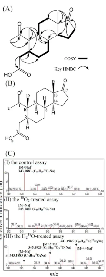
(1H), 1.55 (1H), and 1.42 (1H)] (Table S2). The connectivity of its C-8 and C-9 was approached by the HMBC spectrum (Figure S4D), in which the cross-peaks of H-8/C-9 and H-9/C-8 were observed. Together, the interpretations of distinctive cross-peaks in the COSY and HMBC spectra (Figure 4A) allowed the assignment of compound1as a 2,3-seco-androstane skeleton with one hydroxyl at C-3 and three ketone functionalities at C-1, C-3, and C-17, respectively. Accordingly, compound1 was characterized as the shown structure, and named as 1,17-dioxo-2,3-seco -androstan-3-oic acid (Figure 4B).
A Hydrolytic Ring Cleavage Mechanism Is Adopted byS. denitrificansto Degrade Cholesterol
In the classical 9,10-seco-pathway, oxygenases catalyze oxyge-nolytic ring fission using molecular oxygen as the co-substrate. To determine the origin of the oxygen atoms at C-1 and C-3 of 1,17-dioxo-2,3-seco-androstan-3-oic acid (DSAO), we conducted three
in vitrotransformation assays using 1-testosterone (which has two
16
O atoms at C-3 and C-17) as the substrate: (i) an H218O-treated
assay contained approximately 65% H2 18
O (mole/mole) in the reaction mixture, (ii) an18O2-treated assay, and (iii) a control assay
without the introduction of H218O or18O2. Similar to the DSAO
purified from the control assay (Figure 4CI), no additional18 O-isotopic sodium adduct ion appeared in the ESI-mass spectrum of the DSAO purified from the18O2-treated assay (Figure 4CII). In
contrast, the ESI-mass spectrum of DSAO purified from the H218O-treated assay showed three dominant sodium adduct ions
([M+Na]+
, m/z =343.1883, 345.1928, and 347.1963;
Table 1.UPLC-HRMS and UV absorption behavior of the intermediates involved in aerobic cholesterol catabolism byGordonia cholesterolivoransDSMZ 45229.
Compound ID
UPLC behavior (RTa,min)
Molecular formula/ predicted molecular massb Dominantion peaks Identification of
product ions Mode observed UV Absorption maximum
Cholesterol 11.72 C27H46O/386.3537 369.3531 [M-H2O+H]+ APCI and ESI ,210
Cholest-4-en-3-one 11.02 C27H44O/384.3381 385.3469 367.3365
[M+H]+
[M-H2O+H]+ APCI and ESI APCI and ESI
238
26-hydroxycholest-4-en-3-one 9.40 C27H44O2/400.3330 401.3431 383.3314
[M+H]+[M-H
2O+H]+ APCI and ESI APCI and ESI
241
Cholest-4-en-3-one-26-oic acid 7.39 C27H42O3/414.3123 415.3212 397.3107 379.3001 437.3065
[M+H]+
[M-H2O+H]+ [M-2H2O+H]+ [M+Na]+
APCI and ESI APCI and ESI APCI and ESI ESI
243
Cholest-4-en-3-one-24-oic acid 5.83 C24H36O3/372.2655 373.2743 [M+H]+ APCI and ESI 243
Pregn-4-en-3-one-20-carboxylic acid
4.94 C22H32O3/344.2343 345.2430 327.2324 309.2218
[M+H]+
[M-H2O+H]+ [M-2H2O+H]+
APCI and ESI APCI and ESI APCI
241
Androst-4-en-3,17-dione 3.62 C19H26O2/286.1926 287.2011 269.1905 309.1931
[M+H]+[M-H 2O+H]+ [M+Na]+
APCI and ESI APCI and ESI ESI
238
Androsta-1,4-diene-3,17-dione 3.18 C19H24O2/284.1770 285.1855 267.1749 307.1674
[M+H]+[M-H 2O+H]+ [M+Na]+
APCI and ESI APCI and ESI ESI
242
aRT, retention time.
Figure 4CIII). Their elemental compositions were respectively calculated as C19H2816O4Na, C19H2816O318ONa, and
C19H2816O218O2Na using MassLynxTMMass Spectrometry
Soft-ware (Waters). These data indicate that in the alternative pathway, after the activation of the A-ring through a hydration reaction, the cleavage of the core ring system of cholesterol begins with the A-ring by a hydrolysis reaction.
25-Hydroxycholest-4-en-3-one is Transformed to 26-Hydroxycholest-4-en-3-one inS. denitrificans
In the 9,10-seco-pathway, 26-hydroxycholest-4-en-3-one is produced from cholest-4-en-3-one by steroid C26-hydroxylase, which requires oxygen and NADH as the co-substrate and the electron donor, respectively [28]. Steroid C26-hydroxylase activity was not detected in aerobically cholesterol-grown S. denitrificans
cells (Table S1). In an 18O-labeled water-treated assay, the anaerobic reaction mixture (1 ml) containing ,65% H218O
(mole/mole), soluble protein fraction precipitated at 25% ammo-nia sulfate saturation (1.2 mg), 5% (w/v) hydroxypropyl-b -cyclodextrin, K3[Fe(CN)6] (5 mM), and cholest-4-en-3-one
(2 mM) was incubated for 16 h. The ethyl acetate-extractable sample was then analyzed using UPLC- atmospheric pressure chemical ionization (APCI)-HRMS. The two constitutional isomers (25-hydroxycholest-4-en-3-one and 26-hydroxycholest-4-en-3-one) can be distinguished easily because they exhibited different UPLC behavior (Figure 5). In the mass spectrum of 25-hydroxycholest-4-en-3-one, around two thirds of the product showed an18O signal which originated from H2
18
O (Figure 5C).
18
O-incorporation signal was also observed in 26-hydroxycholest-4-en-3-one produced from the 18O-labeled water-treated assay (Figure 5F). In contrast, no 18O-incorporation signals were detected in the hydroxylated steroids produced in the control assay (Figures 5A and 5D) and18O2-treated assay (Figures 5B and
5E).
Discussion
An Alternative Cholesterol Catabolic Pathway is Present inS. denitrificans
In this study, we used 13C-labeled cholesterol as a tracer to identify the downstream C27,C19 intermediates (from
26-hydroxycholest-4-en-3-one to DSAO, see Figure 1B for their structures) of the aerobic cholesterol catabolic pathway. According to the13C-metabolomics and18O-incorporation data, we demon-strate thatS. denitrificansadopts an oxygenase-independent strategy to degrade cholesterol under oxic conditions. In the proposed pathway, 1,17-dioxo-2,3-seco-androstan-3-oic acid (the product of core ring cleavage) serves as the key intermediate. This compound
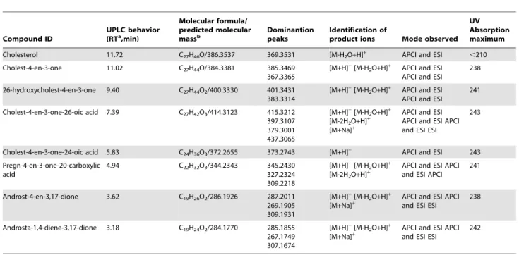
Figure 2. Aerobic cholesterol catabolic pathway byS denitrifi-canswas inhibited bytert-butyl alcohol but not bya,a9-D.(A)
UPLC-HRMS analysis of ethyl-acetate extracts of cholesterol-grown bacterial cells with or withouta,a9-D. (AI)G.cholesterolivoransDSMZ
45229 grown with cholesterol (2 mM), (AII)G.cholesterolivoransgrown with cholesterol anda,a9-D (5 mM), (AIII)S. denitrificansDSMZ 13999
grown with cholesterol, and (AIV)S. denitrificansgrown with cholesterol and a,a9-D. (B) UPLC-HRMS analysis of ethyl-acetate extracts of cholesterol-grownS. denitrificanscells with different concentrations of
tert-butyl alcohol. (BI) The aerobic growth withouttert-butyl alcohol,
(BII) in the presence of 2.5% (v/v)tert-butyl alcohol, and (BIII) in the presence of 5%tert-butyl alcohol. Abbreviations: TIC, total ion current;
26-OH, 26-hydroxycholest-4-en-3-one; 25-OH, 25-hydroxycholest-4-en-3-one; PCA, pregn-4-en-3-one-20-carboxylic acid; AD, androst-4-en-3,17-dione; ADD, androsta-1,4-diene-3,17-androst-4-en-3,17-dione; *, unidentified nitrogen compounds.
has never been reported to be involved in aerobic steroid catabolism. Since the 9,10-seco-pathway was established thirty years ago [6], this is the first documented case that clearly demonstrates the existence of an alternative cholesterol catabolic pathway in bacteria. The proposed alternative pathway is significantly different from the 9,10-seco-pathway.
Comparison with the Classical 9,10-seco-pathway
In the alternative catabolic pathway, the side-chain degradation also precedes the core ring cleavage (Figure 1B). However, the alternative pathway differs from the established 9,10-seco-pathway in the mechanisms of substrate activation and core ring cleavage. In the in vivo[4C-13C]cholesterol biotransformation assay, both
13
C-labeled 25-hydroxycholest-4-en-3-one and 26-hydroxycholest-4-en-3-one could be detected. The NADH-dependent steroid C26-hydroxylase was purified from Rhodococcus jostii [28] and
Mycobacterium tuberculosis[34]. However, neither the corresponding gene [30] nor the enzyme activity (this study) of steroid C26-hydroxylase could be detected inS. denitrificanscells. Moreover, we confirmed that the terminal hydroxyl group of 26-hydroxycholest-4-en-3-one produced byS. denitrificansoriginates from water. These data indicate that in the aerobic cholesterol catabolism by S. denitrificans, 26-hydroxycholest-4-en-3-one may be produced from 25-hydroxycholest-4-en-3-one through a novel isomerization reaction. Similar isomerization reactions of hydroxyl groups occur in monoterpene metabolism [35,36]. Researchers have recently isolated and characterized linalool dehydratase-isomerase [37]. This enzyme catalyzes the migration of a hydroxyl group from a tertiary carbon to a primary one. A similar enzyme might catalyze the isomerization of 25-hydroxycholest-4-en-3-one to a 26-hydroxyl structure.
After substrate activation by adding a hydroxyl group at C-26, the aliphatic side-chain of the C27steroid substrates is degraded
through a series of retro-aldol andb-oxidation reactions to form C24, C22 acidic intermediates and C19 androgens, which were
observed in cholesterol-grownS. denitrificanscultures. These side-chain-degrading reactions do not require oxygenases. In contrast to the 9,10-seco-pathway, the proposed catabolic route applies no oxygenases for ring fission (Figure 1B). The core ring structure opens first at the A-ring through a hydrolytic mechanism. The
18
O-incorporation experiments corroborate the proposed hydro-lytic ring cleavage mechanism.
Recently, new pathways for the degradation of aromatic compounds under oxic conditions were unraveled [38,39]. These pathways operate primarily in facultative anaerobes and use a hydrolytic mechanism to open the ring of the substrates. However, these aerobic pathways still employ monooxygenases to introduce hydroxyl groups into the aromatic ring for substrate activation. In
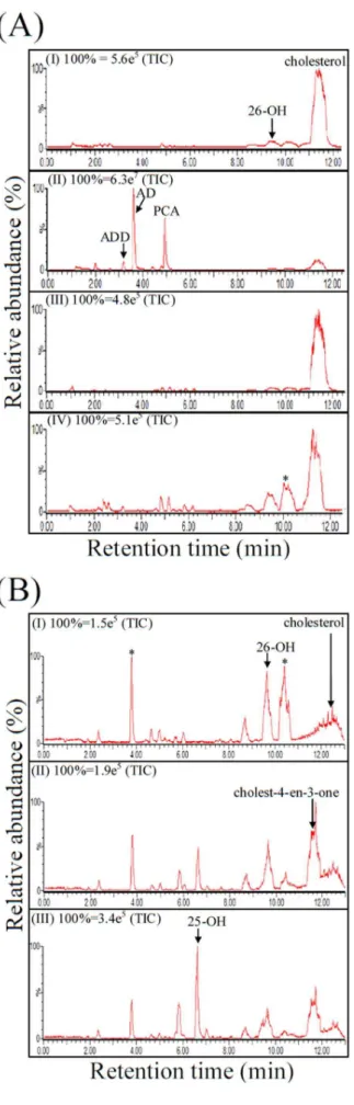
Figure 3. Aerobic cholesterol catabolism by S. denitrificans
DSMZ 13999. (A) Time course of cholesterol consumption and intermediate production in a S. denitrificans fed-batch culture.
[4C-13C]cholesterol (1 mM) was fed to a starvedS. denitrificansculture
(OD600nm= 0.9) after 2 mM cholesterol was exhausted. 0.1 mM of
estrone was added as the internal control. The culture was incubation at 28uC for 30 hours with shaking (180 rpm). The steroids sampled at different time intervals were extracted with ethyl acetate three times, and the dominant intermediates were then separated and quantified using a reverse-phase HPLC system. Data are averages of three determinations (standard deviations ,0.1). (B) High-resolution mass spectra of the dominant13C-labeled intermediates detected in ethyl-acetate extracts ofS. denitrificanscells grown on [4C-13C]cholesterol
(1 mM). *The predicted elemental composition of individual interme-diates was calculated using MassLynxTMMass Spectrometry Software
(Waters). See Figure S1 for other detected13C-labeled intermediates
derived from [4C-13C]cholesterol.
contrast, the proposed aerobic cholesterol degradation pathway does not require any oxygenases-catalyzed reactions till the stage at which the steroidal A-ring opens.
Potential Ecological Significance of the Proposed Cholesterol Degradation Pathway
S. denitrificanscan degrade cholesterol regardless of the presence of oxygen. Several lines of evidence suggest that very similar metabolic strategies may be adopted byS. denitrficansto degrade cholesterol under oxic and anoxic conditions. First, previous proteome analyses revealed no apparent differences in soluble protein patterns of anaerobically and aerobically grown S. denitrificans cells [33]. Second, the steroid-transforming enzymes involved in the initial steps of anaerobic cholesterol metabolism by
S. denitrificansare not oxygen-labilein vivo[30,32]. Third, the O2
-dependent steroid-transforming enzymes, including steroid C26-hydroxylase and 3-ketosteroid 9a-hydroxylase, are not detected in aerobically cholesterol-grownS. denitrificanscells (this study).
It is tempting to speculate that S. denitrificans have developed efficient mechanisms to profit from the available carbon sources regardless of the prevailing redox state. The first adaptive mechanism could be the ability to initiate the degradation of steroid substrates under both oxic and anoxic conditions via similar reactions and intermediates [33]. The second mechanism involves the adoption of oxygenase-independent aerobic catabolic pathways. Both mechanisms would enhance the metabolic competence of these organisms because they can switch quickly between aerobic and anaerobic metabolic modes. Moreover, under micro-aerobic conditions, when the oxygen tension becomes insufficient, the organisms can channel the oxygen flux to the respiratory electron transport chain, and still profit from the steroid substrate through oxygenase-independent catabolic path-ways that do not consume molecular oxygen.
Recently, very similar ring cleavage mechanisms were observed in anaerobic testosterone degradation by a c-proteobacterium,
Steroidobacter denitrificans DSMZ 18526 [40]. Interestingly, the bacterial strain uses the 9,10-seco-pathway to degrade testosterone when oxygen is available. These data indicated that bacteria adopt the oxygenase-independent 2,3-seco-pathway to degrade steroids not only under anaerobic conditions [40], but also under aerobic conditions (at least in this case). So far, less is known about the enzymes (especially the A-ring-cleavage enzyme) and their corresponding genes involved in the 2,3-seco-pathway. Therefore,
in situ 13C-metabolomics seems to be a feasible approach to investigate the contribution of the 2,3-seco-pathway in the degradation of cholesterol and other steroids in natural environ-ments and engineered systems.
Conclusions
The results of this study demonstrate that microbial degradation of one substrate can proceed via different mechanisms under the same conditions. The cholesterol degradation pathway proposed in this study further underpins the diversity of microbial catabolism of organic compounds. It also broadens our under-standing of the strategies that microorganisms use to cope with and adapt to environmental conditions and challenging inert substrates such as steroids.
Figure 4. The structure elucidation and the investigation of the ring cleavage mechanism of compound 1 (1,17-dioxo-2,3-seco -androstan-3-oic acid, DSAO).(A) The interpretations of COSY and key HMBC spectra of compound 1. (B) The chemical structure of compound1. *The oxygen atoms were labeled with18O in the H
218
O-incorporation assay. (C) ESI-mass spectra (positive ion mode) of DSAO. (CI) DSAO purified from the anaerobic control assay. (CII) DSAO purified
from the18O2-treated assay. (CIII) DSAO purified from the18O-labeled
H2O-treated assay. For detailed NMR spectral data of compound1, see
Table S2.
Materials and Methods
Chemicals and Bacterial Strains
The [4C-13C]cholesterol, 18O-labeled water (97 atom%), and 18O
2 (99 atom%) were purchased from Sigma-Aldrich.
[2,3,4C-13C]testosterone was obtained from Isosciences. The
chemicals were of analytical grade and were purchased from Fluka, Mallinckrodt Baker, Merck, or Sigma-Aldrich. Sterolibacter-ium denitrificansDSMZ 13999 andGordonia cholesterolivoransDSMZ 45229 were obtained from the Deutsche Sammlung fu¨r Mikroor-ganismen und Zellkulturen (Braunschweig, Germany).
The Preparation of Steroid Intermediates
25-Hydroxycholest-4-en-3-one was producedin vitroand puri-fied as described elsewhere [32]. 1-Testosterone, androst-1-en-3,17-dione, and 1-hydroxysteroids (C19) were produced and
purified as mentioned [41].
Fed-batch Growth ofS. denitrificanswith Unlabeled Cholesterol
In this study, 0.5% of hydroxypropyl-b-cyclodextrin was always added to the bacterial cultures to improve the solubility of cholesterol in media. S. denitrificans was grown in phosphate-buffered shake-flask cultures (500 ml in 2 l Erlenmeyer flasks) containing 2 mM cholesterol. The culture was incubated at 28uC in an orbital shaker (180 rpm). In 1 l of distilled water, the medium contained the following: 0.77 g cholesterol, 5 g hydro-xypropyl-b-cyclodextrin, 1.0 g NH4Cl, 0.5 g MgSO4?7 H2O, and
0.1 g CaCl2?2H2O. After autoclaving, sterile 50 ml KH2PO4
-K2HPO4buffer solution (1 M, pH 7.0), vitamins (1 ml l21) [42],
EDTA-chelated mixture of trace elements (1 ml l21) [43], and selenite and tungstate solution (1 ml l21) [44] were added. The amounts of residual cholesterol in the cultures were monitored using HPLC. After the consumption of 1.5 mM cholesterol, the pH of the cultures was adjusted to pH,2 using 5M HCl. The acid-treated cultures were extracted 3 times with the same volume of ethyl acetate to recover the residual cholesterol and its
Figure 5. APCI-mass spectra (positive mode) of UPLC-separated 25-hydroxycholest-4-en-3-one (25-OH) and 26-hydroxycholest-4-en-3-one (26-OH).(A) 25-OH produced in the anoxic control assay. (B) 25-OH produced in the18O
2-treated assay. (C) 25-OH produced in the H218
O-treated assay. (D) 26-OH produced in the anoxic control assay. (E) 26-OH produced in the18O2-treated assay. (F) 26-OH produced in the H218O-treated
assay.
derivatives from the aqueous phase. The separation of ethyl acetate extracts was performed using silica gel chromatography, TLC, and HPLC.
Cholesterol Catabolism byS. denitrificansin the Presence ofa,a9-D
S. denitrificanswas grown in two phosphate-buffered shake-flask cultures (50 ml in 250 ml-Erlenmeyer flasks) containing 2 mM cholesterol at 28uC with shaking. After the consumption of 1 mM cholesterol, 5 mM of a,a9-D (an inhibitor of 3-ketosteroid 9a -hydroxylase [12–15]) was added to one of the culture, and the incubation of both cultures continued for 16 h. The pH of the cultures was subsequently adjusted to pH ,2, and ethyl acetate was used to extract cholesterol-derived neutral and acidic intermediates. Cholesterol metabolism by G. cholesterolivorans
DSMZ 45229 was also studied using the same procedure for comparison. The four ethyl acetate extracts were analyzed using UPLC-HRMS.
Effect oftert-Butyl Alcohol on Cholesterol Catabolism of S. denitrificans
S. denitrificanswas aerobically grown in three phosphate-buffered shake-flask cultures (50 ml) containing 2.5 mM cholesterol. After the consumption of 2 mM cholesterol, 2.5% and 5% (v/v) tert -butyl alcohol (an analog of 25-hydroxycholest-4-en-3-one) was individually added to two cultures. 2-Propanol was then added to three cultures to bring the final alcohol concentration to 5% (v/v) in all cultures. The incubation of the three cultures continued further 16 h. The pH of the cultures was adjusted to pH,2, and ethyl acetate was used to extract cholesterol-derived intermediates. The three ethyl acetate extracts were analyzed using UPLC-HRMS.
Fed-batch Growth ofS. denitrificans with [4C-13C]Cholesterol
AS. denitrificansculture (500 ml) was first grown with 2 mM of unlabeled cholesterol in a 2 l Erlenmeyer flask. After the unlabeled cholesterol was completely consumed, 50 ml of the stock culture was transferred into a sterile 250-ml Erlenmeyer flask. The S. denitrificans cells were subsequently fed with 1 mM [4C-13 C]cho-lesterol and incubated at 28uC with shaking (180 rpm). Estrone (0.1 mM) which cannot be utilized by S. denitrificansas a carbon and energy source was added as an internal control. Samples (3 ml) were withdrawn every two hours. Culture samples (0.1 ml
63) were centrifuged at 10,0006gfor 10 min to harvest the S. denitrificanscells. The protein content in the pellet was determined using bicinchoninic acid (BCA) assay. The residual culture samples (2.7 ml) were acidified to pH,2, and extracted three times with the same volume of ethyl acetate to recover cholesterol-derived intermediates. The ethyl acetate fractions were combined, the solvent was evaporated, and the residue was re-dissolved in 300ml of methanol. The [4C-13C]cholesterol-derived intermediates in 60ml samples were identified using ultra-performance liquid chromatography - high-resolution mass spectrometry (UPLC-HRMS). The amount of residual cholesterol and cholesterol-derived intermediates in the samples (80ml63) was determined
using HPLC.
Fed-batch Growth ofS. denitrificans with [2,3,4C-13C]Testosterone
In another 50 mlin vivobiotransformation assay,S. denitrificans
(50 ml culture in a 250-ml Erlenmeyer flask) transferred from the same stock culture was fed with 1 mM [2,3,4C-13C]testosterone.
The samples (3 ml) were withdrawn after 16h incubation. The pH of the culture samples was adjusted to pH,2 using 5M HCl. The ethyl acetate -extractable samples were analyzed using UPLC-HRMS.
Preparation of Cell Extracts
TheS. denitrificanscultures (500 ml in 2 l Erlenmeyer flasks) were grown with 2 mM cholesterol with shaking (180 rpm). Cells were harvested by centrifugation in the exponential growth phase at OD600of 0.8,1.0 (optical path 1 cm) and the cell pellet was then
stored at280uC. All steps used for preparation of cell extracts were performed at 4uC. Frozen cells were suspended in twice the volume of 150 mM Tris-HCl buffer (pH 7) containing 0.1 mg of DNase I ml21. Cells were broken by passing the cell suspension through a French pressure cell (Thermo Fisher Scientific) twice at 137 MPa. The cell lysate was fractionated using two steps of centrifugation: the first step involved centrifugation for 30 min at 20,0006gto remove the cell debris, unbroken cells and residual cholesterol. The supernatant (crude cell extract) was then centrifuged at 100,0006g for 1.5 h to separate soluble proteins from membrane-bound proteins.
In vitro18O-Incorporation Assays for the Production of 1,17-Dioxo-2,3-seco-androstan-3-oic Acid
To determine the origins of the oxygen atoms at C-1 and C-3 of 1,17-dioxo-2,3-seco-androstan-3-oic acid, threein vitroassays were performed in an anaerobic chamber containing 95% N2and 5%
H2(1atm). The three reaction mixtures (3 ml for each assay) were
incubated at 30uC for 16 h with shaking. After the acidic treatment, the steroid products were extracted from the assays using ethyl acetate, and the extracts were analyzed using UPLC-APCI-mass spectrometry.
(1) Control assay. A 3-ml reaction mixture containing 50 mM Tris-HCl buffer (pH 7) and soluble proteins (15 mg) of
S. denitrificanswere sealed in a 10-ml glass bottle with a rubber stopper. The reaction was started by adding 200ml of 67.5 mM 1-testosterone solution (in 2-propanol) to the assay. The final concentration of the steroid substrate in the reaction mixture was 4.5 mM. The final 2-propanol content was 6.67%.
(2) 18O2-Treated assay. 1.8 ml of 18O2 gas (99 atom %)
was introduced into an anaerobic glass bottle containing 7 ml of headspace (95% N2and 5% H2, 1atm) and 3 ml reaction mixture
containing the same components as the control assay. The final
18
O2concentration in the headspace was,20%.
(3) 18O-Labeled Water-treated assay. A 2.0 ml sample of
18
O-labeled water (97 atom %) was added to 1.0 ml of 150 mM Tris-HCl buffer (pH 7) containing soluble proteins ofS. denitrificans
(15 mg). The final18O-water content was approximately 64.7%. The reaction was started by adding 4.5 mM of 1-testosterone to the anoxic assay. The 2-propanol content was also 6.67%.
Activity Assays for Steroid C26-Hydroxylase (Cyp125)
The Cyp125 activity ofG. cholesterolivoransandS. denitrificanswas measured by monitoring the product (26-hydroxycholest-4-en-3-one) concentration using a Hitachi HPLC module. The reaction mixture (1 ml) contained an air-saturated 100 mM potassium phosphate buffer (pH 7.0), 5 mM NADH, 0.5 mM cholest-4-en-3-one, 5% (w/v) 2-hydroxypropyl-b-cyclodextrin, and soluble proteins (20 mg) precipitated at 50% ammonium sulfate satura-tion. In the anaerobic assays, 2 mM 1,4-dithiothreitol was added to remove residual O2 present in the reaction mixture (1 ml),
which was prepared in an anaerobic chamber containing 95% N2
incubated at 30uC for 16 h with shaking. The reaction was stopped by the addition of 20ml of 25% HCl, and the steroids were extracted using ethyl acetate.
In vitro18O-Incorporation Assays for the Production of 26-Hydroxycholest-4-en-3-one
To determine the origins of the oxygen atoms at C-26 of 26-hydroxycholest-4-en-3-one, threein vitroassays were performed in an anaerobic chamber. In all assays, 20ml of 100 mM cholest-4-en-3-one solution (in 2-propanol) was added to empty glass bottles (3-ml). After complete evaporation of the solvent, the reaction mixture (1 ml) was dispensed anaerobically. The three reaction mixtures (1 ml for each assay) were incubated at 30uC for 16 h with shaking. The ethyl acetate extracts were analyzed using UPLC-APCI-mass spectrometry.
(1) Control Assay. In a 3-ml glass bottle sealed with a rubber stopper, the 1-ml reaction mixture contained 50 mM Tris-HCl buffer (pH 8), 2 mM cholest-4-en-3-one (pre-coated on the bottle wall), 5 mM K3[Fe(CN)6], 5% (w/v)
hydroxypro-pyl-b-cyclodextrin, and solubleS. denitrificansproteins (1.2 mg) precipitated at 25% ammonia sulfate saturation.
(2) 18O2-Treated Assay. 0.5 ml of 18O2 gas (99 atom %) was
introduced into an anaerobic glass bottle containing 2 ml of headspace (95% N2 and 5% H2, 1atm) and 1 ml reaction
mixture containing the same components as the control assay. The final18O2concentration in the headspace was,20%.
(3) 18O-Labeled Water-Treated Assay. A total of 0.67 ml of 18
O-labeled water (97 atom %) was added to 0.33 ml of 150 mM Tris-HCl buffer (pH 8) containing soluble proteins (1.2 mg) precipitated at 25% ammonia sulfate saturation, 15 mM K3[Fe(CN)6], 15% (w/v) hydroxypropyl-b-cyclodextrin. The
final18O-water content was approximately 65%. 2 mM (the final concentration) of cholest-4-en-3-one was pre-coated on the bottle wall as mentioned.
Silica Gel Chromatography
A 385 ml silica gel column (5563 cm; SiliaFlashHP60, Silicycle) was equilibrated with 2 bed volumes ofn-hexane - ethyl acetate (65:35, v/v). The ethyl acetate extract (approximately 400 mg dissolved in 3 ml ethyl acetate) containing cholesterol-derived intermediates was loaded into the column and eluted with the equilibrium solvent system at a flow rate of 2 ml min21. The eluate was collected in 5-ml fractions, and a 0.5 ml sample was taken from each fraction. The solvent was evaporated to dryness, and the residue was re-dissolved in 10ml of methanol. The samples were analyzed using TLC. The fractions containing the same compounds were pooled and evaporated to dryness, and 200ml of methanol was used to dissolve the residue. Further purification of cholesterol-derived intermediates was performed using TLC.
Thin Layer Chromatography (TLC)
The steroid products were then separated on silica gel aluminum TLC plates (ALUGRAMH Xtra SIL G/UVF254,
thickness, 0.2 mm, 20620 cm; Macherey-Nagel) using the
following developing solvent system: dichloromethane - ethyl acetate - methanol (14:4:1, v/v). The steroid compounds were visualized under UV light at 254 nm or by spraying the TLC plates with 30% (v/v) H2SO4.
High-Performance Liquid Chromatography (HPLC)
A reversed-phase Hitachi HPLC system was used for the final separation. The separation was achieved on an analytical RP-C18
column (Luna 18(2), 5mm, 15064.6 mm; Phenomenex) incubated at 35uC. The mobile phase included a mixture of two solvents: A (0.1% aqueous trifluoroacetic acid) and B (methanol containing 0.1% trifluoroacetic acid). The C27steroids were eluted at a flow
rate of 1.0 ml/min with a gradient from 80%–90% B over 5 min, followed by isocratic elution at 90% B for 10 min, a gradient from 90%–100% B for 5 min, and further isocratic elution for 20 min. The separation of C19 steroids were performed at a flow rate of
1.0 ml/min with a gradient from 40%–65% B within 50 min. The steroid products were detected in the range of 200–300 nm using a photodiode array detector. The structures of HPLC-purified intermediates were elucidated using NMR spectroscopy and mass spectrometry. In addition, HPLC was used for the quantification of some steroid substrates and intermediates present in the S. denitrificans cultures. The quantification of steroids (cholesterol, cholest-4-en-3-one, ADD, androstan-1,3,17-trione, and 1,17-dioxo-2,3-seco-androstan-3-oic acid) was calculated from their respective peak areas using a standard curve of individual standards. The R2 values for the standard curves were .0.98. Data are averages of three measurements.
Ultra-Performance Liquid Chromatography–Atmospheric Pressure Chemical Ionization–High-Resolution Mass Spectrometry (UPLC-APCI-HRMS)
The ethyl acetate extractable samples or HPLC-purified steroid intermediates were analyzed using UPLC-MS with UPLC coupled to an APCI-mass spectrometer. Mass spectral data were obtained using a Waters HDMS-QTOF synapt mass spectrometer (Waters) equipped with a standard APCI source operating in the positive ion mode. Separation was achieved on a reversed-phase C18
column (Acquity UPLCH BEH C18, 1.7mm, 10062.1 mm;
Waters) with a flow rate of 0.3 ml min21at 50uC (column oven temperature). The mobile phase comprised a mixture of two solvents: Solvent A (2% (v/v) acetonitrile containing 0.1% formic acid to enable excellent ionization in the APCI) and Solvent B (90% isopropanol containing 0.1% formic acid). Separation was achieved with a linear gradient of Solvent B from 30% to 90% in 12 min. In APCI-MS analysis, the temperature of the ion source was maintained at 100uC. Nitrogen desolvation gas was set at a flow rate of 500 l h–1 and the probe was heated to 400uC. Nitrogen served as the APCI nebulizer gas. The corona current was maintained at 20mA, and the electron multiplier voltage was set to1700 eV. The parent scan was in the range of 50–500m/z. The predicted elemental composition of individual intermediates was calculated using MassLynxTM Mass Spectrometry Software (Waters).
Ultra-Performance Liquid Chromatography–Electrospray Ionization–High-Resolution Mass Spectrometry (UPLC-ESI-HRMS)
NMR Spectroscopy
The1H- and13C-NMR spectra were recorded at 27uC using a Bruker AV600_GRC 600MHz NMR. Chemical shifts (d) were recorded and shown as ppm values with deuterated methanol (99.8%,1H:d= 3.31 ppm;13C:d= 49.0 ppm) as the solvent and internal reference.
Supporting Information
Figure S1 High-resolution mass spectra of other 13 C-labeled intermediates detected in ethyl-acetate extracts of S. denitrificans cells grown on [4C-13C]cholesterol (1 mM). *The predicted elemental composition of individual intermediates was calculated using MassLynxTMMass Spectrom-etry Software (Waters).
(TIF)
Figure S2 High-resolution mass spectra of 13C-labeled intermediates detected in ethyl-acetate extracts of S. denitrificans cells grown on [2,3,4C-13C]testosterone (1 mM). *The predicted elemental composition of individual intermediates was calculated using MassLynxTMMass Spectrom-etry Software (Waters).
(TIF)
Figure S3 ESI- (A) and (B) APCI-mass spectra of HPLC-purified compound 1.The predicted elemental composition of
the product ions was calculated using MassLynxTM Mass Spectrometry Software (Waters).
(TIF)
Figure S4 1H-NMR (A), 13C-NMR (B), 1H-1H COSY (C), HMBC (D), and HSQC (E) spectra of HPLC-purified compound 1 (600 MHz, CD3OD).
(PDF)
Table S1 Steroid C26-hydroxylase activity was detected in G. cholesterolivorans, but not inS. denitrificanscells.
(DOC)
Table S2 1H, 13C, COSY, and HMBC interpretations of compound1[din ppm, multi. (Jin Hz)].
(PDF)
Acknowledgments
We thank the Small Molecule Metabolomics Core Facility sponsored by the Institute of Plant and Microbial Biology, Academia Sinica for UPLC-MS analyses. NMR analysis was performed by the NMR Laboratory sponsored by the Genomics Research Center, Academia Sinica, Taiwan.
Author Contributions
Conceived and designed the experiments: Y-RC P-HW. Performed the experiments: P-HW C-YT C-WL Y-WT. Analyzed the data: P-HW T-HL Y-RC. Contributed reagents/materials/analysis tools: T-HL. Wrote the paper: Y-RC WI.
References
1. Johnson DF, Bennett RD, Heftmann E (1963) Cholesterol in Higher Plants. Science 140: 198–199.
2. Kochhar SP (1983) Influence of processing on sterols of edible vegetable oils. Prog Lipid Res 22: 161–188.
3. Weete JD (1989) Structure and function of sterols in fungi. Adv Lipid Res 23: 484–491.
4. Weete JD, Abril M, Blackwell M (2010) Phylogenetic distribution of fungal sterols. PLoS One 5: e10899.
5. Ismail W, Chiang YR (2011) Oxic and anoxic metabolism of steroids by bacteria. J Bioremed Biodegrad S1: 001. doi:10.4172/2155–6199.
6. Kieslich K (1985) Microbial side-chain degradation of sterols. J Basic Microbiol 25: 461–474.
7. Horinouchi M, Hayashi T, Kudo T (2012) Steroid degradation inComamonas testosteroni. J Steroid Biochem Mol Biol 129: 4–14.
8. Fernandes P, Cruz A, Angelova B, Pinheiro HM, Cabral JMS (2003) Microbial conversion of steroid compounds: recent developments. Enzyme Microb Tec 32: 688–705.
9. Doukyu N (2009) Characteristics and biotechnological applications of microbial cholesterol oxidases. Appl Microbiol Biotechnol 83: 825–837.
10. Tak JD (1942) On bacteria decomposing cholesterol. Antonie van Leeuwenhoek 8: 32–40.
11. Whitmarsh JM (1964) Intermediates of microbiological metabolism of cholesterol. Biochem J 90: 23–24.
12. Arima K, Nagasawa M, Bae M, Tamura G (1969) Microbial transformation of sterols. Part I. Decomposition of cholesterol by microorganisms. Agric Biol Chem 33: 1636–1634.
13. Nagasawa M, Bae M, Tamura G, Arima K (1969) Microbial transformation of sterols. Part II. Cleavage of sterol side chains by microorganisms. Agric Biol Chem 33: 1644–1650.
14. Nagasawa M, Watanabe N, Hashiba H, Murakami M, Bae M, et al. (1970) Microbial transformation of sterols. Part V. Inhibitors of microbial degradation of cholesterol. Agric Biol Chem 34: 838–844.
15. Owen RW, Mason AN, Bilton RF (1983) The degradation of cholesterol by Pseudomonassp. NCIB 10590 under aerobic conditions. J Lipid Res 24: 1500– 1511.
16. Sih CJ, Tai HH, Tsong YY (1967) The mechanism of microbial conversion of cholesterol into 17-keto steroids. J Am Chem Soc 89: 1957–1958.
17. Sih CJ, Tai HH, Tsong YY, Lee SS, Coombe RG (1968) Mechanisms of steroid oxidation by microorganisms. XIV. pathway of cholesterol side-chain degrada-tion. Biochemistry 7: 808–818.
18. Sih CJ, Wang KC, Gibson DT, Whitlock HWJ (1965) On the mechanism of ring A cleavage in the degradation of 9,10-seco steroids by microorganisms. J Am Chem Soc 87: 1386–1387.
19. Sih CJ, Lee SS, Tsong YY, Wang KC (1965) 3,4-Dihydroxy-9,10-secoandrosta-1,3,5(10)-triene-9,17-dione. An intermediate in the microbiological degradation of ring A of androst-4-ene-3,17-dione. J Am Chem Soc 87: 1385–1386.
20. Sih CJ, Lee SS, Tsong YY, Wang KC (1966) Mechanisms of steroid oxidation by microorganisms. VIII. 3,4-Dihydroxy-9,10-secoandrosta-1,3,5(10)-triene-9,17-dione, an intermediate in the microbiological degradation of ring A of androst-4-ene-3,17-dione. J Biol Chem 241: 540–550.
21. Fahrbach M, Kuever J, Meinke R, Ka¨mpfer P, Hollender J (2006)Denitratisoma oestradiolicum gen. nov., sp. nov., a 17beta-oestradiol-degrading, denitrifying betaproteobacterium. Int J Syst Evol Microbiol 56: 1547–1552.
22. Horinouchi M, Hayashi T, Yamamoto T, Kudo T (2003) A new bacterial steroid degradation gene cluster inComamonas testosteroniTA441 which consists of aromatic-compound degradation genes for seco-steroids and 3-ketosteroid dehydrogenase genes. Appl Environ Microbiol 69: 4421–4430.
23. Van der Geize R, Yam K, Heuser T, Wilbrink MH, Hara H, et al. (2007) A gene cluster encoding cholesterol catabolism in a soil actinomycete provides insight intoMycobacterium tuberculosissurvival in macrophages. Proc Natl Acad Sci USA 104: 1947–1952.
24. Pandey AK, Sassetti CM (2008) Mycobacterial persistence requires the utilization of host cholesterol. Proc Natl Acad Sci USA 105: 4376–4380. 25. Van der Geize R, Hessels GI, van Gerwen R, van der Meijden P, Dijkhuizen L
(2002) Molecular and functional characterization of kshA and kshB, encoding two components of 3-ketosteroid 9alpha-hydroxylase, a class IA monooxygenase, inRhodococcus erythropolisstrain SQ1. Mol Microbiol 45: 1007–1018. 26. Capyk JK, D’Angelo I, Strynadka NC, Eltis LD (2009) Characterization of
3-ketosteroid 9{alpha}-hydroxylase, a Rieske oxygenase in the cholesterol degradation pathway ofMycobacterium tuberculosis. J Biol Chem 284: 9937–9946. 27. Petrusma M, Dijkhuizen L, van der Geize R (2009)Rhodococcus rhodochrousDSM 43269 3-ketosteroid 9alpha-hydroxylase, a two-component iron-sulfur-contain-ing monooxygenase with subtle steroid substrate specificity. Appl Environ Microbiol 75: 5300–5307.
28. Rosloniec KZ, Wilbrink MH, Capyk JK, Mohn WW, Ostendorf M, et al. (2009) Cytochrome P450 125 (CYP125) catalyses C26-hydroxylation to initiate sterol side-chain degradation inRhodococcus jostiiRHA1. Mol Microbiol 74: 1031–1043. 29. Drzyzga O, Ferna´ndez de las Heras L, Morales V, Navarro Llorens JM, Perera J (2011) Cholesterol degradation by Gordonia cholesterolivorans. Appl Environ Microbiol 77: 4802–4810.
30. Dermer J, Fuchs G (2012) Molybdoenzyme that catalyzes the anaerobic hydroxylation of a tertiary carbon atom in the side chain of cholesterol. J Biol Chem 287: 36905–36916.
31. Tarlera S, Denner EB (2003)Sterolibacterium denitrificansgen. nov., sp. nov., a novel cholesterol-oxidizing, denitrifying member of the beta-Proteobacteria. Int J Syst Evol Microbiol 53: 1085–1091.
32. Chiang YR, Ismail W, Mu¨ller M, Fuchs G (2007) Initial steps in the anoxic metabolism of cholesterol by the denitrifyingSterolibacterium denitrificans. J Biol Chem 282: 13240–13249.
34. Capyk JK, Kalscheuer R, Stewart GR, Liu J, Kwon H, et al. (2009) Mycobacterial cytochrome p450 125 (cyp125) catalyzes the terminal hydroxyl-ation of C27steroids. J Biol Chem 284: 35534–35542.
35. Hylemon PB, Harder J (1998) Biotransformation of monoterpenes, bile acids, and other isoprenoids in anaerobic ecosystems. FEMS Microbiol Rev 22: 475– 488.
36. Heyen U, Harder J (2000) Geranic acid formation, an initial reaction of anaerobic monoterpene metabolism in denitrifyingAlcaligenes defragrans. Appl Environ Microbiol 66: 3004–3009.
37. Brodkorb D, Gottschall M, Marmulla R, Lu¨ddeke F, Harder J (2010). Linalool dehydratase-isomerase, a bifunctional enzyme in the anaerobic degradation of monoterpenes. J Biol Chem 285:30436–30442.
38. Fuchs G, Boll M, Heider J (2011) Microbial degradation of aromatic compounds - from one strategy to four. Nat Rev Microbiol 9: 803–816.
39. Ismail W, Gescher J (2012) Epoxy coenzyme a thioester pathways for degradation of aromatic compounds. Appl Environ Microbiol 78: 5043–5051. 40. Wang PH, Leu YL, Ismail W, Tang SL, Tsai CY, et al. (2013) The anaerobic
and aerobic cleavage of the steroid core ring structure bySteroidobacter denitrificans. J Lipid Res 54: 1493–1504.
41. Leu YL, Wang PH, Shiao MS, Ismail W, Chiang YR (2011) A novel testosterone catabolic pathway in bacteria. J Bacteriol 193: 4447–4455.
42. Pfenning N (1978)Rhodocyclus purpureusgen. nov. and sp. nov., a ring-shaped, vitamin B12-requiring member of the family Rhodospirillaceae. Int J Syst bacteriol 28: 283–288.
43. Rabus R, Widdel F (1995) Anaerobic degradation of ethylbenzene and other aromatic hydrocarbons by new denitrifying bacteria. Arch Microbiol 163: 96– 103.