In Vivo Binding of the Cry11Bb Toxin of
Bacillus thuringiensis
subsp.
medellin
to the Midgut of Mosquito Larvae
(Diptera: Culicidae)
Lina María Ruiz, César Segura/*, Judith Trujillo**, Sergio Orduz/***/
+Unidad de Biotecnología y Control Biológico, Corporación para Investigaciones Biológicas, Medellín, Colombia *Facultad de Medicina, Universidad de Antioquia, Medellín, Colombia **Facultad de Medicina, Universidad Pontificia Bolivariana, Medellín,
Colombia ***Universidad de Pamplona, Pamplona (NS), Colombia
Bacillus thuringiensis subsp. medellin produces numerous proteins among which 94 kDa known as Cry11Bb, has mosquitocidal activity. The mode of action of the Cry11 proteins has been described as similar to those of the Cry1 toxins, nevertheless, the mechanism of action is still not clear. In this study we investigated the in vivo binding of the Cry11Bb toxin to the midgut of the insect species Anopheles albimanus, Aedes aegypti, and Culex quinquefasciatus
by immunohistochemical analysis. Spodoptera frugiperda was included as negative control.
The Cry11Bb protein was detected on the apical microvilli of the midgut epithelial cells, mostly on the posterior midgut and gastric caeca of the three mosquito species. Additionally, the toxin was detected in the Malpighian tubules of An. albimanus, Ae. aegypti, Cx. quinquefasciatus, and in the basal membrane of the epithelial cells of Ae. aegypti midgut. No toxin accumulation was observed in the peritrophic membrane of any of the mosquito species studied. These results confirm that the primary site of action of the Cry11 toxins is the apical membrane of the midgut epithelial cells of mosquito larvae.
Key words: Diptera - immunohistochemistry - in vivo binding - Bacillus thuringiensis - Cry11Bb toxin
Mosquitoes are vectors of important tropical diseases such as malaria, yellow fever, and dengue. Control of mosquito vectors has been accomplished with bioinsec-ticides developed with the bacteria Bacillus thuringiensis
subsp. israelensis and B. sphaericus. B. thuringiensis
subsp. israelensis produces Cry4Aa, Cry4Ba, Cry10Aa, Cry11Aa, and Cyt1Aa toxins, while B. sphaericus pro-duces a binary toxin. Resistance of several populations of Culex mosquitoesto B. sphaericus toxin in different regions of the world has been reported (Rao et al. 1995, Regis et al. 1995). Resistance to B. thuringiensis subsp.
israelensis has not been reported in the field, although resistant mosquitoes populations have thus been selected in the laboratory (Georghiou & Wirth 1997, Wirth et al. 1998). For these reasons and due to the severe impact on public health of the diseases transmitted by mosquitoes, interest on identification of new strains, and active toxins against mosquitoes has increased resulting in the dis-covery of new toxins (Schnepf et al. 1998). The Cry11Bb toxin produced by B. thuringiensis subsp. medellin is active against different species of mosquito larvae (Orduz et al. 1994, 1998), and could represent a new alternative for mosquito larvae control.
Financial support: Comité para el Desarrollo de la Investigación, Universidad de Antioquia, Colciencias, and Corporación para Investigaciones Biológicas
+Corresponding author. Fax: +574-441.5514. E-mail: [email protected]
Received 14 July 2003 Accepted 7 January 2004
The Cry11Bb protein requires a proteolytic process-ing of the 94 kDa protoxin in order to produce the 30 and 35 kDa active fragments, through an intermediate 68 kDa carried-out by intestinal proteases of the target insect (Segura et al. 2000). The mode of action of Cry proteins has been described based mainly on B. thuringiensis lepi-dopteran active toxins. The active fragments specifically interact with brush border membranes of the midgut’s epi-thelial cells (Van Rie et al. 1990), with the irreversible bind-ing bebind-ing fundamental for toxicity (Rajamohan & Charles 1995, Abdul-Rauf & Ellar 1999, Aronson & Shai 2001). Later on in the process, pores are formed, that possibly require the intermolecular interaction among several mono-mers of the toxin (Aronson et al. 1999, Soberon et al. 2000). These pores alter the permeability of the membrane (Luo et al. 1999), cause inhibition of amino acid transport (Parenti et al. 1995), with cellular death occurring due to an os-motic lysis mechanism.
The analyses of the in vivo and in vitro binding of the Cry4Aa, Cry4Ba, and Cry11Aa B. thuringiensis subsp.
membrane vesicles (BBMV), showed a specific and satu-rable interaction of the intermediate 125I -68 kDa with the BBMV of Ae. aegypti. On the other hand, ligand blot as-says results with the 30 and 35 kDa fragments to BBMV of Cx. quinquefasciatus, Ae. aegypti, and An. albimanus
indicate that there is no binding, whereas the 94 kDa and 68 kDa fragments showed binding to the mosquitoes BBMV (Segura 2001).
In this study, we investigated the in vivo binding of the Cry11Bb toxin to the mosquito larvae midgut, through immunohistochemical methods in order to determine the primary site of action of this toxin.
MATERIALS AND METHODS
Insects - Larvae from the three mosquito species used
(Ae. aegypti, An. albimanus, Cx. quinquefasciatus)and the lepidopteran Spodoptera frugiperda were maintained under laboratory conditions at 30 ± 2oC under a 12:12 (light:dark) photoperiod in the insectary of the Corporación para Investigaciones Biológicas, Medellín, Colombia. Mosquitoes were fed with soybean powder. S. frugiperda
was fed with an artificial diet based on bean (Arango et al. 2002), and included as a negative control in which bind-ing of Cry11Bb protein is not expected to occur.
Solubilization and purification of the Cry11Bb δ -en-dotoxin - The Cry11Bb protoxin was obtained from the acrystalliferous recombinant strain SPL-407 of B. thuringiensis subsp. thuriengiensis that had been trans-formed with the plasmid pSOB and contained a 3.1-kb insert of DNA encoding the Cry11Bb protoxin (Orduz et al. 1998). These cells were grown in 250 ml of M-one me-dium (Restrepo et al. 1997) supplemented with erythro-mycin (25 µg/ml) for 48 h at 30°C; and then transferred to a 20 l fermentor containing 10 l of culture medium; the fermentation conditions have already been published (Vallejo et al. 1999). The final whole culture was harvested by centrifugation, the pellet was treated with 1 M NaCl for 1 h with shaking at 300 rpm, at 30°C; the salt was removed by washing twice in water supplemented with 10 mM of phenylmethylsulfonylflouride (PMSF) (Sigma) and 10 mM ethylenediaminetetraacetic acid (EDTA) at 4°C. The re-sulting pellet was treated with 1 M NaCl/1% Triton-X100 at 4°C, salt and detergent were removed with extensive washes by centrifugation at 9000 g for 15 min in distilled water supplemented with 1 mM PMSF and 100 mM EDTA at 4°C, and the pellet was resuspended in 50 ml of 50 mM cyclohexylaminopropane sulfonic acid (CAPS) pH 10.6/ 0.05% β-mercaptoethanol, and left in the shaker for 1 h at 37°C, at 300 rpm. The supernatant containing the soluble toxin was centrifuged at 15,000 g for 1 h at 4°C, and pro-tein concentration was determined by the Bradford method with bovine serum albumin-BSA as standard (Bradford 1976).
Purification of the Cry11Bb toxin was performed through ionic exchange chromatography in a fast perfor-mance liquid chromatograph (FPLC - Bio-Rad, BioLogic LP) using an anionic exchange column Econopack High Q (Bio-Rad), previously balanced with 50 mM CAPS, pH 10.6. The soluble protein (50 mg) was filtered through a 0.22 µm filter and applied to a 5 ml bed of a Q-sepharose column. The toxin was eluted from the column with a NaCl
gradient from 0 to 1 M in 50 mM CAPS (pH 10.6) during 40 min. The collected fractions were analyzed through so-dium dodecyl sulfate 10% polyacrilamide gel electrophore-sis (SDS-PAGE), and fractions were finally aliquoted and stored at –20°C until used.
Cry11Bb treatments - Early fourth instars of the mos-quito species under starvation for 20 h were treated with 21.5 µg/ml, which corresponded to approximately 500-fold the half lethal concentration for the mosquitoes for vari-able periods of time, 0.25, 0.5, 0.75, 1, and 2 h, this concen-tration had been previously used by Orduz et al. (1994). When intoxication and mortality were observed, larvae were removed to a petri dish with distilled water to wash the toxin excess. Fifth instar S. frugiperda larvae were fed with a micro-syringe with 10 µl of Cry11Bb toxin solution containing 10 µg/ml while control larvae were treated with 10 µl of 1% BSA.
Preparation and sectioning of insect tissues - After exposure to the Cry11Bb treatments, mosquito larvae were placed in neutral formaldehyde. After feeding S. frugiperda
larvae with the toxin, guts were dissected at 4°C and the midgut was fixed in neutral formaldehyde. Mosquito lar-vae and their midguts were dehydrated in increasing iso-propyl alcohol concentrations, rinsed in xylol and included in paraffin. Five µm sections were obtained and placed in carriers loaded with 2% 3-aminopropyltriethoxy-silane (Bravo et al. 1992b).
Antibodies - Mice polyclonal antibody against Cry11Bb (94 kDa) was prepared as indicated by Harlow and Lane (1999). Sensibilization and specificity were evaluated by ELISA and Western blot with 1:100, 1:1000 dilutions, respectively, of the mice sera using standard techniques (Voller et al. 1980). The immunodetection was performed in combination with a peroxidase-conjugated goat IgG fraction to mouse IgG (whole molecule) (ICN Pharmaceuticals, Eappel).
in imidazole-HCl buffer pH 7.5 containing hydrogen per-oxide and an antimicrobial agent (Liquid DAB large vol-ume substrate-chromogen system DAKO) for 3 min at room temperature. The counterstaining of the tissue sec-tions was performed with Harris hematoxilin for 10 s and ammoniacal water for 10 s; the tissues were dehydrated in increasing concentrations of ethanol and clarified in xy-lol; finally, the sections were covered with entellan (Merk) mounting resin and analyzed by light microscopy.
RESULTS
Polyclonal antibodies against Cry11Bb were able to detect the 94 kDa protoxin, the 68 kDa intermediate and the 35 kDa form of the Cry11Bb toxin - Production of polyclonal antibodies anti-Cry11Bb1 was assessed by ELISA, obtaining a titre of 3.56 (log10). These polyclonal antibodies obtained in mice recognized the 94 kDa protoxin, as well as the products of the proteolytic pro-cessing, including the 68 kDa intermediate and the 35 kDa active fragment; however, the 30 kDa active fragment was only weakly recognized by Western blot (Fig. 1).
Toxicological effects of the Cry11Bb toxin - In the treatments containing 21.5 µg/ml of Cry11Bb, approxi-mately 500-fold the LC50 of the Cry11Bb for Ae. aegypti, An. albimanus, and Cx. quinquefasciatus, mosquito lar-vae intoxication was observed 30 min after exposure to the toxin and mortality increased 30 min later on for Ae. aegypti and Cx. quinquefasciatus. The external toxico-logical symptoms were seen only 2 h after treatment in
An. albimanus larvae. Toxic effects on S. frugiperda lar-vae were not observed after treatment with the Cry11Bb toxin.
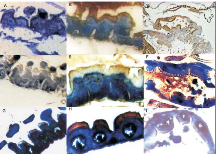
Immunohistochemical localization of the Cry11Bb toxin on the apical microvilli of the midgut ephitelial cells - Binding of the Cry11Bb toxin to mosquito larvae tissues was observed after 15 min exposure to the Cry11Bb toxin by a brownish staining at the apical microvilli, with
the staining intensity remaining unchanged from this time onwards.Control larvae not exposed to the Cry11Bb toxin or those in which the polyclonal antibody against Cry11Bb was omitted, did not show the brownish coloration (Figs 2A, D, G) in none of the mosquito species tested. The Cry11Bb toxin was also detected on the apical microvilli and in the basal membrane of the posterior midgut epithe-lium cells of Ae. aegypti, An. albimanus,and Cx. quin-quefasciatus after 15 min exposure to the toxin (Figs 2B, E, H, respectively). In the gastric caeca of the three mos-quito species, strong signals were also observed (Figs 2C, F, I), while in anterior midgut the signal was weak (data not shown).
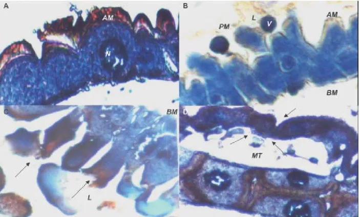
Histopathological effects of the Cry11Bb toxin in mosquito larvae - The general histopathological changes induced by the Cry11Bb toxin in the mosquito larvae gut epithelia included vacuolization of the cytoplasm, hyper-trophy of the epithelial cells and their nucleus, brush bor-der membrane impairment, and disintegration of the cells. After 2 h exposure to the Cry11Bb toxin, vacuolization of the cytoplasm and swollen nuclei were observed (Fig. 3A), midgut columnar cells of the mosquito larvae were elon-gated (Fig. 3B), the cells were disrupted at the apical re-gion with vesicle formation, lysis and leakage of cyto-plasm material into the gut lumen (Figs 3B, C). Addition-ally, in Ae. aegypti and An. albimanus larvae, the Cry11Bb toxin was detected in the Malpighian tubules 15 and 45 min after treatment with the toxin, respectively, while in
Cx. quinquefasciatus larvae, the Cry11Bb toxin was only detected in the Malpighian tubule microvilli junctions 1 h after treatment, and 1 h later these structures had shrunken (Fig. 3D).
Cry11Bb protein was detected in a diffuse way inside the cells and in the apical and basal parts of S. frugiperda
gut epithelium (Fig. 4B), in spite of the faint binding, the midgut of S. frugiperda cells had no histopathological changes (Fig. 4B).
DISCUSSION
Present in vivo experiments showed that theCry11Bb toxin bound preferentially to the posterior midgut apical microvilli and the gastric caeca of the evaluated mosquito species; and therefore, the epithelial cells of the midgut could be considered as the main target of this toxin. The Cry11Bb toxin preferred localization in gastric caeca and the posterior midgut of mosquito larvae may be due to a higher concentration of receptor molecules or of molecules with higher binding affinity. Differential binding in the anterior and posterior midgut of dipterans has already been observed in mosquito larvae treated with B. thuringiensis or B. sphaericus toxins(Ravoahangimalala et al. 1993, Ravoahangimalala & Charles 1995). Toxico-logical effects of the Cry11Bb toxin in mosquito larvae were in agreement with the data reported by Orduz et al. (1994).
The immunolocalization of the Cry11Bb toxin observed in the basal membrane of Ae. aegypti and An. albimanus
larvae was possibly due to the toxin’s leakage or at least part of it from the midgut lumen to the basal area of the epithelium after disruption of the cellular integrity of the midgut tissue. The deterioration of the intercellular junc-Fig. 1A: sodium dodecyl sulfate-polyacrilamide gel electrophoresis;
tions could be a consequence of the damage caused by the Cry11Bb toxin at the cellular membrane level, as this situation has also been observed in Heliothis virescens
midgut after 3 h exposure to the Cry1Ac (Forcada et al. 1999).
Additionally, the Cry11Bb toxin was observed in Cx. quinquefasciatus larval midgut 15 min after treatment, and 1 h after exposure to the toxin on the apical microvilli of the Malpighian tubules. It is unknown if the binding of the Cry11Bb toxin to the Malpighian tubules contributes to mortality of mosquito larvae. The binding sites of sev-eral Cry toxins have been located on the Malpighian tu-bule epithelium of some insect species; however, their role in toxicity has not been fully characterized. Although Maddrell et al. (1989) reported important changes in the trans-epithelial potential difference of Rhodnius prolixus
Malpighian tubules after treatment with B. thuringiensis
toxin and Reisner et al. (1989) located B. thuringiensis kurstaki δ-endotoxins on the Malpighian tubules of
Calpodes ethlius larvae anddescribed its effect as inhibi-tory of fluid secretion causing cytological alterations, which ended-up in cellular lysis and epithelial damage, Denolf et al. (1993) reported binding of Cry1Ab1, Cry1Ac1, and Cry1Ba1 toxins to the Malpighian tubules of Ostrinia
nubilalis, but did not suggest a relevant role in the mor-tality of this lepidopteran. It is possible that the damage caused by the Cry11Bb toxin in the mosquito larvae mid-gut intercellular junctions, as seen in Fig. 3B, could have permitted toxin leakage into the hemolymph to reach the Malpighian tubules, producing shrinking of their epithe-lium (Fig. 3D), and perhaps impairing their osmoregula-tory functions before larval death.
Although S. frugiperda was not susceptible to the toxin, the weak localization of the Cry11Bb protein in its midgut, could be the result of non-specific binging or to low affinity binding to a putative receptor with some de-gree of homology to the mosquito larvae receptors. How-ever, this binding does not guarantee toxicity (Ferré et al. 1991, Bravo et al. 1992a, Feldman et al. 1995).
Some studies have suggested that Cry1 toxins could have more than one binding site in Heliothis sp.,
Spodoptera sp., and Manduca sexta (Oddu et al. 1993, Masson et al. 1995). It is assumed that the mode of action of the Cry11 toxins is similar to that of Cry1 toxins; there-fore, it is possible that the Cry11Bb toxin could have more than one receptor in the midgut apical microvilli of the mosquitoes evaluated as it has been suggested by Segura (2001).
Fig. 3: hispathological effects of the Cry11Bb toxin Bacillus thuringiensis subsp. medellin in mosquito larvae after 2 h of exposure to the Cry11Bb toxin. A: vacuolization of the cytoplasm and hypertrophy of the Culex quinquefasciatus epithelial cells and their nuclei; B: vesicle formation in the apical region of cell towards the midgut lumen of the Anopheles albimanus; C:arrows indicates lysis of columnar cells of Cx. quinquefasciatus; D:localization of the protein Cry11Bb on the apical microvilli of the Malpighian tubules of Cx. quinquefasciatus larvae2 h after exposure to the toxin. The arrow indicates the shrunken tubules. AM: apical microvilli; BM: basal membrane; L: midgut lumen; PM: peritrophic membrane; V: vesicles; N: nucleus. Magnification 40X
Fig. 4. immunohistochemical localization of Bacillus thuringiensis subsp. medellin protein Cry11Bb on the apical microvilli of epithelial cells of the midgut of Spodoptera frugiperda larvae. Omitting primary antibody control (A). Midgut with faint signals on the apical microvilli of the midgut and basal part of the epithelial cells (B). Magnification 100X
Previous in vitro binding studies have shown that the Cry11Bb protoxin (94 kDa) and the 68 kDa intermediate form interact with Ae. aegypti brush border membrane vesicles, while the 30/35 kDa toxin does not (Segura 2001). The in vivobinding of Cry11Bb to the apical microvilli of mosquito larvae, probably corresponds to the 68 kDa form, since the 94 kDa protoxin disappears in the midgut lumen
10 min after treating the larvae (Segura et al. 2000), and the binding pattern seen as the brownish staining was similar in all the analyzed times of the present study.
More-over, the Cry11Bb toxin was localized in the Malphigian tubules of Ae. aegypti An. albimanus, and Cx. quinque-fasciatus larvae, which may indicate a possible relation-ship with the mode of action of this toxin. Further research that includes in vivo or in vitro homologous and heter-ologous competition assays will permit to know if the Cry11Aa and Cry11Bb toxins share a common receptor. This type of research will contribute to the design of more effective biological control agents.
REFERENCES
Abdul-Rauf M, Ellar D 1999. Toxicity and receptor binding properties of a Bacillus thuringiensis CryIC toxin active against both Lepidoptera and Diptera. J Invertebr Pathol 73: 52-58.
Arango JA, Romero M, Orduz S 2002. Diversity of Bacillus thuringiensis strains from Colombia with insecticidal activ-ity against Spodoptera frugiperda (Lepidoptera:Noctuidae).
J Appl Microbiol92: 466-474.
Aronson A, Shai Y 2001. Why Bacillus thuringiensis insecti-cidal toxins are so effective: unique features of their mode of action. FEMS Microbiol Lett195: 1-8.
Aronson A, Geng C, Wu L 1999. Aggregation of Bacillus thuringiensis Cry1A toxins upon binding to target insect larval midgut vesicles. Appl Microbiol Biotechnol65: 2503-2507.
Bradford MM 1976. A rapid and sensitive method for the quantitation of microgram quantities of protein utilizing the principle of protein-dye binding. An Biochem72: 248-254.
Bravo A, Hendricks K, Jansens S, Peferoen M 1992a. Immuno-cytochemical analysis of specific binding of Bacillus thuringiensis insecticidal crystal proteins to lepidopteran and coleopteran midgut membranes. J Invertebr Pathol60: 247-253.
Bravo A, Jansens S, Peferoen M 1992b. Immunocytochemical localization of Bacillus thuringiensis insecticidal crystal proteins in intoxicated insects. J Invertebr Pathol60: 237-246.
Denolf P, Jansens S, Peferoen M, Degheele D, Van Rie J 1993. Two different Bacillus thuringiensis delta-endotoxin recep-tors in the midgut brush border membrane of the european corn borer, Ostrinia nubilalis (Hübner) (Lepidoptera: Pyralidae). Appl Microbiol Biotechnol59: 1828-1837. Feldmann F, Dullemans A, Waalwijh C 1995. Binding of the
CryIVD toxin of Bacillus thuringiensis subsp. israelensis
to larval dipteran midgut proteins. Appl Microbiol Biotechnol 61: 2601-2605.
Ferré J, Real MD, Van Rie J, Jansens S, Peferoen M 1991. Resistance to the Bacillus thuringiensis bioinsecticide in a field population of Plutella xylostella is due to a change in a midgut membrane receptor. Proc Natl Acad Sci USA88: 5119-5123.
Forcada C, Alcácer E, Garcerá MD, Tato A, Martínez R 1999. Resistance to Bacillus thuringiensis Cry1Ac toxin in three strains of Heliothis virescens: Proteolytic and SEM study of the larval midgut. Arch Insect Biochem Physiol42: 51-63.
Georghiou G, Wirth MC 1997. Influence of exposure to single versus multiple toxins of Bacillus thuringiensis subsp.
israelensis on development of resistance in the mosquito
Culex quinquefasciatus (Diptera: Culicidae). Appl Environ Microbiol63: 1095-1101.
Harlow E, Lane D 1999. Using Antibodies: A Laboratory Manual, Cold Spring Harbor Laboratory Press, New York, 495 pp.
Luo K, Banks D, Adang M 1999. Toxicity, binding, and perme-ability analyses of four Bacillus thuringiensis Cry1 δ -en-dotoxins using brush border membrane vesicles of
Spodoptera exigua and Spodoptera frugiperda. Appl Microbiol Biotechnol 60: 457-464.
Maddrell SH, Overton JA, Ellar DJ, Knowles BH 1989. Action of activated 27.000 Mr toxin from Bacillus thuringiensis
var. israelensis on Malpighian tubules of the insect, Rhodnius prolixus. J Cell Sci94: 601-608.
Masson L, Lu Y-J, Mazza A, Brousseau R, Adang M 1995. The Cry1A(c) receptor purified form Manduca sexta displays multiple specificities. J Biol Chem270: 20309-20315. Oddou P, Hartmann H, Radecke F, Geiser M 1993.
Immuno-logically unrelated Heliothis sp. and Spodoptera sp. midgut membrane-proteins bind Bacillus thuringiensis CryIA(b)
δ-endotoxins. Eur J Biochem212: 145-150.
Orduz S, Diaz T, Thiéry I, Charles J-F, Rojas W 1994. Crystal proteins from Bacillus thuringiensis serovar. medellin. Appl Microbiol Biotechnol40: 794-799.
Orduz S, Realpe M, Arango R, Murillo L, Delécluse A 1998. Sequence of the cry11Bb1 gene from Bacillus thuringiensis
subsp. medellin and toxicity analysis of its encoded pro-tein. Biochim Biophys Acta1388: 267-272.
Parenti P, Villa M, Hanozet G, Tasca M, Giordana B 1995. Interaction of the insecticidal crystal protein CryIA from
Bacillus thuringiensis with amino acid transport into brush border membranes from Bombyx mori larval midgut. J Invertebr Pathol 65: 35-42.
Rajamohan F, Alcantara E, Lee MK, Chen XJ, Curtis A, Dean D 1995. Single amino acid changes in domain II of Bacillus thuringiensis CryIAb δ-endotoxin affect irreversible bind-ing to Manduca sexta midgut membrane vesicles. J Bacteriol 177: 2276-2282.
Rao DR, Mani TR, Rajendran R, Joseph AS, Gajanana A, Reuben R 1995. Development of high level of resistance to
Bacillus sphaericus in a field population of Culex quin-quefasciatus from Kochi, India. J Amer Mosq Control Assoc 11: 1-5.
Ravoahangimalala O, Charles J-F 1995. In vitro binding of Ba-cillus thuringiensis var. israelensis individual toxins to mid-gut cells of Anopheles gambiae larvae (Diptera: Culicidae).
FEBS Lett362: 111-115.
Ravoahangimalala O, Charles J-F, Shoeller-Raccaud J 1993. Immunological localization of Bacillus thuringiensis serovar
israelensis toxins in midgut cells of intoxicated Anopheles gambiae larvae (Diptera: Culicidae). Res Microbiol 144: 271-278.
Regis L, Silva-Filha MHNL, de Oliveria CMF, Rios EM, da Silva SB, Furtado A 1995. Integrated control measures against Culex quinquefasciatus, the vector of filariasis in Recife. Mem Inst Oswaldo Cruz90: 115-120.
Reisner W, Feir D, Lavrik P, Ryerse J 1989. Effect of Bacillus thuringiensis kurstaki d-endotoxin on insect Malpighian tubule structure and function. J Invertebr Pathol54: 175-190.
Restrepo N, Gutiérrez D, Patiño MM, Thiéry I, Delécluse A, Orduz S 1997. Cloning, expression and toxicity of a mosquitocidal toxin gene of Bacillus thuringiensis subsp.
medellin. Mem Inst Oswaldo Cruz92: 257-262.
Schnepf E, Crickmore N, Van Rie J, Lereclus D, Baum J, Feitelson J, Zeigler DR, Dean DH 1998. Bacillus thu-ringiensis and its pesticidal crystal proteins. Microbiol Mol Biol Rev 62: 775-806.
Segura C 2001. Study on the Mode of Action of Bacillus thuringiensis subsp. medellin toxins, PhD Thesis, Univer-sity of Antioquia, Medellin, Colombia.
thuringiensis subsp. medellin. J Invertebr Pathol76: 56-62.
Soberón M, Pérez R, Núñez-Valdéz M, Lorence A, Gómez I, Sánchez J, Bravo A 2000. Evidence for intermolecular inter-action as a necessary step for pore-formation activity and toxicity of Bacillus thuringiensis Cry1Ab toxin. FEMS Microbiol Lett 191: 221-225.
Vallejo F, Gonzalez A, Posada A, Restrepo A, Orduz S 1999. Production of Bacillus thuringiensis subsp medellin by batch and fed-batch culture. Biotechnol Tech 13: 279-281. Van Rie J, Jansens S, Höfte H, Degheele D, Van Mellaert H
1990. Receptors on the brush border membrane of the
in-sect midgut as determinants of the specificity of Bacillus thuringiensis delta-endotoxins. Appl Microbiol Biotechnol 56: 1378-1385.
Voller A, Bidwell D, Burek C 1980. An enzyme-linked immunosorbent assay (ELISA) for antibodies to thyroglo-bulin. Proc Soc Exp Biol Med163: 402-405.
Wirth MC, Delecluse A, Federici BA, Walton WE 1998. Vari-able cross-resistance to Cry11B from Bacillus thuringiensis