ABSTRACT
Newly forming bone graft: a novel surgical
approach to the treatment of denuded roots
Adriana Campos Passanezi SANT’ANA1, Bruna F. Rahal FERRAZ2, Maria Lúcia Rubo de REZENDE1, Sebastião Luiz Aguiar GREGHI1, Carla Andreotti DAMANTE3, Euloir PASSANEZI4
1- DDS, PhD, Associate Professor, Discipline of Periodontics, Department of Prosthodontics, Bauru School of Dentistry, University of São Paulo, Bauru, SP, Brazil. 2- DDS, MSD, Graduate student, Discipline of Periodontics, Department of Prosthodontics, Bauru School of Dentistry, University of São Paulo, Bauru, SP, Brazil. 3- DDS, PhD, Assistant Professor, Discipline of Periodontics, Department of Prosthodontics, Bauru School of Dentistry, University of São Paulo. Bauru, SP, Brazil. 4- DDS, PhD, Head of the Discipline of Periodontics, Department of Prosthodontics, Bauru School of Dentistry, University of São Paulo, Bauru, SP, Brazil.
Corresponding address: Adriana Campos Passanezi Sant’Ana - Faculdade de Odontologia de Bauru - USP - Departamento de Prótese - Disciplina de Periodontia - Al. Otávio Pinheiro Brisolla 9-75 - 17012-901 - Bauru - SP - Brazil - Phone: 00 55 (14) 3235-8278 - Fax: 00 55 (14) 3227-5105 - e-mail: [email protected]
Received: October 13, 2010 - Modiication: July 14, 2011 - Accepted: August 15, 2011
M
any techniques have been proposed for root coverage. However, none of them presents predictable results in deep and wide recessions. Objective: The aim of this case series report is to describe an alternative technique for root coverage at sites showing deep recessions and attachment loss >4 mm at buccal sites. Material and Methods: Four patients presenting deep recession defects at buccal sites (≥4 mm) were treated by the newly forming bone graft technique, which consists in the creation of an alveolar socket at edentulous ridge and transferring of granulation tissue present in this socket to the recession defect after 21 days. Clinical periodontal parameters, including recession depth (RD), probing depth (PD), clinical attachment level (CAL), bleeding on probing (BOP), plaque index (PI) and keratinized gingiva width (KGW) were evaluated by a single examiner immediately before surgery and at 1, 3, 6 and 9 months postoperatively. Results: All cases showed reduction in RD and PD, along with CAL gain, although no increase in KGW could be observed. These indings suggest that the technique could favor periodontal regeneration along with root coverage, especially in areas showing deep recessions and attachment loss.Key words: Gingival recession. Guided tissue regeneration. Citric acid. Grafts.
INTRODUCTION
Coverage of denuded roots has been a main concern for both professionals and patients. Many surgical techniques have been proposed to cover denuded roots, showing varying rates of success depending on factors related to the surgical technique and anatomical features of the lesions13,14,17,26. A greater reduction in recession
depth and width as well as a greater increase in the width of keratinized gingiva are obtained by subepithelial connective tissue graft (SCTG) associated to coronally positioned flap5,13,19.
However, the success of such technique is limited
in wide recession defects presenting interproximal bone and soft tissue loss17.
To overcome these problems, the use of barrier membranes positioned over exposed root
surface (GTR) warranting a space for regeneration to occur along with root coverage has been suggested7,20,23,26,27. This technique resulted in gain
of attachment level, reduction of probing depth, bleeding on probing and recession depth19,23,26
in single, large, deep, localized marginal tissue recessions showing >5 mm of depth27, in the
presence of hypersensitivity7, cervical caries
lesions or restorations23,28. Histological studies
demonstrated that wound healing proceeded with the formation of new cementum, periodontal ligament and alveolar bone coronal to the pre-existent level, suggesting that root coverage was accomplished by regeneration of periodontal tissues7,20. In turn, it is contra-indicated for the
treatment of multiple and shallow recessions18,30 or
in areas of thin gingival tissue11.
root coverage was proposed8,12,24,25 to support
the membrane and to act as an osteoinductive/ osteoconductive biomaterial, resulting in improved reduction of recession and probing depth and gain of attachment level compared to the use of barrier membranes alone8,12, as well as a slightly smaller
- but not signiicant - reduction in recession depth
compared with subepithelial connective tissue graft24.
Recently, the use of growth factors or stem cells under membranes has been proposed to treat denuded areas15 or missing papillae16, with
promising results. Passanezi, et al.21 (1989)
proposed a surgical technique based on the transfer of osteogenic cells to treat infrabony periodontal defects with high rates of clinical success. The technique consists in the transfer of healing bone from a surgically created alveolar socket to infrabony periodontal defects approximately 21 days after. A substantial quantity of a relatively mature newly forming bone containing a vast amount of osteoblasts with osteogenic potential is observed in alveolar socket 4-12 weeks after tooth extraction9. This material shows positive staining
against collagen I, osteocalcin, bone sialoprotein and alkaline phosphatase activity22, which are
considered as markers of mesenchymal stem cells3,6, resulting in regeneration of periodontal
tissues in animal and human studies21.
The treatment of wide recession defects by the newly forming bone graft (NFBG) technique would then be favored by the formation of a new periodontal attachment apparatus, even in the presence of attachment loss or thin keratinized tissue. Considering that, the aim of this case series report is to propose a new technique for coverage of denuded roots in order to achieve root coverage along with successful regeneration of bone, cementum and periodontal ligament, especially at deep and wide recession defects.
CASE SERIES REPORT
Case 1
Patient #1 was a 44-year-old systemically healthy never-smoker female who presented for treatment at the Clinics of the Discipline of Periodontics at Bauru School of Dentistry, University of São Paulo, Brazil. Patient reported no use of antibiotics or other medicines in the previous 6-month period. Treatment plan involved extraction of the mandibular left central incisor and root coverage of the mandibular right second premolar, which showed a 5 mm-deep and 4 mm-wide recession at baseline examination. No tooth mobility was present. Clinical and radiographic examination suggested a slight loss of soft and hard tissues at distal sites compatible with a Miller class III
recession defect, since the mandibular right second premolar was adjacent to an edentulous ridge.
Case 2
Patient #2 presented for treatment at the Periodontics Clinics complaining of recession at the
mandibular left irst premolar. Clinical examination
revealed a 5 mm-deep recession defect. Patient reported no systemic disease, no smoking, no use of antibiotics or any other medications or periodontal treatment in the 6-month period previous to baseline examination. No tooth mobility was present. Clinical and radiographic examination suggested a Miller’s class III recession defect.
Case 3
Patient #3, a 35 year-old female, presented a deep recession defect (5 mm) at the mandibular right second premolar. Patient was systemically healthy, never smoker, and reported no use of any medication or periodontal treatment in the 6-month period previous to baseline examination. There was no clinical sign of trauma from occlusion or tooth mobility. Clinical and radiographic examination suggested a Miller’s class III recession defect.
Case 4
Patient #4 was a 32-year-old female with a 4-mm-deep recession defect at the mandibular right second premolar. No tooth mobility was present, and there was no clinical sign of trauma from occlusion. Patient reported to be systemically healthy and never smoker. Additionally, patient reported no regular use of medications or periodontal treatment during the past 6 months. Clinical and radiographic examination suggested a Miller’s class III recession defect.
Phase I therapy
All patients were submitted to a phase I therapy, which included removal of caries and endodontic lesions, oral hygiene instruction and scaling and root planning aiming at plaque control. Surgical treatment was performed after active treatment,
when resolution of inlammation was achieved, as
observed by absence of bleeding on probing and clinical signs of gingival health.
Clinical examination
Clinical examinations were performed by a single trained examiner immediately before surgery and at 1, 3, 6 and 9 months post-operatively. Depth of marginal tissue recession (DR), probing depth (PD), clinical attachment level (CAL), bleeding on probing (BOP) and keratinized gingiva width (KGW) were determined using a millimeter manual probe
(HuFriedy, Chicago, IL, USA). DR was determined
gingival margin. PD was determined by the distance from gingival margin to the bottom of the sulcus.
CAL was deined as the distance from
cementum-enamel junction to the bottom of sulcus (DR+PD). KGW was determined by the distance from gingival margin to the mucogingival junction. The presence of bleeding upon probing was recorded as 1 and its absence as 0. Plaque index was recorded as the presence (1) or absence (0) of plaque after staining of tooth surface with a plaque-evidencing solution. Percentage of root coverage was determined by the application of the formula11:
% root coverage=(RDinitial-RDinal)x100/RDinitial
Description of NFBg for root coverage
-Surgical creation of the alveolar socket
The alveolar socket was created by perforation of alveolar bone ridge with a diamond bur, as previously described21 (Figure 1A-B). A bovine type
1 collagen membrane (GenDerm, Baumer, Bauru, SP, Brazil) was positioned over the socket (Figure 1C) to prevent ingrowth of connective tissue or epithelial cells. Flaps were sutured without tension with silk 4-0 (ethicon, Johnson & Johnson, São Paulo, SP, Brazil), as shown in Figure 1D. Patients were prescribed antibiotics (Amoxicilin, 1500 mg,
t.i.d, 7 days) and non-steroidal anti-inlammatory
(Nimesulide, 100 mg, b.i.d., 3 days) and instructed
not to rinse during the irst 48 h. Sutures were
removed after 7 days, with clinical evidence of uneventful wound healing.
-Surgical technique for root coverage
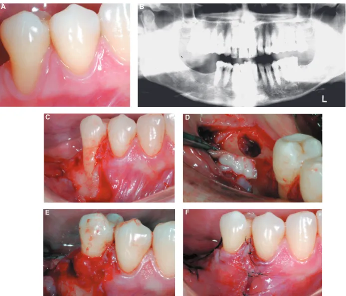
Root coverage procedure was carried out 21 days after (Figure 2A, B). After appropriate anesthesia, a
trapezoidal lap7 was performed and a full thickness
lap was raised (Figure 2C). After debridement
and scaling and root planning, root surface was
conditioned with a citric acid (pH 1) and a 50%
tetracycline solution (Discipline of Biochemistry, School of Dentistry at Bauru-USP) during 3 minutes, followed by vigorous rinsing with saline solution.
Afterwards, a full thickness lap was raised at donor
site (Figure 2D). The healing tissue was removed from the alveolus with a Lucas curette (Chinelatto, Ribeirão Preto, SP, Brazil) and transferred to the receptor site (Figure 2e). Flaps were displaced coronally and sutured at cementoenamel junction (Figure 2F). A periodontal dressing (CoePack, GC America INC, Alsip, IL, U.S.A) protected the area for 14 days. Patient was prescribed a
non-steroidal anti-inlammatory and instructed not to rinse during the irst 48 h. Sutures were removed
after 14 days. Patient was advised to carry out
routine oral hygiene procedures with dental loss
and soft brush for the next 30 days. Postoperative controls were performed at 1, 3, 6 and 9 months and included instruction of oral hygiene and supra and subgingival plaque control, when necessary.
Figure 1- Prepare of donor site. (A) Occlusal view from edentulous ridge selected for the creation of a surgical alveolus; (B) Rising of a full thickness lap and creation of a surgical socket by perforation of alveolar ridge with a diamond bur in high speed with vigorous irrigation; (C) Bovine type 1 collagen barrier membrane trimmed to overlap defects margins in 2-3 mm positioned over the defect; (D) Primary closure of the laps without tension with 4-0 silk
A
B
C
RESULTS
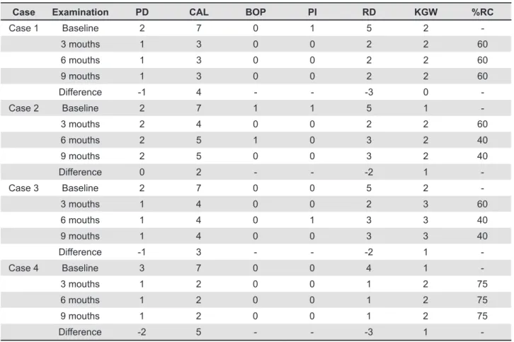
Table 1 describes RD, PD, CAL, BOP, PI, KGW and the percentage of root coverage observed at buccal sites at baseline and at the postoperative examinations for all cases described. A reduction in RD was observed for all cases after 9 months, although Cases 2 and 3 showed a slight relapse from 3-month to 6- and 9-month examinations. All cases resulted in CAL gain and reduction or stability of PD measures, as well as absence of plaque accumulation and BOP. Cases 2, 3 and 4 showed a slight increase in KGW, while no change was observed in Case 1. Figure 3 shows the results obtained in Case 1, in which CAL gain was observed at interproximal and buccal sites, along
C D
B A
Figure 2- Treatment of recession defect by the newly forming bone technique. A: Buccal view of the 5-mm-deep and 4-mm wide recession defect at the mandibular right second premolar; B: Panoramic x-ray view of the area. A slight loss of interproximal bone at distal site of the mandibular right second premolar can be noticed; C: Trapezoidal full thickness lap at receptor site 21 days after surgical creation of alveolar socket; D: Occlusal view of donor site containing the healing tissue after rising of a full-thickness lap; E: Positioning of the newly forming bone at the receptor site. A slight compression with saline solution embedded gauze warranted a close contact between the graft material and the root surface; F: Flaps displaced coronally and sutured at the level of cementoenamel junction without tension
E F
with reduction of RD from 5 mm at baseline to 2 mm after 9 months.
DISCUSSION
The aim of this case series report is to present a new surgical technique that could favor both root coverage and periodontal regeneration, especially at sites showing deep recession defects and attachment loss. The primary results obtained showed reduction of PD, BOP and plaque index and gain of CAL after 9 months of follow-up. A slight increase in KGW was observed after treatment.
These results are in agreement with other studies1,5,7,8,19,23,24-29 which suggested reduction
of probing depth and gain of attachment level in wide and deep recession defects treated by GTR
associated to a coronal positioned lap. Similar results were obtained by the use of β-TCP and 0.3
ng/mL rhPDGF-BB under a collagen membrane for treatment of denuded roots, resulting in the formation of new cementum, periodontal ligament and alveolar bone along with root coverage15.
The main advantage of this novel technique is the use of autograft material containing osteogenic cells9,22 capable of regenerating periodontal tissues21
even in horizontal defects. Disadvantages of the
technique include the necessity of performing two surgical procedures in a short period of time, as well as the existence of two surgical sites at the graft surgery. Besides, patient is required to present at least one condemned tooth or an alveolar ridge that allows the surgical creation of an alveolar socket.
An important issue was the dificulty in stabilizing
the graft on the root surface, due to the convexity of roots and the absence of remaining walls to support the graft. This was at least partially solved
by performing suture of apical portions of the laps
prior to positioning of the graft, which allowed more stability of the graft.
The aim of the present technique is to achieve periodontal regeneration coronal to the preexistent level along with root coverage. This is especially important at sites showing deep recession and dehiscence defects, when conventional soft tissue grafts show limited results23,29. Many studies1,5,19,29
indicate that subepithelial connective tissue results in greater complete and mean root coverage, and a greater increase in KGW than GTR techniques, with
long-term stability of the results obtained. However,
at sites showing interproximal bone loss or deep dehiscence defects and severe attachment loss, regeneration of periodontal tissues may be required to improve attachment support and, consequently,
Case Examination PD CAL BOP PI RD KGW %RC
Case 1 Baseline 2 7 0 1 5 2
-3 mouths 1 3 0 0 2 2 60
6 mouths 1 3 0 0 2 2 60
9 mouths 1 3 0 0 2 2 60
Difference -1 4 - - -3 0
-Case 2 Baseline 2 7 1 1 5 1
-3 mouths 2 4 0 0 2 2 60
6 mouths 2 5 1 0 3 2 40
9 mouths 2 5 0 0 3 2 40
Difference 0 2 - - -2 1
-Case 3 Baseline 2 7 0 0 5 2
-3 mouths 1 4 0 0 2 3 60
6 mouths 1 4 0 1 3 3 40
9 mouths 1 4 0 0 3 3 40
Difference -1 3 - - -2 1
-Case 4 Baseline 3 7 0 0 4 1
-3 mouths 1 2 0 0 1 2 75
6 mouths 1 2 0 0 1 2 75
9 mouths 1 2 0 0 1 2 75
Difference -2 5 - - -3 1
reduce recession depth7,26,29 without the formation
of a long junctional epithelium, as observed when defects with such features are treated by SCTGs10.
Periodontal regeneration can only be evaluated at the histologic level, which was not performed at
the present study. However, previous histological
studies performed in animal models showed that NFBG is capable of forming new alveolar bone, cementum and periodontal ligament coronal to the base of the pre-existent defect21.
Since the healing tissue present at fresh alveolar sockets shows positive staining for collagen I, osteonectin, bone sialoprotein and alkaline phosphatase activity9, it is possible
that mesenchymal stem cells are available at the material, as suggested by other studies investigating the presence of mesenchymal stem cells in periodontal ligament by using the same panel of markers3,6. Considering that one of the
main features of mesenchymal stem cells is plasticity3 and the healing tissue removed from
alveolar sockets and transferred to periodontal defects are able to form bone, periodontal ligament and cementum21, it seems reasonable to believe
that mesenchymal stem cells are present at the newly forming bone granulation tissue, which could explain its osteogenic properties9,21,22.
The use of regenerative techniques to achieve root coverage has been used for many years26.
Trombelli, et al.27 (1995) showed that areas of
4-6 mm recessions treated by guided tissue regeneration resulted in greater reduction of recession and probing depth, as well as greater gain of attachment level than sites treated by
free gingival grafts and coronally advanced lap.
Other researchers have also demonstrated that the treatment of large recession defects by GTR results in the formation of new alveolar bone, cementum and periodontal ligament coronal to the base of the defect5,7,13,27,28, although unsatisfactory results were
observed in shallow recession defects18.
The percentage of root coverage obtained by GTR varies from 16.7%-100%4,5,7,8,20,23,26,27, which
are in agreement with results presented in this case report. In comparison with subepithelial connective tissue grafts, GTR shows better results in reduction of probing depth and gain of attachment level, especially in areas showing >5 mm of recession, and similar or slightly smaller reduction of recession1,19,24,29.The main factors associated
with incomplete root coverage are related to early exposition of the membrane and wide recessions2,4.
These problems are overcome with the present technique, since the use of barrier membranes is unnecessary due to the osteogenic properties of the material21.
The use of allografts in combination with barrier membranes improves the results obtained by the
use of barrier membranes alone8,12 or subepithelial
connective graft1,24,29 in wide recession defects.
Cases treated by bone grafts and GTR showed decreased probing depth at post-operative evaluations, suggesting a reconstitution of biological width, without the formation of long junctional epithelium1,8,12,24,29. Considering that in
the present case a histological evaluation was not performed, reduction of probing depth and gain of attachment are suggestive of regeneration of periodontal tissues.
The primary results obtained in the present case series suggest that the technique is able to cover denuded areas, along with reconstitution of biological width and gain of attachment level, especially in wide recession areas. Predictable and long-term well succeeded cases of root coverage by the NFBT require a strict selection of patients and sites to be treated. Further studies are necessary to compare the results obtained by the proposed technique with other conventional reconstructive periodontal plastic surgeries, such as subepithelial connective tissue grafts and guided bone regeneration.
CONCLUSIONS
The results obtained in the present clinical case series suggest that NFBT can be an alternative to the treatment of deep and wide recession defects.
REFERENCES
1- Al-Hamdan K, Eber R, Sarment D, Kowalski C, Wang HL.
Guided tissue regeneration-based root coverage: meta-analysis. J Periodontol. 2003;74:1520-33.
2- Baldi C, Pini-Prato G, Pagliaro U, Nieri M, Saletta D, Muzzi L, et
al. Coronally advanced lap procedure for root coverage. Is lap
thickness a relevant predictor to achieve root coverage? A 19-case series. J Periodontol. 1999;70:1077-84.
3- Bartold PM, Shi S, Gronthos S. Stem cells and periodontal regeneration. Periodontol 2000. 2006;40:164-72.
4- Boltchi FE, Allen EP, Hallmon WW. The use of a bioabsorbable
barrier for regenerative management of marginal tissue recession. I. Report of 100 consecutively treated teeth. J Periodontol. 2000;71:1641-53.
5- Chambrone L, Sukekava F, Araújo MG, Pustiglione Fe, Chambrone LA, Lima LA. Root coverage procedures for the treatment of localized recession-type defects: a Cochrane systematic review. J Periodontol. 2010;81:452-78.
6- Chen SC, Marino V, Gronthos S, Bartold PM. Location of putative stem cells in human periodontal ligament. J Periodontal Res. 2006;41:547-53.
7- Cortellini P, Clauser C, Pini-Prato GP. Histologic assessment
of new attachment following the treatment of a human buccal recession by means of a guided tissue regeneration procedure. J Periodontol. 1993;64:387-91.
8- Dodge JR, Greenwell H, Drisko C, Wittwer JW, Yancey J,
Rebitski G. Improved bone regeneration and root coverage using a resorbable membrane with physically assisted cell migration and DFDBA. Int J Periodontics Restorative Dent. 2000;20:398-411.
9- Evian CI, Rosenberg ES, Coslet JG, Corn H. The osteogenic
10- Goldstein M, Boyan BD, Cochran DL, Schwarz Z. Human
histology of new attachment after root coverage using subepithelial connective tissue graft. J Clin Periodontol. 2001;28:657-62.
11- Harris RJ. A comparative study of root coverage obtained with
guided tissue regeneration utilizing a bioabsorbable membrane versus the connective tissue with partial-thickness double pedicle graft. J Periodontol. 1997;68:779-90.
12- Kimble KM, Eber RM, Soehren S, Shyr Y, Wang HL. Treatment
of gingival recession using a collagen membrane with or without the use of demineralized freeze-dried bone allograft for space maintenance. J Periodontol. 2004;75:210-20.
13- Langer B, Langer L. Subepithelial connective tissue graft technique for root coverage. J Periodontol. 1985;56:715-20.
14- Matter J. Free gingival graft and coronally repositioned lap.
A 2-year follow-up report. J Clin Periodontol. 1979;6:437-42. 15- McGuire MK, Scheyer eT, Schupbach P. Growth factor-mediated treatment of recession defects: a randomized controlled trial and histologic and microcomputed tomography examination. J Periodontol. 2009;80:550-64.
16- McGuire MK, Scheyer eT. A randomized, double-blind,
placebo-controlled study to determine the safety and eficacy of cultured and expanded autologous ibroblast injections for the treatment of interdental papillary insuficiency associated with the papilla
priming procedure. J Periodontol. 2007;78:4-17.
17- Miller PD Jr. A classiication of marginal tissue recession. Int
J Periodontics Restorative Dent. 1985;5:8-13.
18- Müller HP, Stahl M, Eger T. Failure of root coverage of shallow
gingival recessions employing GTR and a bioresorbable membrane. Int J Periodontics Restorative Dent. 2001;21:171-81.
19- Nickels K, Ratka-Krüger P, Neukranz E, Raetzke P, Eickholz P.
Ten-year results after connective tissue grafts and guided tissue regeneration for root coverage. J Periodontol. 2010;81:827-36.
20- Parma-Benfenati S, Tinti C. Histologic evaluation of new
attachment utilizing a titanium-reinforced barrier membrane in a mucogingival recession defect. A case report. J Periodontol. 1998;69:834-9.
21- Passanezi e, Janson WA, Nahas D, Campos Júnior A. Newly forming bone autografts to treat periodontal infrabony pockets: clinical and histological events. Int J Periodontics Restorative Dent. 1989;9:141-53.
22- Penteado R, Romito GA, Pustiglioni Fe, Marques MM. Morphological and proliferative analysis of the healing tissue in human alveolar sockets covered or not by an e-PTFe membrane: a preliminary immunohistochemical and ultrastructural study. Braz J Oral Sci. 2005;4:664-9.
23- Pini Prato GP, Tinti C, Vincenzi G, Magnani C, Cortellini P, Clauser C. Guided tissue regeneration versus mucogingival surgery in the treatment of human buccal recession. J Periodontol. 1992;63:919-28.
24- Rosetti eP, Marcantonio RA, Rossa C Jr, Chaves, eS, Goissis G, Marcantonio e Jr. Treatment of gingival recession: comparative study between subepithelial connective tissue graft and guided tissue regeneration. J Periodontol. 2000;71:1441-7.
25- Shih SD, Allen eP. Use of guided tissue regeneration to treat a mucogingival defect associated with interdental bone loss: a case report. Int J Periodontics Restorative Dent. 1994;14:552-61. 26- Tinti C, Vicenzi G, Cortellini P, Pini Prato G, Clauser C. Guided tissue regeneration in the treatment of human facial recession. A 12-case report. J Periodontol. 1992;63:554-60.
27- Trombelli L, Schincaglia GP, Scapoli C, Calura G. Healing
response of buccal gingival recessions treated with expanded
polytetraluoroethylene membranes. A retrospective report. J
Periodontol. 1995;66:14-22.
28- Urbani G, Lombardo G, Castellarin M, Santi e, Abtibol T. Surgical correction of gingival recessions associated with radicular carious lesions. Compend Contin educ Dent. 1996;17:330-2.
29- Wang H-L, Bunyaratavej P, Labadie M, Shyr Y, MacNeil RL.
Comparison of 2 techniques for treatment of gingival recession. J. Periodontol. 2001;72:1301-11.