Prevalence assessment of root dilaceration in permanent incisors
Beatriz Feitosa da Silva1, Luciana Ellen Dantas Costa2, Ricardo Villar Beltrão3, Tânia Lemos Rodrigues4,
Ricardo Lombardi de Farias5,Rejane Targino Soares Beltrão6
Introduction: Root dilaceration is a dental anomaly characterized by an abnormal curvature of the dental root, which can cause problems during eruption and complications in the orthodontic and endodontic treatment or extraction. Objective:
The aim of this study was to determine the prevalence of root dilaceration in permanent incisors in an oral radiology clinic in the city of João Pessoa / PB, Brazil. Methods: The sample consisted of 548 patients who allowed assessments of their den-tal radiographs, a toden-tal of 3,948 examined teeth. Each tooth was examined according to the occurrence of root dilaceration, regarding type (mild, moderate or severe), root third that it was in and direction of the root. The angle formed by the root deviation in relation to the long tooth axis was obtained by means of a diagram printed on transparent acetate, placed over the radiograph. Results: The results were analyzed with SPSS software (Statistical Package for Social Sciences) version 13.0, performing an exploratory data analysis. The prevalence of root dilaceration in the studied sample was 1.03% (41 cases), with higher incidence in males (65.8%), being the superior lateral incisor the most affected teeth (78%). The most prevalent type of root dilaceration was the mild one (73.1%), occurring more in the apical third (90.2%) and with distal direction of the root (95.1%). Conclusion: Considering these results, it is emphasized the importance of performing a diagnostic radiographic ex-amination prior to planning a treatment in permanent anterior teeth.
Keywords: Incisor. Tooth root. Tooth abnormalities. Dental radiograph. Prevalence.
How to cite this article: Silva BF, Costa LED, Beltrão RV, Rodrigues TL, Farias RL, Beltrão RTS. Prevalence assessment of root dilaceration in permanent incisors. Den-tal Press J Orthod. 2012 Nov-Dec;17(6):97-102.
» The author reports no commercial, proprietary or financial interest in the products or companies described in this article.
Contact address: Luciana Ellen Dantas Costa
Rua Eurico Uchoa, 100 – Bairro dos Estados – João Pessoa/PB – Brazil CEP: 58031-150 – E-mail: [email protected] 1 Graduated in Dentistry, UFPB.
2 MSc in Pediatric and Preventive Dentistry, UFPB. Professor of Public Health, UFCG. 3 PhD in Radiology, UFBA. Professor of Radiology, UFPB.
4 PhD in Oral and Maxillofacial Surgery, UFRJ. Professor of Surgery, UFPB. 5 PhD in Orthodontics. Faculty of Dentistry of Araraquara. Professor of
Orthodontics, UFPB.
6 PhD in Orthodontics and Dental Public Health, USP. Professor of Orthodontics, UFPB.
Submitted: March 18, 2009 - Revised and accepted: March 29, 2010
Introdução: a dilaceração radicular é uma anomalia dentária caracterizada por uma curvatura anormal na raiz do dente que pode causar problemas na erupção e complicações durante movimentação ortodôntica, tratamento endodôntico ou exodon-tia. Objetivo: o objetivo do presente trabalho foi verificar a prevalência da dilaceração radicular em incisivos permanentes, em uma clínica de radiologia odontológica da cidade de João Pessoa/PB. Método: a amostra foi constituída de 548 pacientes que possibilitaram avaliações em suas radiografias dentárias, totalizando 3948 dentes examinados. Examinou-se cada dente segundo a ocorrência de dilaceração radicular, com relação ao tipo (discreta, moderada ou severa), o terço radicular em que se encontrava e a direção da raiz. A angulação formada pelo desvio da raiz em relação ao longo eixo do dente foi obtida por meio de um diagrama impresso em acetato transparente, colocado sobre a radiografia. Resultados: os resultados foram analisados no programa SPSS, efetuando-se uma análise exploratória dos dados. A prevalência da dilaceração radicular na amostra estu-dada foi de 1,03% (41 casos), com maior incidência no sexo masculino (65,8%), sendo os incisivos laterais superiores os dentes mais acometidos (78%). O tipo de dilaceração radicular mais prevalente foi a discreta (73,1%), ocorrendo mais no terço apical (90,2%) e com direção distal da raiz (95,1%). Conclusão: diante desses resultados, ressaltam-se a importância da realização prévia de um exame radiográfico de diagnóstico ao planejar um tratamento em dentes permanentes anteriores.
IntROduCtIOn
It is relatively common, in clinical experience, that the non-eruption of permanent incisors is due to prolonged retention of its deciduous cor-responding tooth, to the absence of the permanent succeeding tooth or the installation of develop-mental abnormalities; which is noticed from the age of eight, in the first transitional period of the mixed dentition.3
With the exception of trauma, root dilaceration is a dental anomaly in which the tooth root presents a curvature of varied angle, caused by the displace-ment of the crown from the rest of the root dur-ing its early development.19 For Chohayeb,7 a root deviation can be considered root dilaceration when the angle between the root and the long axis of the tooth is equal to or greater than 20°.
The cause of root dilaceration is controver-sial, being attributed to a strong relation with the trauma in the primary dentition — as intrusive luxation or avulsion of its deciduous corresponding tooth.3,14,17,22,29 However, there are reports that asso-ciate to this anomaly, hereditary factors,19 the pres-ence of cysts or tumors,5,8 supernumerary teeth25 and ectopic development of the upper incisor tooth germ, with the root following the curvature of the palate, presenting the alteration.15
Brin et al3 and Laskaris14 explained their possible theories to justify such a relation with dental trau-ma. Due to proximity between the deciduous root apex with the permanent germ, the occurrence of injuries during odontogenesis may cause displace-ment of the calcified portion of the rest of the tooth, which continues its development in this new posi-tion, creating an unusual angle.3,14
Gonçalves et al12 associated the premature loss of deciduous teeth to the local formation of a fibrotic scar which prevents normal development of the per-manent tooth, the same occurs in the presence of any obstacle to normal development of tooth germ, causing a progressive angulation crown/root.13
The apical root dilaceration may affect approxi-mately 1 to 4.9% of all permanent teeth, with a higher frequency in female patients,10,21,28 and it may, occasionally, involve deciduous teeth as a result of previous trauma due to neonatal laryngoscopy or endotracheal intubation.4
According to Silva Filho et al,25 of all permanent incisors, the upper centrals are the most affected (70.6%) followed by the laterals (20.6%) and low-er incisors (8.8%). While Erlich, Plow-ereira, Panella10 when examining all permanent teeth, except the third molars, said to be the permanent upper lateral incisors the most affected, followed by the upper second premolars and lower first premolars.
The diagnosis of root dilaceration takes place, usual-ly by radiographic examination.10 Clinically, the failed eruption of permanent incisors, especially the upper ones, leads professionals to suspect of that anomaly. 4
The radiograph is essential to observe the stage of root formation and the degree of dilaceration, being important to determine the morphology and spatial position of the tooth in the bone.12 Making use of the periapical, occlusal, and panoramic radiographs or computed tomography, which provides precisely, the real location of the dental and bone structures.4
Depending on the degree of root angulation, the treatment and prognosis of teeth with root dilacera-tion varies.9,15 In mild cases, treatment is not neces-sary, however, there are cases where the tooth must be exposed surgically and orthodontically moved, and in more severe cases, due to treatment impos-sibility, the surgical removal followed by prosthetic rehabilitation is indicated.6,15,20
The studies on prevalence of root dilaceration in-clude, mostly, all permanent teeth,10,16,21,28 not being observed studies regarding only central and lateral permanent incisors.
It is also observed that these studies10,16,21,28 gen-erally evaluate radiographs of patients from educa-tional institutions, which can provide a biased re-sult, that do not correspond to reality, since the most complicated cases are usually referred to these insti-tutions. To eliminate this bias, a oral radiology clinic in the city of João Pessoa/PB (Brazil), was selected by convenience, in order to refer epidemiological data in an untainted population, seeking detailed and real information in relation to a group of teeth.
MAtERIAL And MEtHOdS
In this cross-sectional, descriptive study, we ob-tained a sample of 3948 central and lateral per-manent incisors, observed in 548 radiographs from an oral radiology clinic in the city of João Pessoa, Paraíba (Brazil), evaluated within the period from October to December, 2008.
From the evaluated radiographs, 382 were pan-oramic and 166 were periapical, belonging to 238 female individuals and 310 to males.
All exams were standardized, obtained by the same operator and machine. Panoramic and peri-apical radiographs and teleradiographs (only for the cases of labial-palatal dilaceration) that showed maximum sharpness, medium contrast and density, were included in the sample. The X-ray examina-tions of patients under the age of seven years and permanent incisors with less than two thirds of root formed were excluded from the sample.
The radiographs were interpreted in the dark with the aid of a magnifier (2x magnifying capacity) on a light box with black cardboard masks, the pan-oramic measuring 15 x 30 cm and the periapicals 2.5 x 5.0 cm.
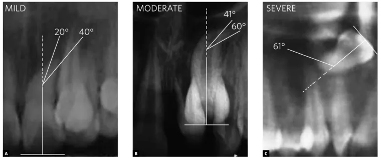
Each tooth was examined for the occurrence of root dilaceration, according to Santana, Consolaro and Tavano,21 in mild (20-40°), moderate (41-60°),
severe curvature degree (≥ 61°), root third in which it was present (cervical, middle or apical), and root direction (mesial, distal, buccal or lingual). The de-viations greater than 20° formed by the roots in re-lation to the long axis of the tooth were considered as dilaceration.7
To classify the dilaceration in accordance with the degree of curvature, in mild, moderate or se-vere, a method similar to the one by Schneider,24 and Erlich, Pereira and Panella10 was used which measured the angle formed by the midlines of the tooth long axis and the deviated segment. In this study a diagram was made on AutoCAD software (version 2006), containing the angles and indexes in
MILD MODERATE SEVERE 40°
60° 61°
20° 41°
Figure 2 - Mild, moderate and severe root dilaceration type.
Figure 1 - Diagram showing rates - classification of dilaceration in mild,
moderate or severe. Source: Santana, Consolaro and Tavano21,
Schnei-der24, and Erlich, Pereira and Panella.10
A B C
MILD
20°
41°
40°
60°
61°
transparent acetate (Fig 1). To measure the values, the radiograph was superimposed on the diagram and evaluated on the light box (Fig 2).
If the patient did not present root dilaceration, only the identification data were noted (gender and age) and number of examined teeth.
The data were classified in SPSS v. 13.0 (Statisti-cal Package for Social Sciences) an exploratory data analysis was performed with the construction of single frequency and double entry tables, as well as statistical result graphs.
The study was previously approved by the person in charge of the oral radiology clinic and the Com-mittee on Ethics in Research involving human be-ings, of the Center for Health Sciences, Federal Uni-versity of Paraíba, with protocol No. 0141.
RESuLtS
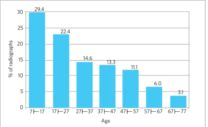
Five hundred and forty eight radiographs of up-per central and lateral up-permanent incisors, aged from 7 to 77 years of age were evaluated (Fig 3). Pan-oramic radiographs (69.7%) and periapical (30.3%)
belonged to 238 (43.4%) female patients and 310 (56.6%) males.
The prevalence of root dilaceration in the sample was 1.03%, affecting the maxilla (82.9%) more than the mandible (17.1%). The upper lateral incisors were the most affected (32 - 3.35%), followed by the lower lateral incisors (6 - 0.6%), upper central (2 - 0.2%) and lower central (1 - 0,1%) (Table 1). Twenty-seven cases (65.8%) of dilaceration in males and 14 (34.2%) in females was observed.
The permanent incisors with root dilaceration presented a mild curvature in 73.1% (30) of the sample, followed by the moderate type (7 - 17.0%) and severe (4 - 9.7%). As for the root third where they were located: 90.2% (37) of dilacerations were in the apical third, 7.3% (3) in the middle third and only 1 (2.4%) in the cervical third. The dilacerated roots were more frequently facing distal in 95.1% (39 teeth), only one was mesial and another one was buccal (2.4%).
dISCuSSIOn
Root dilaceration is a dental anomaly of shape characterized by a change in the root or crown an-gulation of the formed tooth.4 Although its etiology is associated, in most cases, to trauma in the decidu-ous dentition,2,18,29 factors such as abnormal devel-opment of the root due to the presence of cysts or adjacent tumors;5,18 the development of the ectopic tooth germ and hereditary factors, may be associated with such anomaly.15,26
According to the literature, root dilaceration is relatively rare, with a prevalence of approximately 1 to 4.9% for all dental groups.10,16,28 However, no studies were found related to root dilaceration re-stricted to the incisor region, subject-matter of this work, which showed a prevalence of 1.03%.
Santana, Consolaro and Tavano21 observed root dilacerations in 19 central incisors and upper perma-nent lateral out of the 203 teeth affected with this anomaly, with a prevalence of 0.3% for the incisors. Malcic et al16 observed a prevalence of 1.01% of root dilaceration in incisors while examining all teeth.
From the root dilaceration observed, the group most frequently affected was the upper lateral in-cisors (78%), followed by lower lateral (14.6%), upper central (4.9%) and lower central (2.5%)
Figure 3 - Distribution of the percentage of examined radiographs ac-cording to the age of individuals. João Pessoa / PB (Brazil), 2008.
30 29.4
22.4
14.6 13.3
11.1
6.0 3.1 25
20
7 17 17 27 27 37 37 47 47 57 57 67 67 77 15
10
Age
% of r
adiogr
aphs
5
0
Teeth UCI LCI ULI LLI
Number of examined teeth 1001 993 955 999
Total number of dilaceration 2 1 32 6
Prevalence of dilaceration (%) 0,2 0,1 3,35 0,6
Table 1 - Prevalence of root dilaceration in permanent incisors. João Pes-soa / PB (Brazil), 2008.
agreeing with Erlich, Pereira and Panella10 by claim-ing that from all incisors, the upper lateral were the most affected by dilaceration (22.1%). However, the results of Silva Filho et al25 contradict those in this study, since the central incisors were the most affected (70.6%), followed by the lateral (20.6%) and lower incisors (8.8%). It is noteworthy that Santana, Consolaro and Tavano21 did not observe root dilaceration in lower incisors, studying 6,443 permanent incisors.
The root dilaceration in incisors was more fre-quent in the maxilla (82.8%) than in the mandi-ble (17.2%). This value was similar to the results found by Malcic et al16 (83.3% of the cases stud-ied in maxillary periapical radiographs and 72.2% of maxillary cases, in panoramic films); and Erlich, Pereira and Panella10 (58.1%). Perhaps, this result is a consequence of trauma factor, affecting with some frequency the maxillary region of children in the deciduous dentition phase and especially the upper incisors, due to its location.2
Regarding gender, root dilaceration was more frequent in males (65.8%) than in females (34.2%). Opposed to these results, Erlich, Pereira and Pan-ella,10 Vicci and Capeloza28 reported this to occur more in females than in males, 68.88% and 59.6% of the samples, respectively.
The most prevalent type of dilaceration in this sample was the mild one (73.1%), followed by moderate (17%) and severe (9.7%) confirming the findings of Santana, Consolaro and Tavano,21 Eh-rlich, Pereira and Panella10 who observed, respec-tively, that 69.4% and 84.5% were dilacerations of the mild type, 27.9% and 13.8% moderate and 2.9% and 1.66% severe.
As for the root third where they were located, 90.2% of dilacerations were in the apical third, which agrees with the results of Malcic et al;16 San-tana, Consolaro and Tavano21 and Erlich, Pereira and Panella.10 In the middle and apical third, the preva-lence was 7.3% and 2.4%, confirming the findings of Malcic et al16 of 13% and 2.3% respectively. Santana, Consolaro and Tavano,21 studying 19 upper incisors with root dilaceration, did not observe a curvature in the cervical third.
Regarding the direction of the anomaly, the roots were distally directed in 95.1% of the incisors, in
agreement with the study of Santana, Consolaro and Tavano21 who observed that 89% of the roots were focused to distal.
The root dilaceration of the mild type, involving most of the sample, usually does not result in tooth loss during orthodontic mechanics, not even in com-plications during endodontic or surgical treatment.16 In cases of root dilaceration of severe type, there is a need for specialized follow up and greater clini-cal and radiographic control during orthodontic me-chanics, especially in traction, resulting in the need for root canal treatment and apicectomy to finish traction and obtain a favorable6 prognosis, in some cases. Thus, the patient in the beginning of ment must be alerted to the need for further treat-ment, or even the possibility of tooth loss depend-ing on the position where the tooth is located, while in the mild type of root dilaceration this possibility does not exist.16
The panoramic and periapical radiographs are important tools and widely used by clinicians in daily practice as a complementary test for diagnosis. In the case of root dilacerations, these exams have a fundamental role in diagnosis, as well as to obtain the prevalence studies. As in the study by Malcic et al,16 periapical and panoramic radiographs were evaluated in this study to determine the prevalence of root dilaceration.
Faerman et al11 emphasized that early diag-nosis of the dilaceration involves performing ra-diographs on all children in the mixed dentition phase, with a good clinical examination, avoiding functional, esthetic, phonetic and psychological problems to patients.
In cases where there is the need for surgery for orthodontic traction, extraction or endodon-tic treatment, of a dilacerated tooth, it is essential to obtain the position of the unerupted tooth, for surgical planning and evaluation of degree of diffi-culty, thus avoiding unexpected events during sur-gery.6,9,11,16
COnCLuSIOnS
1. Andreasen JO, Andreasen FM. Textbook and color atlas of traumatic injures to the teeth. 3th ed. St. Louis: CV Mosby; 1994.
2. Artun J, Behbehani F, Al-Jame B, Kerosuo H. Incisor trauma in an adolescent Arab population: Prevalence, severity, and occlusal risk factors. Am J Orthod Dentofacial Orthop. 2005;128(3):347-52.
3. Brin I, Fuks A, Bassat YB, Zilberman Y. Trauma to primary incisors and its effect on the permanent successors. Pediatric Dent. 1984;6(2):78-82.
4. Casati-Alvares L, Tavano O. Aspectos radiográficos das anomalias dentárias e maxilares. In: Freitas A, Rosa JE, Souza IF. Radiologia odontológica. 6ª ed. São Paulo: Artes Médicas; 2004.
5. Cardoso LC, Miyahara GI, Magro Filho O, Garcia Júnior IR, Soubhia AMP. Odontoma combinado associado a dentes não-irrompidos: relato de casos clínicos. Rev Odontol Araçatuba. 2003;24(2):47-51.
6. Chew MT, Ong MM. Orthodontic-surgical management of an impacted dilacerated maxillary central incisor: a clinical case report. Pediatric Dent. 2004;26(4):341-4.
7. Chohayeb AA. Dilaceration of permanent upper lateral incisors: frequency, direction and endodontic treatment implication. Oral Surg. 1983;55(5):519-20. 8. Costa LRRS, Correa MSNP, Ribeiro RA. Traumatismo na dentição decídua. In:
Correa MSNP. Odontopediatria na primeira infância. São Paulo: Ed. Santos; 1998. p. 527-47.
9. Costa CS, Medeiros MN, Souza APP, Prado C, Aratani M, Parizotto SPCOL, Moselle O. Dilaceração radicular: tratamento cirúrgico e reabilitação estético-funcional do paciente. Rev Bras Cir Implant. 2001;8(29):76-80.
10. Erlich T, Pereira MF, Panella J. Estudo da prevalência da dilaceração radicular, por meio de exame radiográfico periapical, numa amostra populacional da Grande São Paulo. Rev Pós Grad. 2001;8(2):129-37.
11. Faerman K, Campos V, Souchois MWM, Carneiro MAS, Alexandre GC. A Importância do exame radiográfico na dilaceração radicular de incisivos centrais superiores permanentes após traumatismo dentário. JBP: J Bras Odontopediatr Odontol Bebê. 2002;5(26):328-35.
12. Gonçalves SRJ, Santos AA, Oliveira CCC, Dantas Neta EM, Teles CL, Bonjardim RL. Avulsão traumática anterior na dentição decídua. Odontol Clín-Científ. 2004;3(2):111-6.
13. Hernandes MCCO, Goulart MM, Paleckis LGP. Trauma em dentição decídua e suas conseqüências na dentição permanente: relato de caso clínico. Rev EAP/ APCD. 2003 [acesso 2003 Ago 1]. Disponível em: www.apcd.com.br/rev_art. asp?path=REVISTA&artigo=33.
14. Laskaris G. Anomalias dentárias In: Atlas colorido de doenças bucais da infância e da adolescência. Porto Alegre; São Paulo: Ed. Santos; 2000. p. 2-35.
15. Lin YJ. Treatment of an impacted dilacerated maxillary central incisor. Am J Orthod Dentofacial Orthop. 1999;115(4):406-9.
REFERENCES
16. Malcić A, Jukić S, Brzović V, Miletić I, Pelivan I, Anić I. Prevalence of root dilaceration in adult dental patients in Croatia. Oral Surg Oral Med Oral Pathol Oral Radiol Endod. 2006;102(1):104-9.
17. McNamara T, Wolfe SN, McNamara CM. Controle ortodôntico de um incisivo central superior dilacerado com uma seqüela incomum. Rev Dental Press Ortod Ortop Facial. 1998;3(6):26-8.
18. Neville BW, et al. Alterações dentárias de desenvolvimento. In: Neville BW, Damm DD, Allem CM, Bouquot JE. Patologia oral e maxilofacial. 2ª ed. Rio de Janeiro: Guanabara-Koogan; 2004. p. 68-103.
19. Regezi JA, Sciubba JJ. Anomalias dentárias. In: Patologia bucal: correlações clínicopatológicas. Rio de Janeiro: Guanabara Koogan; 1991. p. 341-62. 20. Rodrigues TL. Tratamento orto-cirúrgico do incisivo central superior incluso com
coroa invertida e dilaceração radicular: um novo enfoque [dissertação]. Rio de Janeiro: Universidade Federal do Rio de Janeiro; 2001.
21. Santana EJB, Consolaro A, Tavano O. Determinação da prevalência e estudo morfológico da dilaceração radicular. Rev Fac Odontol UFBA. 1992-1993;12(13):40-52.
22. Santos SH. Dilaceração radicular, tratamento ortodôntico e estético - relato de um caso clínico. SOTAU: Rev Virtual Odontol. 2007;1(17) [Acesso 2007 Ago 7]. Disponível em: http://sotau.sind.googlepages.com/ SotauRevVirtOdont20071pag17a22.pdf.
23. Shafer WG, Hine MK, Levy BM. Distúrbios de desenvolvimento das estruturas bucais e parabucais. In: Tratado de Patologia bucal. Rio de Janeiro: Guanabara Koogan; 1987. p. 2-79.
24. Schneider SW. A comparison of canal preparations in straight and curved root canals. Oral Surg. 1971;32(2):271-5.
25. Silva Filho OG, Dias JB, Cavassan AO, Carvalho IMM, Navarro MFL, Schuckar M. Distúrbios irruptivos na região ântero-superior: abordagem multidisciplinar. Rev Dental Press Ortod Ortop Maxilar. 1997;2(6):49-62.
26. Tanaka E, Hasegawa T, Hanaoka K, Yoneno K, Matsumoto E, Dalla-Bona D, et al. Severe crowding and a dilacerated maxillary central incisor in an adolescent. Angle Orthod. 2006;76(3):510-8.
27. Vasconcellos RJH, Oliveira DM, Nogueira RVB, Maciel AP; Cordeiro MC. Trauma na dentição decídua: enfoque atual. Rev Cir Traumatol Buco-Maxilo-Facial. 2003;3(2):17-24.
28. Vicci JG, Capelozza ALA. Incidência de lesões dentárias e ósseas evidenciadas através de radiografia panorâmica. Rev Fac Odontol Lins. 2002;14(2):43-6. 29. Viegas CMS, Godoi PFS, Ramos-Jorge ML, Ferreira EF, Zarzar PMPA. Traumatismo
na dentição decídua: prevalência, fatores etiológicos e predisponentes. Arq Odontol. 2006;42(4):257-336.
prevalence of 1.03% of the sample, the lateral inci-sors are the most affected ones (92.6%), particular-ly higher prevalence in the upper incisors (78.0%), occurring more in the apical third (90.2%) and dis-tal direction of the root (95.1%).