outubro de 2013
Universidade do Minho
Escola de Engenharia
Ana Catarina Freitas Salazar de Oliveira
Immobilization of Specific Growth
Factors from Platelet Lysate at the
Surface of Electrospun Nanofibers
for Tissue Engineering Applications
UMinho|20
13
Ana Catarina F
reitas Salazar de Oliv
eir
a
Immobilization of Specific Growt
h F actor s from Platele t L ysate at t he Sur
face of Electrospun Nanofiber
Dissertação de Mestrado
Mestrado Integrado em Engenharia Biomédica
Ramo Biomateriais, Reabilitação e Biomecânica
Trabalho efetuado sob orientação do
Professor Nuno João Meleiro Alves das Neves
outubro de 2013
Ana Catarina Freitas Salazar de Oliveira
Immobilization of Specific Growth
Factors from Platelet Lysate at the
Surface of Electrospun Nanofibers
for Tissue Engineering Applications
DECLARAÇÃO
Nome: Ana Catarina Freitas Salazar de Oliveira Endereço eletrónico: [email protected] Número do Bilhete de Identidade: 13728359
Título dissertação: Immobilization of Specific Growth Factors from Platelet Lysate at the Surface of Electrospun Nanofibers for Tissue Engineering Applications
Ano de conclusão: 2013
Orientador: Professor Doutor Nuno M. Neves
Designação do Mestrado: Mestrado Integrado em Engenharia Biomédica Ramo: Biomateriais, Reabilitação e Biomecânica
É AUTORIZADA A REPRODUÇÃO PARCIAL DESTA DISSERTAÇÃO, APENAS PARA EFEITOS DE INVESTIGAÇÃO, MEDIANTE DECLARAÇÃO ESCRITA DO INTERESSADO, QUE A TAL SE COMPROMETE.
Braga, ____/____/________
III
Acknowledgments
This thesis is the last chapter of the five years I spent in University of Minho. And now is the time to acknowledge all the people, which in one way or another, guided and helped me during all this time.
First I would like to acknowledge my supervisor Professor Nuno Neves for all the help, support, knowledge and availability during this last six months. His enthusiasm about my project and all the ideas and suggestions were a great incitement during all the steps of this work.
I would like to thank Dr. Albino Martins for the supervision, help, suggestions, guidance and all the discussions about the project. Thank you for the patience and knowledge transmitted in areas and subjects I was less familiar. I also want to thank Dr. Ana Rita Pinto for the help during this project especially the time spent with the cell culture experiments. Thank you for your tips, supervision and suggestions.
I would like to thank all the people from 3B’s. I would like to specially thank Nelson Monteiro for the first steps with the electrospinning and PCL meshes and his availability in helping me during my first months in the group. Diana Ribeiro, thank you for your help and always being available for my questions and the endless hours doing all those ELISAs. I would like to thank Dr. Margarida Martins, for the hints about statistical analysis.
I would like to thank the Maxbone and Osteography projects and also QREN for financing this reseach work.
Despite not being part of this thesis I would like to thank Professor João Mano for encouraging us to do ERAMUS, it was a fantastic experience that I would never forget. I would also like to thank Professor Marcel Karperien and Professor Aart van Apeldoorn for welcoming me in the DBE group in Twente. Thank you, Giulia, for helping me trough my first real experience in a lab as well as for introducing me into the cell culture world. All I learned there helped me during all the stages of this thesis. I would also like to thank all the friends I met there, it felt like home.
Thank you to the friends I made during this last five years, for always being there for me and all the moments we spent together.
IV
To my friends from Biomedical Engineering (“Nata da Gata”). I could have not been here if it was not for the people that began this journey with me and that now are the friends that I will never get out of my sight again, we are a real family, that I am proud to be part of. It is amazing to see how we got here, how much we grow and how much our friendship is stronger as time goes by. We could write a book about all the stories and moments we shared together. But I know that much more chapters are about to come, we have a whole life ahead us. All the moments we shared together made me who I am today. Thank you all!
To all the people I had the opportunity to meet during my time in Academic Students Union. I am really proud for all the things we accomplished and done together. It was an amazing experience and the worst part was to say goodbye. When it hurts we know that it was completely worth it. I would like to give a special thank to Luís, Mateus, Pinheiro, Remi, Cléber, André, Ana Rita e Tita. It is really nice to see that the time we spent together turned into an incredible friendship.
To all my friends from Taipas. I know that in the last five years we didn’t spend as much time as I would like together, but I have you always in my mind. Most of you are a part of my life since a long time and it is going to be like that for much more.
Tina, you know how important you are to me, you are my sister and sisters are meant for a lifetime. Since the first day at University we never got separated again. We are always there for each other. For 3ºA, for ERAMUS, for this five years, for the endless talks, for the help, for all the moments, for the wise advices, for the laughs, for all our crazy ideas that no one else would understand, for the future, for our moments, for your unconditional friendship, thank you! I could never have done this without you.
To André, for his patience, support and understanding. Thank you for everything.
To all my family: my grandparents, my uncles, my aunts and my cousins. Thank you for helping me whenever I needed, for being my friends and for believing in me.
To my brother and to my parents. Thank you so much for believing in me unconditionally, for all the support in every step I make, for encouraging me in pursuing my dreams. For always encouraging my work and for giving me all the opportunities. For picking me up whenever I felt down. I could have not done this without you, you are my model and my inspiration. I would never be able to thank you enough.
V
Abstract……….
One of the major problems related with implantable biomaterials is the limited bioactivity and suboptimal integration with the host tissue. The functionalization of biomaterial substrates to insert biological cues by the immobilization of biomolecules at their surface has been proposed as effective to overcome some of these limitations. Different immobilization strategies can be followed (such as adsorption or covalent immobilization) however, a critical aspect to have in consideration is to keep the bioactivity of molecule functionality. Therefore, the work developed in this thesis aims to activate and functionalize the surface of electrospun polycaprolactone (PCL) nanofiber meshes by the insertion of chemical groups (i.e. amine groups) to achieve a covalent immobilization of antibodies. The immobilization of the defined antibodies will allow for the selective binding of growth factors (GFs), either recombinant or derived from a biological fluid such as Platelet Lysates (PLs) that are known to have high concentrations of autologous GFs.
We determined the maximum immobilization capacity of the defined antibodies (i.e. TGF-β1, bFGF and VEGF) in the mentioned nanofibrous surface. The GFs of interest were further incubated into the corresponding biofunctionalized substrate, assessing the maximum binding capacity as well as the selective binding of the GFs from a pool of different proteins present in human platelet lysate samples. The bioactivity of the bound VEGF was further assessed by seeding and culturing a specific endothelial cell line (HPMEC-ST1.6R) over the biofunctionalized substrate. The biological data demonstrates that the immobilization strategy does not compromise the availability of the antibody neither the functionality of the bounded-GF. The combination of two antibodies (i.e. bFGF and VEGF) was tested in a mixed experiment or in separate regions of the same mesh in a side-by-side configuration. For the mix design, the biofunctionalized nanofibrous substrate was able to selectively bind two different GFs from the studied biological fluid. For the side-by-side a watertight chamber was developed to physically separate the substrate into two different areas, each one with a defined antibody just to validate the concept.
Our results confirm the efficiency of the immobilization method as well as the bioactivity of the bound GFs, showing a promising potential for the immobilization of different antibodies and corresponding GFs depending on the intended application. This strategy will enable designing advanced autologous therapies since both GFs and cells could be from the same donor, allowing the implementation of very effective and personalized therapies.
VII
Resumo
Um dos grandes problemas relacionados com a implantação de biomateriais é a limitada bioatividade e integração com o tecido local. A funcionalização de substratos de forma a inserir sinais biológicos através da imobilização de biomoléculas na sua superfície pode ser uma tentativa para ultrapassar estas limitações. Diferentes técnicas de imobilização podem ser realizadas (por exemplo, absorção e imobilização covalente); contudo, um fator importante a ter em consideração é manter a bioatividade da molécula. Nesse sentido, o trabalho desenvolvido nesta tese tem como objetivo a ativação e funcionalização da superfície de nano fibras produzidas por electrospinning através da inserção de grupos amina de modo a conseguir uma imobilização covalente dos anticorpos. A imobilização de anticorpos específicos permitirá uma ligação seletiva de fatores de crescimento (FC), que podem ser recombinantes, ou retirados a partir de um fluído biológico, neste caso o Lisado de Plaquetas, que é conhecido por apresentar grandes concentrações de fatores de crescimento.
Ao longo deste projeto, diferentes ensaios foram realizados para determinar a capacidade máxima de imobilização de anticorpos (TGF-β1, bFGF e VEGF) no substrato acima mencionado. Os fatores de crescimento referidos foram então incubados no substrato nanofibroso correspondente, determinando a máxima capacidade de ligação, assim como a ligação específica dos fatores de crescimento a partir das diferentes proteínas presentes no lisado de plaquetas. A bioatividade do VEGF previamente ligado ao anticorpo foi determinada através de uma linha celular endotelial (HPMEC-ST1.6R). Os dados biológicos confirmaram que a estratégia de imobilização adotada não afetou a disponibilidade e funcionalidade do fator de crescimento. A combinação de dois anticorpos (bFGF e VEGF) foi testada misturando-os numa só solução ou então imobilizando-os lado a lado em áreas específicas da malha. No primeiro caso, através da mistura e consequente imobilização dos dois anticorpos, foi possível ao substrato biofuncionalizado selecionar dois fatores de crescimento distintos do Lisado de Plaquetas. Para a imobilização dos fatores lado a lado foi desenvolvido um sistema capaz de separar o substrato em duas áreas distintas, assegurando que as duas soluções não se misturavam.
Estes resultados confirmaram a eficiência do método de imobilização, assim como a bioatividade dos FC. Com esta estratégia será possível selecionar diferentes fatores de crescimento tendo em vista a aplicação pretendida, bem como a implementação de uma terapia autóloga possibilitando o desenvolvimento de tratamentos mais efetivos e personalizados.
IX Table of Contents Acknowledgments ... III Abstract………. ... V Resumo ... VII Table of Contents ... IX List of Abbreviations………..………..……….XIII List of Figures ... XVII List of Tables.. ... XIX
Chapter 1 Introduction ... 1
1.1 Abstract ... 3
1.2 Tissue Engineering and Regenerative Medicine... 4
1.2.1 Requirements of a biomaterial scaffold ... 5
1.2.2 Cell sources ... 6
1.2.3 Bioactive molecules: growth factors ... 7
1.2.3.1 Growth factors and the healing cascade ... 8
1.2.3.2 Growth factors: properties and roles ... 9
1.3 Growth factors sources ...11
1.4 Immobilization methods and strategies ...13
1.4.1 Non-covalent immobilization ...14
1.4.2 Covalent immobilization strategies ...15
1.5 Applications of immobilized growth factors ...17
1.5.1 Angiogenesis ...17
1.5.2 Other relevant applications: dermal healing, cartilage, bone and stem cells differentiation ...19
1.6 Final Remarks ...20
1.7 References ...21
Chapter 2 Materials and Methods ...27
X
2.2 Methods ...30
2.2.1 Scaffold Fabrication and (Bio)Functionalization ...30
2.2.1.1 The processing technique Electrospinning ...30
2.2.1.2 Surface Functionalization of Electrospun Nanofibers ...31
2.2.2 Antibodies Immobilization Strategy ...32
2.2.2.1 EDC/NHS ratio and concentration optimization ...35
2.2.2.2 Optimization of Single Antibody Immobilization and Determination of the Standard Curves .36 2.2.2.3 Mixed immobilization of two antibodies (VEGF and bFGF) ...36
2.2.2.4 Side-by-side immobilization of two antibodies ...37
2.2.2.5 Laser Scanning Confocal Microscopy ...38
2.2.3 Recombinant and PL-derived growth factor quantification ...38
2.2.3.1 Platelets Lysate: Preparation and Activation ...38
2.2.3.2 Fluorescence-Linked immunosorbent Assay (FLISA) ...39
2.2.3.3 Enzyme-Linked Immunosorbent Assay (ELISA) ...39
2.2.4 Biological Part ...40
2.2.4.1 Cell culture and seeding ...40
2.2.4.2 Cell Viability ...41
2.2.4.3 Cell Proliferation ...42
2.2.4.4 Total Protein synthesis ...42
2.2.4.5 Statistical analysis ...43
2.3 References ...43
Chapter 3 ...45
Biofunctional Nanofibrous substrate comprising immobilized antibodies and selective binding of autologous growth factors ...47
Abstract ...47
3.1 Introduction ...48
3.2 Materials and Methods ...49
3.2.1 Materials ...49
XI
3.2.2.1 Electrospinning of nanofiber meshes ...50
3.2.2.2 Ultraviolet-Ozone irradiation and aminolysis ...50
3.2.2.3 Antibodies immobilization ...50
3.2.2.3.1 EDC/NHS ratio and concentrations optimization ...50
3.2.2.3.2 Optimization of Single Antibody Immobilization and Determination of the Standard Curves ...51
3.2.2.3.3 Mixed immobilization of two antibodies ...51
3.2.2.3.4 Side-by-side immobilization of two antibodies ...52
3.2.2.4 Laser Scanning Confocal Microscopy ...52
3.2.2.5 Recombinant and PL-derived growth factor quantification ...53
3.2.2.5.1 Platelets Lysates: preparation and activation ...53
3.2.2.5.2 Fluorescence-Linked Immunosorbent Assay (FLISA) ...53
3.2.2.5.3 Enzyme-Linked Immunosorbent Assay (ELISA) ...54
3.2.2.6 Biological Assays ...54
3.2.2.6.1 Cell culture and seeding ...54
3.2.2.6.2 Cell Viability ...55
3.2.2.6.3 Cell proliferation ...55
3.2.2.6.4 Total Protein synthesis...56
3.2.2.6.5 Statistical analysis ...56
3.3 Results and Discussion ...56
3.3.1 Optimization of Antibodies Immobilization ...57
3.3.1.1 EDC/NHS ratio and concentrations...57
3.3.2 Single antibody immobilization at the Nanofibrous surface ...59
3.3.2.1 Antibodies immobilization efficiency ...59
3.3.2.2 Primary antibodies standard curve ...60
3.3.2.3 Spatial distribution of antibodies at the surface of electrospun nanofibers ...61
3.3.3 Growth Factors binding capacity to the biofunctionalized nanofibrous substrate ...62
XII
3.3.3.2 Quantification of bound PL-derived growth factors ...64
3.3.4 VEGF biological activity ...65
3.3.5 Immobilization of multiple antibodies in different spatial configurations ...68
3.3.5.1 Mixed immobilization of two different GFs ...68
3.3.5.2 Side-by-side immobilization of two distinct antibodies ...70
3.4 Conclusions ...71
3.5 References ...72
Chapter 4 General Conclusions and Future Work ...75
4.1 General Conclusions ...77
XIII
List of abbreviations A
Ab – antibody B
BMP – Bone Morphogenetic Protein BSA – Bovine Serum Albumin C
cm – centimeter D
3D - Three dimensional DNA - Deoxyribonucleic acid DMF - Dimethylformamide E
EGF – Epidermal Growth Factor ECM – Extra Cellular Matrix
EDC - 1-Ethyl-3-(3-dimethylaminopropyl)carbodiimide ECGS – Endothelial cell growth supplement
F
bFGF – Basic Fibroblast Growth Factor FBS – Fetal Bovine Serum
G
GFs – Growth Factors H
h – hour
XIV
I
IGF – Insulin Growth Factor M M – Molar mM - Milimolar mm – millimeter µl – microliter µm – micrometer min – minutes mL – mililiter MTS – 3-(4,5-dimethylthiazol-2-yl)-5-(3-carboxymethoxyphenyl)- 2-(4-sulfophenyl)-2H-tetrazolium MES – 2-(N-Morpholino)ethanesulfonic acid
N NFMs- Nanofiber Meshes NHS - hydroxysuccinimide nm – nanometer P PCL- Polycaprolactone
PBS - Phosphate buffered saline solution PL – Platelet Lysate
PRP – Platelet Rich Plasma PEG – Poly (ethylene glycol) PLGA – Poly (lactic-co-glycolic acid) PLLA – Poly (L-Lactic Acid)
R
RT – Room Temperature RM – Regenerative Medicine
XV
S
SEM – Scanning Electron Microscope T
TGF-1 – Transforming Growth Factor
TE- Tissue Engineering U
UV- UltraViolet V
VEGF – Vascular Endothelial Growth Factor v/v – Volume/volume
W
XVII
List of Figures
Chapter 1 – Introduction
Figure 1.1 The three main fundamental components of a TE strategy: a) biomaterial scaffold; b) cells and c) bioactive molecules.
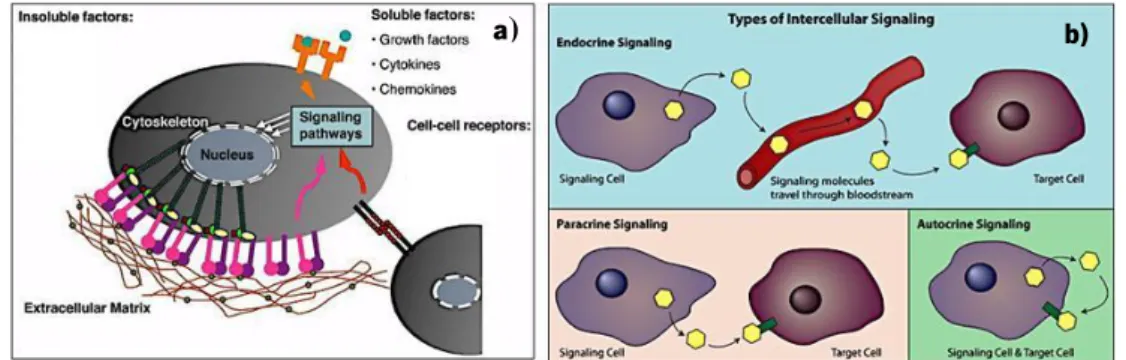
Figure 1.2 The extracellular microenvironment (a) and cell signaling trough soluble factors.
Adapted from 28.
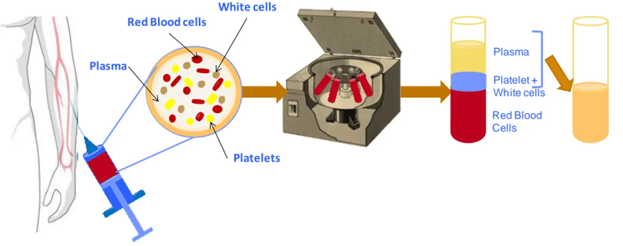
Figure 1.3 Schematic representation of PRP established process. In this procedure about 30 to 60 mL of blood is taken from the donor and centrifuged during 15 minutes and 3200 rpm. With this centrifugation step it is possible to separate the different constituents of the human blood. PRP can be storage until further use and upon activation PRP facilitates the local release of different GFs for tissue engineering therapies.
Chapter 2 – Materials and Methods
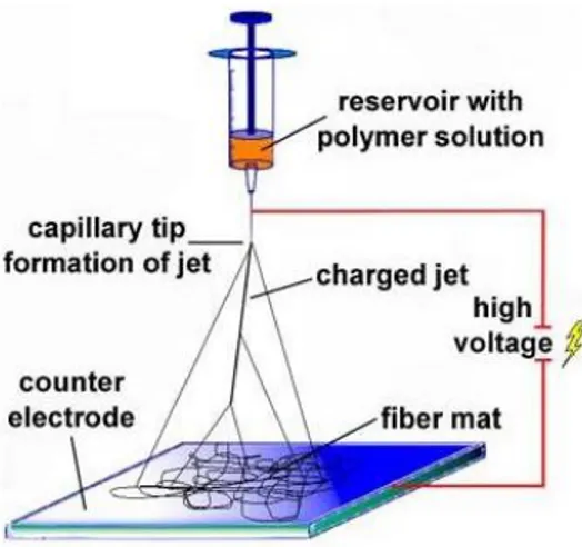
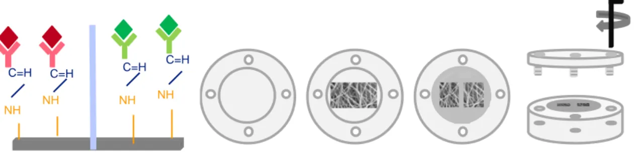
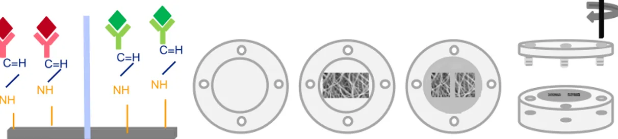
Figure 2.1 Electrospinning setup composed by an electric power supply, a syringe pump and collector.
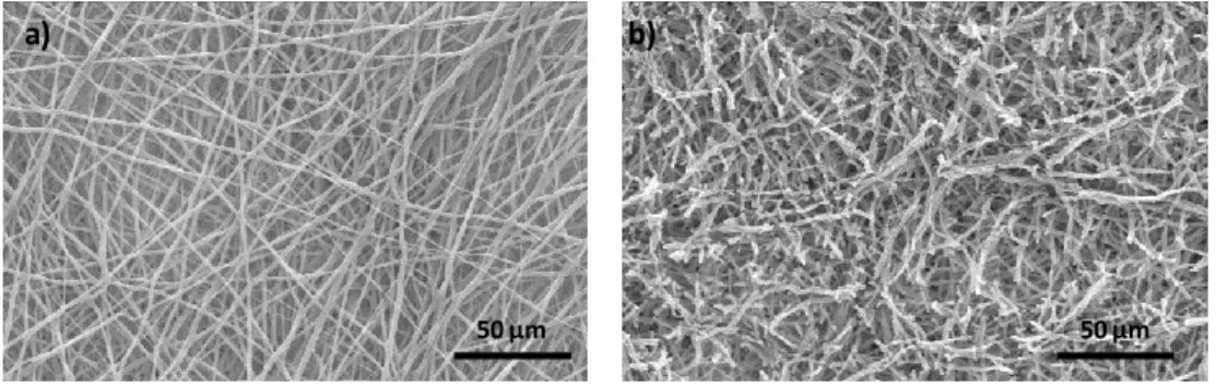
Figure 2.2 SEM analysis of an electrospun PCL NFM at 500X magnification: a) not subjected to any physicochemical treatment and b) after UV-ozone and aminolysis functionalization
Figure 2.3 The antibody structure, representing the variable and the non-variable region, as well as the carboxyl group at the end of the former region.
Figure 2.4 Antibody and antigen immobilization strategy, applied to the electrospun PCL NFMs, for further detection by the fluorescence reading method (sequence of steps required) Figure 2.5 EDC reacts preferentially with the carboxyl groups forming O-acylisourea, an unstable reactive ester that with the combination of NHS forms a semi-stable amine-reactive ester. This NHS ester can readily react with the available amine groups at the surface of electrospun nanofibers.
Figure 2.6 Schematic representation of the compartment watertight device, that allows the simultaneous immobilization of two different antibodies in two areas of a single mesh.
XVIII
Chapter 3
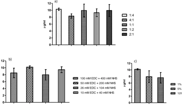
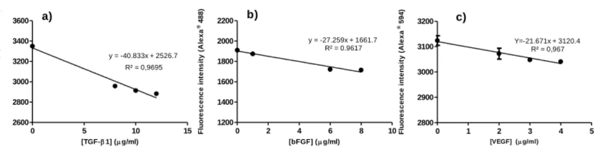
Figure 3.1 Schematic representation of the compartmental watertight device that allows the simultaneous immobilization of two distinct antibodies in two areas of a single mesh. Figure 3.2 EDC/NHS ratios and concentrations optimization. (a) optimization of the coupling agents EDC/NHS ratio; (b) optimization of the individual EDC and NHS concentrations, maintaining the previously optimized ratio 1:4; (c) optimization of the final concentration of the EDC/NHS mixture (50 mM EDC + 200 mM NHS, optimized before) in the antibody solution.
Figure 3.3 Maximum immobilization capacity of a single antibody at the surface of
activated and functionalized electrospun nanofiber: a) immobilization of anti-TGF-1, b)
immobilization of anti-bFGF and c) immobilization of anti-VEGF.
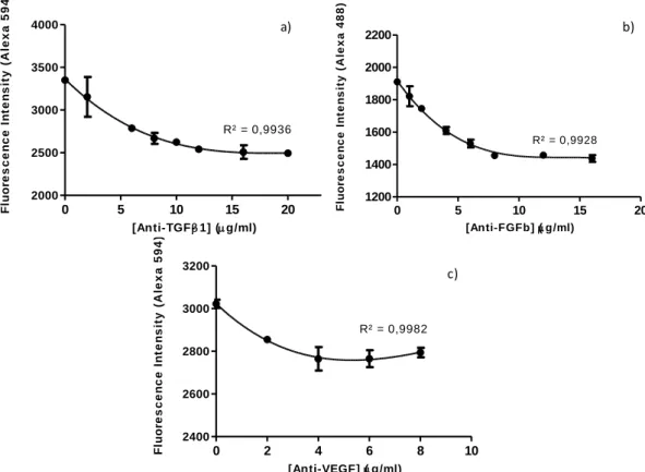
Figure 3.4 Standard curves for single antibody immobilization at the surface of activated
and functionalized electrospun nanofibers. a) TGF-1 antibody standard curve varying
between 0 g/mL and the maximum concentration that can be immobilized (i.e. 12
g/mL). b) bFGF antibody standard curve ranges from 0 g/mL to 8 g/mL; and c) VEGF
antibody standard curve varies between 0 g/mL and 4 g/mL.
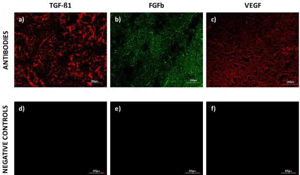
Figure 3.5 Spatial distribution of immobilized primary antibodies at the surface of activated and functionalized electrospun nanofibers. Primary antibodies were immobilized at the
previous optimized concentrations: a) 12 μg/mL of anti-TGF-1, b) 8 μg/mL of anti-bFGF
and c) 4 μg/mL of anti-VEGF. In the case of the TGF-1 and VEGF antibodies, the
secondary antibody Alexa Fluor ® 594 was used, whereas the secondary antibody Alexa
Fluor® 488 was used for the bFGF antibody. The negative controls d), e) and f) were subjected to all the steps except the incubation with the primary antibodies.
Figure 3.6 Capability of the biofunctionalized nanofibrous substrate to bind different
concentrations of the recombinant protein: a) TGF-1, b) bFGF and c) VEGF.
Figure 3.7 Biochemical performance of the endothelial cell line cultured on unmodified electrospun PCL NFM, NFM with immobilized VEGF antibody (NFM_Ab1), both in
XIX
supplemented medium (ECGS) ; NFM with immobilized VEGF antibody (NFM_Ab2), NFM
with bound recombinant VEGF (NFM+VEGFRec) PL-derived VEGF (NFM+VEGFPL) with non
supplemented media a) Cell Proliferation, b) cell viability, c) total protein synthesis and d) intracellular VEGF synthesis. Statistical analysis was performed for the five different conditions comparing each time point (Day 1, Day 3 and day 7). Data was considered statistical different for p values < 0.05. (*) denote significant differences when compared to
NFM condition, (+) when compared to NFM_Ab1 supplemented media, (x) when compared to
NFM_Ab2, (#) when compared to NFM_VEGF and (&) when compared to NFM_PL.
Figure 3.8 (a) Quantification of mixed immobilized bFGF and VEGF antibodies. (b) Relative quantification of bound GFs (i.e. VEGF and bFGF) derived from PL.
Figure 3.9 Spatial distribution of the mixed immobilized primary antibodies at the surface of a single activated and functionalized Nanofibrous substrates. The bFGF and VEGF antibodies were simultaneously immobilized in the same mesh, at the previously optimized
concentrations. a) Alexa Fluor ® 448 was used as the secondary antibody for the anti-bFGF);
b) the Alexa Fluor ® 594 was used for the anti-VEGF; c) the spatial distribution of the two
primary antibodies (merge view); and d) activated and functionalized Nanofibrous substrates without primary antibodies immobilization.
Figure 3.10 Laser scanning confocal microcopy image demonstrating the side-by-side antibodies immobilization over the same activated and functionalized Nanofibrous substrate. List of Tables
Chapter 1
Table 1.1 Most commonly used Growth Factors: biological role and targets
Chapter 3
Table 3.1 Quantification of the growth factors of interest derived from two human PL samples
1
3
Chapter 1
Introduction
Growth factors immobilization for tissue engineering and regenerative medicine therapies
1.1 Abstract
To achieve a more effective and faster regeneration of tissues and organs, the immobilization of bioactive molecules at the surface of biomaterial substrates has attracted tremendous interest as a promising Tissue Engineering and Regenerative Medicine approach. Growth factors as bioactive molecules play a pivotal role in wound healing cascade and have a significant role in a wide range of cellular events, such as proliferation, migration, cell signaling and differentiation. There are different immobilization strategies that can be followed to functionalize the biomaterial substrates with growth factors. However, a critical issue is the preservation of the bioactive molecule functionality after immobilization. Covalent immobilization is able to accomplish those requirements, leading to the development of devices with high functionality and to successfully achieve localized and sustained growth factor availability. Biological samples, like platelet rich plasma (PRP), have gained special interest mainly due to the easy assessability, variety and autologous source of growth factors envisioning personalized therapies. In this introduction we will discuss the various growth factors immobilization strategies available and its applications.
Key Words: Growth Factors, Immobilization, Biomaterials, Platelet Rich Plasma, Regenerative Medicine, Tissue Engineering
4
1.2 Tissue Engineering and Regenerative Medicine
Injuries and diseases can affect tissues and organs which may result in the partial or total loss of function. When no medical intervention is conducted, the physiological response of the body is restricted and is mostly confined to the auto-regenerative process. This might be an effective response to small injuries, but does not lead to the restoration of the normal
structure and function of large defects 1–3. In the last decades, the conventional treatment
modalities relies on the replacement of the affected organ or tissue by synthetic implantable
devices that can restore the tissues’ or organs’ function 1,4. Current clinical therapies for
restoring tissue structure and function largely rely on the: transplantation of organs (such as kidney, liver, heart, lung, pancreas); on the use of tissue transplants (such as autografts, allografts or xenografts), on the administration of growth factors (GFs) and on the
implantation of artificial devices (metal-alloys, ceramics or other prosthesis) 1,5. Regrettably,
these strategies not always have the desired outcomes mostly caused by immune rejection, insufficient biocompatibility, the required mechanical and physical properties, complicated surgical procedures, chronic inflammation and lack of clinical predictability. With artificial implantable devices, the patient needs to face problems often related with the reliability and the fitting of the device into the defect site.
Due to the increased incidence of injuries and diseases, and the medical need to create more effective therapies for improving the outcome of current types of tissue loss, the field of Tissue Engineering and Regenerative Medicine (TERM) has proposed alternative
solutions and strategies that can overcome some of those limitations 3,6. The increasing
knowledge on wound healing and tissue formation physiology, as well as the advances made
in materials science and cell biology, is essential to the development of effective strategies 1,3.
The TERM research aims at replicate these physiological processes, in order to develop more
efficient hybrid systems for a complete restoration of damaged organ or tissue 1,7–9.
Tissue Engineering (TE) aims to create, replace and facilitate the regeneration of damaged or diseased tissue with the combination of three different fundamental factors: biomaterials (scaffolds), cells and bioactive molecules (in most cases GFs) (Figure 1.1). The final purpose of TE is to create a tissue construct that upon transplantation will give raise to similar tissue found in the body.
5
TE approaches that may be followed are: (a) the delivery of tissue-inducing molecules that stimulate host cells to function normally; and (b) the development of a 3D matrix or
scaffold biomaterial in which cells grow to create a living 3D tissue substitute 7–9. The former
strategy is the most commonly followed in the field of TE, where living cells are seeded in a natural and/or synthetic substrate in order to create implantable devices able to restore the function of tissues. In this strategy, the required bioactive molecules (such as GFs, cytokines and proteins) are often supplied in the culture medium or may be released by drug delivery
system 7,9,10.
1.2.2 Requirements of a biomaterial scaffold
The scaffold plays a unique role in the TE strategies since it is designed to serve as a temporary support for the cell proliferation, migration and differentiation, in order to form a
hybrid tissue construct in vitro 4. During the past two decades, many studies have been
conducted in the development of biomaterials scaffold with potential applicability in clinical TE strategies. For that reason, scaffolds should have the following properties: (a) promote
cell-biomaterial and cell-cell interactions, cell adhesion and ECM deposition 7,11,12; (b) facilitate
a sufficient transport of gases, nutrients and other soluble factors, to allow cell survival, Figure 1.1 The three fundamental components of a Tissue Engineering strategy: a) biomaterial
6
proliferation and differentiation 7,13; (c) biodegradability in a rate compatible to the kinetics of
tissue growth 14; (d) act as delivery vehicles for biomolecules and bioactive factors 13; (e)
support the stress developed at the implant site and retain mechanical strength after
implantation, 15; (f) the porosity and the pore size should allow the cell ingrowth, an uniform
cell distribution and migration, an improved ECM deposition and facilitate neovascularization
of the construct 7,16,17; (g) provoke a minimal degree of inflammation or toxicity in vivo 18.
1.2.2 Cell sources
The cell is the basic structural and functional unit of a living organism. Multicellular
organisms are made up of many different cell types with specialized functions 19. These
specialized cells enable performing specific functions. Depending on the final application and the tissue to be repaired, different cell sources can be selected and expanded in vitro such as
the hematopoietic, endothelial, chondrocytes, osteoblasts, epithelial cells 20.
When cells are used for TE approaches, a biopsy of a donor tissue is dissociated into individual cells. Indeed cells source can be xenogenic (such as bovine or porcine), allogenic (donor from the same species but from a different individual) or autologous (from the patient itself). The preferred cell source to use in a TE strategy is the autologous cells, where a biopsy of a donor tissue is obtained, followed by the cells dissociation and expansion in vitro,
and, finally, their implantation into the host 10. One of the limitations of applying cell-based
regenerative medicine therapies to organ replacement is the difficulty of obtaining sufficient number of cells in relevant therapeutically quantity, as well as the protocols of in vitro cell
manipulation are not completely elucidated 21.
While several tissues remain important cell sources of therapeutic relevant differentiated
cells, stem cells have emerged as a very strong alternative. Stem cells have the remarkable
potential to differentiate into different cell types in the body. They serve as an endogenous repair system, able to participate in the permanent homeostasis and maintenance of the organs dying cells. Stem cells are distinguished from other cell types by two important characteristics: they are unspecialized cells capable of renewing themselves through cell division (the self-renewal capacity) and they can give rise to specialized cell types under
7
when a stem cell divides, each new cell has the potential either to remain as a stem cell or to become another cell type with a more specialized and differentiated function.
Stem cells can be characterized as totipotent (ability to differentiate into the cells of all the tissues of an entire organism) pluripotent (the potential to differentiate into most specialized cells in the body, but not all the tissues of an organism) and multipotent (the
ability to form multiple cell types). 23 There are different types of stem cells: the embryonic
stem cells (ESCs) 25, responsible for embryonic and fetal development and growth; the adult
stem cells (ASCs)26, responsible for the growth, tissue maintenance, regeneration and repair
of diseased or damaged tissue.
1.2.3 Bioactive molecules: growth factors
Different signals from the extracellular microenvironment can play significant roles over the cellular performance, namely insoluble extracellular matrix (ECM) macromolecules, diffusible/soluble molecules, and cell–cell receptors (Figure 1.2 a). Although the growth factors (GFs) belong to the category of soluble molecules, they can also be present in the immobilized form within the ECM. The diffusible/soluble molecules (including the GFs) can have different ways of action over cellular activity: autocrine (cell secretes molecules that binds to receptors on that same cell, leading to changes in that same cell), paracrine (cell produces a signal to induce changes in nearby cells) and/or endocrine (communicate a molecule over a long distance; the signals are released from a cell, migrate with the bloodstream and can travel around the entire body) (Figure 1.2 b). Growth factors bind to specific receptors on target cells and regulate the gene expression controlling functions such
as cell growth, tissue morphogenesis, wound healing and regeneration 6,27.
Figure 1.2 The extracellular microenvironment (a) and cell signaling trough soluble factors.
Adapted from 28
8
The bioactive signals (like proteins, GFs, cytokines) involved in tissue repair or function restoration also can play a fundamental role in TERM strategies. A precise control over the levels and sequence of the signaling molecules within a specific location may positively
regulate the regenerative processes 9,27. Proteins, especially GFs, have an important role in
the regulation of a variety of cellular processes, namely in the growth, migration, differentiation and apoptosis of specific cells. Additionally, the activity of GFs is particularly connected when an injury occurs, by coordinating the healing process, until the wound is
completely repaired 6,27. Another important aspect related to the function of GFs is there
crucial role in the exchange of information between different cell populations and their
microenvironment (paracrine effect) 6,29,30.
1.2.3.1 Growth factors and the healing cascade
GFs are known to play a pivotal role in the complex cascade of the physiological repair mechanism by providing the needed signals to the cells and, thereby, leading to an
accelerated functional restoration of damaged or defective tissue 31. Wound healing is a
complex biological process that involves inflammation, mitosis, angiogenesis, synthesis of
proteins and ECM remodeling 32. There are different GFs with different specific targets and
functions that are involved in all the phases of the healing process: Vascular Endothelial Growth Factor (VEGF), Basic Fibroblast Growth Factor (bFGF), Epidermal Growth Factor
(EGF), Insulin Growth Factor (IGF), or Platelet derived growth factor (PDGF) 29,31,33.
In a simple way, the healing cascade involves three different phases: inflammation, a trophic phase (including angiogenesis, proliferation and synthesis of ECM) and remodeling. When a tissue is damaged or injured, the healing cascade begins immediately when platelets come into contact with collagen. After platelet activation, clothing factors, cytokines and
growth factors are released initiating the healing response 31,34.
The inflammatory response is characterized by leucocyte extravasion and
accumulation at the injury site, and monocyte/macrophage activation 35,36. PDGF initiates the
chemotaxis of neutrophiles, macrophages, smooth muscle cells and fibroblast. TGF-β is another important signal for the initiation of the healing cascade by attracting macrophages, stimulating them to produce additional cytokines like FGF that enhances collagen synthesis. TGF-β further enhances collagen expression leading to a strong response of the matrix
9
producing cells to ensure a rapid deposition of new connective tissue at the injury site and
developing the fibrous cloth 37,38.
The trophic phase includes the activation of endothelial cells to initiate the angiogenic process, which is the formation of new blood vessels able to promote blood flow, to support the high metabolic activity of the new tissue (avascular tissue are not comprise in this
angiogenic phase)39. VEGF stimulates this process provinding a new blood supply to the
injured site. As the healing process progresses, several other important responses are activated. The cells migrate into the injury site, using the fibrin matrix as a scaffold, then divide and differentiate, producing collagen, proteoglycans and other components of the
natural ECM 31. Finally, during the remodeling stage, there is a decrease in cell density and,
therefore, on the metabolic activity of the injured tissue. As stated above all these processes are mediated and activated by signaling molecules, like GFs, that limit the duration of each
phase and promote the progression into the next stage 31,40,41.
The basis of TERM research relies on the engineering of a microenvironment able to mimic the critical aspects of the natural healing process, namely the wound healing cascade,
by providing suitable biochemical and physical factors.6 However, due the high complexity of
this process these biological systems are not easy to recreate in vitro 6.
Therefore it is absolutely necessary to provide the cells with a local biochemical and mechanical niche that can mimic the natural environment in which they can proliferate and differentiate efficiently, by creating an artificial ECM and by delivering GFs. In order to induce the regeneration and to accelerate the capability of tissue growth, it is fundamental to create an environment that can mimic the natural wound healing cascade. Due to all these aspects, the integration of GFs and the development of biomaterials that mimic the ECM microenvironment play an important role in the cellular regulation of adhesion, proliferation, differentiation and gene expression.
1.2.3.2 Growth factors: properties and roles
GFs have different mechanisms of action depending on the concentration, on the half-life time, on the phenotype of the target cells as well as on their presentation (soluble or
immobilized in the ECM) 6,30,42. The local and systemic administration of GFs are therapeutic
10
well as the repair and regeneration of tissues in different fields like plastic surgery,
orthopedics and cartilage lesions, muscle injuries and skin 29,30. The different GFs used in
therapeutic applications are presented in Table 1.
To be effective as a therapeutic agent, a GF has to reach the target site without suffering biological degradation and, it has to remain at the target location sufficient time to exert its actions. GFs that are provided exogenously in solution at the site of the injury tend to be not effective because they tend to diffuse away from the wound site, being susceptible to
enzymatic digestion or inactivation 6. In summary, various aspects have a significant effect on
the therapeutic efficacy of GFs, including their short half-life in vivo, the side-effects caused by the administration of multiple or high doses of GFs to reach the desired effect at the target cells, and the possible denaturation of the GFs during manipulation and circulation. All these
issues should be taken into account when designing a successful GF-based therapy 6,43,44.
When designing an implantable system, some specific requirements related to the GFs should be of prior interest: the type of GF to be used and its final application; a feasible preparation method that does not affect the GF bioactivity; a robust system that can restrict the protein conformational mobility and protect the protein from physical and chemical degradation; a high loading efficiency; the ability of the system to retain the GFs at the site of
action; a presentation of the GFs that mimics the temporal profile of the healing process in
11
Table 1 Most commonly used Growth Factors: biological role and targets
GF ROLES TARGETS REFERENCES
TGF-1
Promotes the production of extracellular matrix; Modulates and enhances the proliferation of
fibroblasts;
Increases and stimulates synthesis of collagen type I;
Enhances the proliferation of bone cells.
Bone/ Cartilage
47–50
FGF
Potent inductor of cell proliferation; Promotes angiogenesis and differentiation; Collagen production. Bone/ Cartilage/ Periodontal tissue 51–53 PDGF
Stimulates fibroblast production, angiogenesis and macrophage activation;
Collagen synthesis. Cartilage/ Bone/ Angiogenesis 54,55 EGF
Triggers the expression of genes that leads to DNA synthesis and proliferation;
Promotes mesenchymal and epithelial cell differentiation, angiogenesis and proliferation.
Skin/ Cornea/ Nervous System
56–58
IGF
Chemotactic for fibroblast and stimulates protein synthesis;
Enhances bone formation by the proliferation and differentiation of osteoblasts.
Bone/
Dermal wound healing/ Pancreatic stem cell
differentiation
59–63
VEGF
Promoter of angiogenesis and vasculogenesis; Proliferation of endothelial cells;
Increases microvascular permeability.
Vascularization/ Stem cell differentiation
64–66
1.3 Growth factors sources
Peripheral blood is constituted by different cellular elements like red blood cells, white blood cells and platelets. These components are found in the peripheral circulating blood and are not retained and sequestered by the lymphatic system, spleen, and liver. Certain medical conditions in which the patients lose some of the blood components may require a blood transfusion. Nowadays, there is no need to make a transfusion of the whole
12
blood, being possible to select only the components of interest (like platelets, plasma,
clothing factors) 67,68.
In the past decades, platelet rich plasma (PRP) has been increasingly used in many medical fields, with particular interest in orthopedics and musculoskeletal disorders in
athletes that need a fast recovery and early return to competition 31,69,70. PRP is described as
“a fraction of autologous plasma containing an above baseline platelet concentration and
growth factors which take part in the post-traumatic healing process”71. Therefore, to be
defined as PRP, the platelet count should be at least 1 000 000 platelets/µl, whereas a platelet normal count ranges from 150 000 to 350 000 platelets/µl. Since platelets are a source of GFs, there is a growing interest in the use of PRP as a strategy to optimize the
healing of the tissues 72.
The impact of the discoveries regarding the potential of PRP healing has increased the optimism about autologous based regenerative medicine. PRP is also a cost-effective product, since it is taken from a simple blood samples and, therefore, it is easy to implement this cost-effective source in the current clinical practice. The Figure 1.3 depicts the simplicity of the PRP preparation procedure. Additionally, since PRP is a concentrated extract of lysed platelets, it is also a source of fundamental growth factors that are secreted by the platelets when a wound healing process is initiated. Because it is an autologous source of GFs, less regulatory concerns exist about its biosafety, since the immunogenic reaction and the possibility of disease transmission are eliminated. However, due to its complex composition of proteins, growth factors and cytokines, the mechanisms of action and dosage
13
The possible use of PRP as an alternative biological source of bioactive agents has
gained a special and exciting interest in TERM approaches 31,73. The use of PRP aims to take
advantage of autologous source of bioactive molecules, such as GFs, proteins and cytokines, envisioning an autologous therapy where it is possible to use patient specific therapies. Recent reports suggest that the use of PRP may be an approach to develop clinically relevant materials (GFs+scaffolds) able to deliver GFs and simultaneously to allow cell
culture, and ultimately, integrate the in vitro generated construct into the native tissue
environment 41,74–76. However, there are some controversial works where it is reported that the
use of PRP is not effective 70,77,78. Since the use of PRP is still in an early stage, new
preparation methods and applications need to be explored to maximize its efficiency in TERM strategies. It is also necessary to determine the optimum concentration of the various GFs present in the PRP, which result in more effective PRP-based treatments.
1.4 Immobilization methods and strategies
The GFs released in physiological environments are susceptible to inactivation by degradation, prior to the possibility of reaching the desired target cells. Therefore, high quantities of soluble GFs well above the physiological values may be needed to have the required effects at the cellular level. However, the delivery of high quantities of GFs may lead
Plasma
Red Blood Cells Red Blood cells
White cells
Platelets Plasma
Platelet + White cells
Figure 1.3 Schematic representation of the PRP process. In this procedure about 30 to 60 ml of
blood is taken from the donor and centrifuged during 15 minutes and 3200 rpm. With this centrifugation step it is possible to separate the different constituents of the human blood. PRP can be storage until further use and upon activation PRP facilitates the local release of different GFs for tissue engineering therapies.
14
to undesired cells and tissues side effects related with toxicity. Biomaterial-based systems can be designed to deliver soluble GFs locally, but sometimes they can be ineffective and
costly 6,42,79. To overcome some of these drawbacks on the use of GFs as therapeutic agents,
the biofunctionalization of substrate surfaces by the immobilization of GFs has special interest mainly due to the need to optimize the biological performance of implantable medical
devices 80.
An important requisite of protein immobilization is the biocompatibility and bioactivity of the substrate surface, because it should interact positively with the native structure of the
proteins and biomolecules 81. The substrate chemistry, particularly the availability of reactive
groups, is an important factor to consider when selecting an appropriate immobilization strategy. The location of such reactive groups, relatively to the receptor-binding area of the GF structure, and the dimensions of the substrate are other important aspects to have in consideration. Furthermore, the immobilization of GFs generally requires the use of aqueous-based chemistry, as most of the GFs are either not soluble or may become denaturated in
the presence of organic solvents 6,42,82–84.
This section will mostly be focused on the immobilization strategies that involve the direct binding of the GF to the surface of the biomaterial substrate. Different immobilization methods can be implemented to achieve the biofunctionalization of substrates such as
adsorption, physical entrapment and covalent-immobilization 42,82. However, each
immobilization method results in advantages as well as in limitations. Covalent immobilization is the most reported method, which leads to a strong and stable binding of the bioactive molecule to the substrate. However, the presence of functional groups is required in both the substrate and the molecule to be immobilized. In most of the cases, a linker reagent is necessary. With this method it is also possible to overcome the common problems associated to the adsorption method, namely the deadsorption or the denaturation
of the antibody 82,85.
1.4.1 Non-covalent immobilization
The immobilization of GFs can be divided in two different main categories: non-covalent and non-covalent immobilization. Non-non-covalent immobilization includes physical
15
GFs encapsulation/incorporation into micro-particles or reservoirs. Adsorption of GFs typically takes advantage of direct interactions, such as surface electric -charge or other secondary interactions between the GFs and the substrates surface, or of indirect interaction via
intermediate proteins or other biological molecules 83,86,87. A third approach of non-covalent
immobilization of GFs to a biomaterial is ion complexation. Proteins with different isoelectric points may be used for polyion complexation with charged macromolecules. These methods tend to have some problems associated such as the desadsorption and the irreversible ion
complexation that can cause protein inactivation and denaturation 9,83.
To improve the stability and persistence of the GFs immobilization, and consequent delivery to target cells and tissues, covalent immobilization of GFs to the substrate emerged as an alternative approach. Despite being a more complex process, immobilization strategies can prolong GFs availability than those obtained with weak physical immobilization. Also, the covalent immobilization allows a spatial control over the GF distribution and reduces the amount of GFs required, thereby potentially reducing the cost and increasing the efficacy of various bioactive materials or engineered tissues. Furthermore, the presentation of GF in an immobilized form also has physiological relevance, as both soluble and matrix-bound GFs
perform distinct functions in the in vivo environment 6,42,88.
1.4.2 Covalent immobilization strategies
For the covalent immobilization of bioactive molecules, different chemical and reactive groups are needed in both the substrate and the GFs of interest. For different covalent immobilization methods, different groups are required. The most common reactive groups of
GFs are the amines or the carboxyl groups 84. Whereas in the substrate surfaces, besides the
amines and the carboxyl groups, also double bonds, C-H, N-N and acrylates are frequently
required89. If the substrates do not have the required chemical groups, chemical treatments
can be performed to activate their surface.
Covalent immobilization often requires a linker to achieve a more stable and strong binding of the GF to the substrate. For example, 1-Ethyl-3-(3-dimethylaminopropyl) carbodiimide (EDC) couples the carboxyl groups to the amine groups, resulting in a stable amide bond. In this reaction, EDC reacts with carboxyl groups to form an o-acylisourea
16
intermediate, which further reacts with the amine-containing molecule. However, this intermediate is highly unstable in aqueous solution and susceptible to hydrolysis, which results in re-formation of the carboxylic acid. Thus, sulfo-N-hydroxysuccinimide (sulfo-NHS) or N-hydroxysuccinimide (NHS) may also be added to create a more stable amine-reactive sulfo-NHS or sulfo-NHS ester intermediate, which increases the efficiency of the reaction. This coupling
procedure is simple, inexpensive, effective and can be performed under mild conditions 42,90,91.
Other methods of GFs immobilization involve the use of a photo-initiated reaction,
which allows the binding of the GFs to a substrate 92,93. The first step of this method relies on
the functionalization of the GF with a photo-reactive group and, afterwards, the binding of the modified GF to the substrate surface take place upon exposure to a defined radiation
wavelength (usually, ultra-violet light) 94. One significant advantage of the photo-immobilization
method is the simple development of patterned GFs, which can be easily performed through the use of photomasks or by the precise irradiation of specific areas with laser light sources, leading to a spatial defined distribution of the immobilized GFs. The creation of immobilized GF patterns enables greater control over cell function. Like the GFs immobilization by the use of EDC, photo-immobilization is relatively simple and effective but the use of photomasks frequently involves expensive technologies. The use of crosslinking reagents or the photo-immobilization approaches also may have problems related with the GFs bioactivity, bioavailability and protein configuration. Specifically, the potential caused to the damage GFs
by the ultra violet light is considered the most important drawback 42,82,94.
Functionalization of polymers with acrylate groups is a common method for the activation of biomaterials that can be further cross-linked upon exposure to UV. Crosslinked networks of acrylated polymers are formed via chain-growth polymerization of the acrylates, and this process is initiated by reactive centers, such as radicals, which are generated upon photo-cleavage of the initiator molecules. These free radicals propagate through the unsaturated vinyl bonds on the acrylated polymers, resulting in covalently crosslinked high molecular weight polyacrylate kinetic chains. However, the bioactivity and orientation of the immobilized GFs can be compromised. Because this immobilization strategy depends upon acrylate reaction with another acrylate, the types of substrates that can be used are generally
restricted to acrylated polymers, such as PEG-DA 42,84,85.
By immobilizing GFs onto the surface of biomaterials, they are more protected against cellular inactivation and digestion. As a result, the immobilized GFs have more sustained and
17
longer activity when exposed to physiological environments. Growth factor immobilization may also overcome the diffusion limitations of soluble GFs. While the soluble delivery of GFs elicits responses in the surrounding environment, covalently immobilized GFs may have the additional advantage of inducing local effects within the scaffold where the cells are seeded, maximizing its effect. Maximum interaction with cells in the implant sites facilitates
integration with the host tissues 6,27,88.
1.5 Applications of immobilized growth factors
The effect of soluble GFs over the behavior and signaling of different cell types has been widely described in the last years. However, in what concerns the influence of immobilized GFs, the available information is sparse and the outcomes much less reported. Soluble GFs are recognized by their corresponding cell-surface receptors and, thereafter, are internalized as a complex. In the case of immobilized GFs, cells are not able to internalize those receptor-GFs complexes, leading to a more sustained activation of the intracellular pathways. Consequently, with the immobilization of GFs it is possible to achieve enhanced
and unique cellular responses that cannot be achieved with soluble GFs 27,95. Due to the
improved stability and spatial control offered by the immobilization of GFs, this approach may provide beneficial contributions for different tissue repair strategies. The next sections will focus on the most promising results on the application of immobilized GFs at the surface of substrates. The effects of immobilized GFs have been studied in various areas including angiogenesis, bone repair and regeneration, dermal wound healing, cartilage repair, pancreas and liver, nerves and stem cell differentiation.
1.5.1 Angiogenesis
One of the major problems associated with tissue engineered therapies is the lack of a functional and integrated vascular system. The flow of nutrients and oxygen and the cell metabolites are essential to maintain the viability and functionality of tissues. Particularly, an inadequate vascular system leads to mal-function in mass transport and gases exchange
18
Endothelial cells are the responsible for the capillary vascular network formation (angiogenesis). Almost all tissues depend on a blood supply, and the blood supply depends on endothelial cells, which form the linings of the blood vessels. Endothelial cells have a
remarkable capacity to adjust their number and arrangement to suit local requirements 98.
The formation of new blood vessels can occur during different events, such as wound healing, organ regeneration, and in the formation of the placenta, as well as in several pathological processes (e.g. tumor growth, rheumatoid arthritis, diabetic retinopathy). A switch to the angiogenic phenotype depends on a local change in the balance between angiogenic stimulators and inhibitors. Furthermore, angiogenesis process is regulated by a
complex control system, mediated by soluble molecules, such as VEGF 99. In the in vivo
physiological environment, both matrix-bound and soluble forms of VEGF can be found. Soluble VEGF is believed to induce endothelial cell proliferation, whereas matrix-bound VEGF tends to promote the vascular sprouting and branching associated to neovasculature. Immobilized VEGF has been found to successfully stimulate the proliferation of endothelial
cells 65,99.
Due to the short half-life and the transient effect of soluble VEGF the development of strategies to achieve a more controlled delivery and release of this GF is of utmost importance. Accordingly, the immobilization of VEGF has become a promising strategy to overcome these problems, as a more suitable system to have control over the angiogenic process and endothelial cell function. With this strategy a microvasculature system would be incorporated in some engineered biomaterial substrates and scaffolds leading to more
efficient tissue engineered solutions 42,100. Recent reports demonstrated that the covalent
immobilization of VEGF on different substrates and scaffolds improved the endothelial cells
tubulogenesis as well as stimulates their proliferation 101,102. The release of soluble VEGF
sometimes results in vessels that are not completely functional. For that reason, VEGF was also co-immobilized with other GFs in other to achieve a more efficient and functional angiogenic process. Angiopoetin-1 (a GF related to the vessels stability and maturation) was co-immobilized with VEGF and the biological results demonstrated a significant improved
tubulogenesis, as well as endothelial cell infiltration 103,104. Another example of GF involved in
the angiogenesis process, PDGF-ββ was also immobilized to improve the formation of a
functional engineered microvasculature 105. Despite significant efforts and promising results in
19
improve angiogenic and vessel stability, those strategies still need further demonstration of efficacy.
1.5.2 Other relevant applications: dermal healing, cartilage, bone and stem cells differentiation
The use of GFs has been explored in different tissues, namely in the healing of chronic dermal wounds, overcoming the problems often associated with pain, long-term recovery, high cost and unsuccessful treatment outcome. With the GFs immobilization approach, a more controlled bioavailability of the GFs is possible, overcoming the problems of inadequate delivery of soluble GFs. Also of note is the prolonging bioactivity and half-life of the GFs. EGF immobilization was reported in most studies of dermal wound healing, since it stimulates the migration and proliferation of dermal cells (example: fibroblasts) that play an important role in wound closure. It was demonstrated that lower amounts of immobilized EGF are needed,
when compared to the soluble EGF, to have a positive effect over the cellular response 42,106–108.
Embryonic and adult stem cells have the capacity for self-renewal and also have the ability to differentiate into different cell types. Stem cell differentiation can be achieved in vitro by the application of different molecules and signals. Recent studies evaluated the response of stem cells to immobilized GFs. A wide range of final applications was explored including the immobilization of GFs to promote neural differentiation (PDGF-AA and IFN-ϒ) and VEGF
immobilized to differentiate the stem cells into vascular or hematopoietic cell types 109–112.
Recent efforts have focused on helping the body to restore cartilage, through cell-based and/or biomolecules therapies. A variety of synthetic- and natural-origin polymers were proposed for this purpose, each of it with their benefits and drawbacks. To date, an ideal biomaterial has yet to be created that can promote the functional repair or regenerate the
damaged cartilage 113. However, the addition of signaling molecules such as the GFs seems to
be a promising control to facilitate cartilage regeneration therapies.
TGF-beta was proposed to stimulate chondrogenesis through intracellular pathways 114.
TGF-3 was immobilized into different scaffolds to solve and treat some cartilage injuries and
lesions. Also TGF-1 was immobilized at the surface of different polymers. In both cases the
expression of chondrogenic gene markers was significantly increased, as well as the production of glycosaminoglycans and collagen type II, indicating that neo-cartilage was generated.
20
The gold standard treatment of bone fractures and other orthopedic injuries causing loss of bone tissue rely on the use of autologous bone grafts. However, despite their biocompatibility and exceptional osteoconductive properties, autografts are associated with donor site morbidity. The most widely used ostegenic inducing molecules are the BMPs (namely the BMP-2 and the BMP-7, already approved by the FDA for human use) that induce new bone formation and regeneration at specific sites (used for example
in orthopedic applications such as spinal fusions and oral surgery) 115–117. In these products,
BMPs are delivered to the site of the fracture by being incorporated into a bone implant, and released gradually to promote bone formation. However, the delivery method of BMPs often shows a lack of local retention and the need of high amounts of proteins to achieve the desired biological effects. The immobilization of such GFs may have favorable outcomes in bone tissue engineering strategies, as well as on the osteointegration of orthopedic implants,
with the use of significantly lower amounts of GF to achieve effective osteogenic outcomes 42.
BMPs were immobilized in different substrates such as PLGA scaffolds 118, chitosan
membranes 119 and PCL scaffolds 120 showing that the biological effect of immobilized BMP-2
can significantly increase the expression of osteoblastic differentiation markers, when osteogenic precursor cells are cultured. When comparing the in vivo efficiency of the soluble BMP-2 and the immobilized form, the immobilized approach showed that both the amount
of bone and its maturity have increased 42,118–120.
1.6 Final Remarks
The implementation of chemical immobilization strategies allows the development of highly effective GF delivery systems, promoting the direct interactions between the immobilized GFs and the resident cells and avoiding the potential side effects caused by systemic administration. The biggest challenge of this strategy relies on finding the correct balance between the GFs and the physicochemical properties of the scaffold that can regulate cell behaviors in designing highly effective strategies. By combining biomaterial scaffolds with immobilized GFs at the surface were cells are seeded it will be possible to have a strongest interaction with the host tissues
21
1.7 References
1. Williams, D. F. To engineer is to create: the link between engineering and regeneration. Trends Biotechnol. 24, 4–8 (2006).
2. Langer, R. & Vacanti, J. P. Tissue engineering. Science 260, 920–6 (1993).
3. Fuchs, J. R., Nasseri, B. A. & Vacanti, J. P. Tissue engineering: a 21st century solution to surgical reconstruction. Ann. Thorac. Surg. 72, 577–91 (2001).
4. Saltzman, W. M. Tissue Engineering: Engineering Principles for the Design of Replacement Organs and Tissues. 544 (Oxford University Press, USA, 2004)
5. Yarlagadda, P. K. D. V, Chandrasekharan, M. & Shyan, J. Y. M. Recent advances and current developments in tissue scaffolding. Biomed. Mater. Eng. 15, 159–77 (2005).
6. Chen, F.-M., Zhang, M. & Wu, Z.-F. Toward delivery of multiple growth factors in tissue engineering. Biomaterials 31, 6279–308 (2010).
7. Dhandayuthapani, B., Yoshida, Y., Maekawa, T. & Kumar, D. S. Polymeric Scaffolds in Tissue Engineering Application: A Review. Int. J. Polym. Sci. 2011, 1–19 (2011).
8. Atala, A. Tissue engineering and regenerative medicine: concepts for clinical application.
Rejuvenation Res. 7, 15–31 (2004).
9. Lee, K., Silva, E. A. & Mooney, D. J. Growth factor delivery-based tissue engineering : general approaches and a review of recent developments Growth factor delivery-based tissue engineering : general approaches and a review of recent developments. (2011). doi:10.1098/rsif.2010.0223 10. Atala, A. Engineering tissues , organs and cells. 83–96 (2007). doi:10.1002/term
11. Boyan, B. D., Hummert, T. W., Dean, D. D. & Schwartz, Z. Role of material surfaces in regulating bone and cartilage cell response. Biomaterials 17, 137–46 (1996).
12. Moroni, L. & Elisseeff, J. H. Biomaterials engineered for integration. Mater. Today 11, 44–51 (2008).
13. Garg, T., Singh, O., Arora, S. & Murthy, R. Scaffold: a novel carrier for cell and drug delivery. Crit. Rev. Ther. Drug Carrier Syst. 29, 1–63 (2012).
14. Hutmacher, D. W. Scaffolds in tissue engineering bone and cartilage. Biomaterials 21, 2529–43 (2000).
15. Meyer, U.; Meyer, Th.; Handschel, J.; Wiesmann, H. P. Fundamentals of Tissue Engineering and Regenerative Medicine. (2009).
16. Puppi, D., Chiellini, F., Piras, a. M. & Chiellini, E. Polymeric materials for bone and cartilage repair. Prog. Polym. Sci. 35, 403–440 (2010).
17. Leong, K. F., Cheah, C. M. & Chua, C. K. Solid freeform fabrication of three-dimensional scaffolds for engineering replacement tissues and organs. Biomaterials 24, 2363–2378 (2003).
18. Nair, L. S. & Laurencin, C. T. Biodegradable polymers as biomaterials. Prog. Polym. Sci. 32, 762– 798 (2007).
19. Bruce Alberts, Dennis Bray, Julian Lewis, Martin Raff, Keith Roberts, and J. D. W. From Single Cells to Multicellular Organisms. (1994).
20. Roseti, L., Bassi, A., Grigolo, B. & Fornasari, P. M. Development of Human Chondrocyte – Based Medicinal Products for Autologous Cell Therapy. (2010).
21. Koh, C. J. Tissue Engineering, Stem Cells, and Cloning: Opportunities for Regenerative Medicine.
J. Am. Soc. Nephrol. 15, 1113–1125 (2004).