Differential gene expression profiles in peripheral blood in Northeast Chinese
Han people with acute myocardial infarction
Lin Fan
1*, Heyu Meng
2, Xudong Guo
3, Xiangdong Li
3and Fanbo Meng
3 1China-Japan Union Hospital, Jilin University, Jilin, China.
2Medical College of Yanbian University, Yanji, China.
3
Department of Cardiovascular Medicine, China-Japan Union Hospital of Jilin University, Jilin, China.
Abstract
This study aimed to use gene chips to investigate differential gene expression profiles in the occurrence and devel-opment of acute myocardial infarction (AMI). The study included 12 AMI patients and 12 healthy individuals. Total mRNA of peripheral bloodwas extracted and reversed-transcribed to cDNA for microarray analysis. After establish-ing two pools with three subjects each (3 AMI patients and 3 healthy individuals), the remainestablish-ing samples were used for RT-qPCR to confirm the microarray data. From the microarray results, seven genes were randomly selected for RT-qPCR. RT-qPCR results were analyzed by the 2-DDCt
method. Microarray analysis showed that 228 genes were up- regulated and 271 were down-regulated (p£0.05, |logFC| > 1). Gene ontology showed that these genes belong to 128 cellular components, 521 biological processes, and 151 molecular functions. KEGG pathway analysis showed that these genes are involved in 107 gene pathways. RT-qPCR results for the seven genes showed expres-sion levels consistent with those obtained by microarray. Thus, microarray data could be used to select the patho-genic genes for AMI. Investigating the abnormal expression of these differentially expressed genes might suggest efficient strategies for the prevention, diagnosis, and treatment of AMI.
Keywords: acute myocardial infarction, RNA, differential expression. Received: March 15, 2017; Accepted: September 8, 2017.
Introduction
Acute myocardial infarction (AMI) is one of the dis-eases with high mortality and morbidity globally. Accord-ing to the World Health Organization (WHO), 17.5 million people died of cardiovascular diseases worldwide in 2012, accounting for 46% of the deaths caused by non-commu-nicable diseases, and myocardial infarction was one of the major causes. The global incidence of AMI is rising owing to multiple reasons such as environment, heredity, or life-style. Various complications of AMI, such as cardiac rup-ture, arrhythmia, and ventricular aneurysm, have a great impact on patient life quality, and pose large economic bur-dens on the family and society.
Contemporary clinical epidemiology has shown that hypertension, diabetes, low density lipoprotein (LDL) lipo-proteinemia, smoking, age, and gender are clearly associ-ated with the occurrence of coronary heart disease (CHD), and so the accuracy of early CHD screening has been
greatly improved. However, these traditional risk factors could not fully predict the onset and prognosis of AMI. The existing treatment methods such as anti-platelet aggrega-tion therapy, lipid-lowering therapy, vasodilaaggrega-tion therapy, or coronary stents have achieved great success in improv-ing symptoms, delayimprov-ing disease progression, and reducimprov-ing mortality, but the desired therapeutic effects have not been obtained yet.
Inspired by the obvious characteristic of myocardial infarction (MI), namely familial aggregation, genetic stud-ies on MI have made breakthrough progress in recent years. Studies have found that genetic effects could act as an inde-pendent factor influencing the onset of AMI (Qaseemet al., 2012; Silbigeret al., 2013; Tokatet al., 2013; den Hoedet al., 2015), and genetic polymorphisms are associated with the occurrence of MI (Karahan et al., 2015; Salo et al., 2015). Till date, 152 relevant sites and 320 candidate genes have been associated with increased risk of coronary artery disease (CAD) and AMI (The CARDIoGRAMplusC4D Consortiumet al.,2013; Doet al., 2014; Welteret al., 2014; Barth and Tomaselli, 2015). In studies on gene expression differences in myocardial cells of mice between AMI and sham groupsLOX, POSTN, SPARC, TIMP1, andSFRP2
were differentially expressed, and might have an impact on DOI: http://dx.doi.org/10.1590/1678-4685-GMB-2017-0075
Send correspondence to Fanbo Meng. Department of Cardiovas-cular Medicine, China-Japan Union Hospital of Jilin University, 126 Xiantai Street, 130033 Jilin, China. E-mail: [email protected].
*Current address: Echocardiography Department, First Affiliated Hospital of Soochow University, Suzhou, China.
AMI (Wanget al., 2015). Zhang Yet al.(2015) reported that BNDF, PDGF-AA, and MMP-9 expression was up-regulated in ST-segment elevation myocardial infarc-tion (STMI), and could therefore be used for the assessment of STMI. However, unlike traditional genetic disorders, MI is co-induced by a variety of genetic and environmental factors, as well as the interactions among them. So we have reasons to believe that a comprehensive gene-level analysis might be able to predict cardiovascular risks and make prognosis more accurately (Roberts and Stewart, 2012; Arvindet al., 2015).
In the past decade, AMI onset has been identified to be associated with mutations in a number of genes involved in blood coagulation, the fibrinolytic system, platelet re-ceptors, homocysteine metabolism, endothelial dysfunc-tion, abnormal blood flow, and oxidative stress (Isordia-Salaset al., 2010). A study concerning gene ex-pression in myocardial cells of rats with AMI showed that immune response, chemotaxis, inflammation, cytoskeletal tissues, and other pathways were activated as early re-sponses within 30 min of MI (Erdalet al., 2012). However, most findings regarding myocardial cells are based on ani-mal experiments, as human samples are difficult to obtain. It was previously reported that a polymorphism assay of pe-ripheral blood-associated genes could predict the probabil-ity of CAD occurrence more accurately (Kathiresanet al., 2008). Maciejaket al.(2015) compared the gene expres-sion in peripheral blood of patients with AMI and non-MI CHD and found that some genes showed significantly dif-ferent expression in the acute phase of MI, and the differ-ences gradually disappeared with time. Therefore, in this study, we used peripheral blood as samples, which can be easily obtained and provide accurate results, to determine the genes that were differentially expressed in Han patients with AMI from northeastern China, so as to find new tar-gets for the treatment of MI in Northeast Chinese Han pa-tients by verifying and analyzing these differentially expressed genes.
Materials and Methods
Ethics statement
The contents of this study pertaining to the scope of medical ethics were approved by the Ethics Committee of China-Japan Union Hospital of Jilin University. Collection of samples and information were all approved by the study subjects who provided signed informed consent.
Subjects
In total, 12 AMI patients treated at the China-Japan Union Hospital of Jilin University from June 2012 to Au-gust 2012, as well as 12 healthy individuals, were selected randomly. The diagnostic criteria of AMI were in accor-dance with the guidelines issued by the American Heart As-sociation/American College of Cardiology Foundation in
2013 (O’Gara,et al., 2013). A patient could be diagnosed with MI upon meeting one of the following four standards combined with the changes in typical myocardial necrotic markers (such as troponin T, or troponin I): 1. clear ische-mic symptoms; 2. dynaische-mic electrocardiograph (ECG) changes and pathological Q wave; 3. new ST-T changes in ECG, or new onset of left bundle branch block; 4. segmen-tal wall motion disorder in imaging, or new loss of viable myocardium. The healthy individuals in the control group had no history of myocardial ischemia and had normal ECG, cardiac color Doppler, cardiac enzymes, and tread-mill exercise test results. Patients with diabetes, renal insuf-ficiency, peripheral arterial disease, or stroke were excluded.
Microarray detection and analysis
Three AMI patients and three healthy individuals were randomly selected from all the subjects for microarray analysis: From each subject, 2 ml of peripheral blood was sampled, followed by addition of 2 ml of Trizol and storage at -80 °C before performing the microarray (Beijing Ding-guo Changsheng Biotechnology Co. Ltd., Beijing, China). The microarray platform used in this test was the GPL570 HG-U133 microarray (Affymetrix, USA) with 11,800 pro-bes, and the microarray was performed by Beijing Dingguo Changsheng Biotechnology Co. Ltd. Samples underwent first-strand and second-strand cDNA synthesis, in vitro
transcription, biotin labeling of cRNA, and cRNA frag-mentation, followed by microarray hybridization. Micro-array analysis was performed using an Affymetrix GeneChip® Scanner 3000 (Affymetrix, Santa Clara, CA, USA) according to the manufacturer’s protocol. The raw data (*.CEL files) obtained from Scanner 3000 were nor-malized using significant analysis of microarrays (SAM), and the present or absent calls of each probe set were deter-mined by the MAS5 method using the “affy” package in R. The differentially expressed genes between the AMI and healthy groups were identified by performing a moderated
t-statistic in the “limma” package of R, and the rawpvalues were adjusted by the false discovery rate (FDR) method. The screened genes with differential expression were then subjected to GeneOntology (GO) and pathway analysis. The baseline information of the six subjects was processed for detailed statistics, and the main data were statistically analyzed; there were no statistical differences between the two groups with respect to age, gender, and other parame-ters (Table 1).
cDNA synthesis and real-time PCR
lym-phocytes were then used for RNA extraction using the clas-sical Trizol method performed as specified by the manufacturer’s instructions. The purity and concentration of RNA were evaluated by agarose gel electrophoresis and using a UV spectrophotometer; samples with a A260/280 value ranging from 1.8 to 2.0 were considered qualified samples. According to the instructions in TOYOBO ReverTra Ace kit (TOYOBO, Dalian, China), the obtained RNA was reverse-transcribed to cDNA, which was then stored at -20 °C for RT-qPCR detection. RT-qPCR was performed using the modified TIANGEN fluorescence quantitation pre- mixed reagent kit (Tiangen Biotechnol-ogy Co., Ltd., Beijing, China), and the reaction conditions were: 1 cycle at 95 °C for 15 min followed by 40 cycles of 95 °C for 10 s and 62 °C for 30 s. The reactions were per-formed on an Mx3000P quantitative PCR instrument (Shanghai GeneTimes Technology Inc., Shanghai, China). Certain differentially expressed genes, such as
CYP4F3/TBL1XR1/GBGT1 (up-regulated),
USP25/FDFT1/RORA (down-regulated), and IL13RA1
(not regulated), were randomly selected for RT-qPCR veri-fication.GAPDH was used as the internal (endogenous) control, and the 2-DDCt
method was applied to determine the difference in relative expression. The primer sequences used for each gene are shown in Table 2.
Statistical analysis
SPSS17.0 was used for statistical analysis; the quanti-tative data are expressed as mean± standard deviation, and the intergroup difference was analyzed by thet-test;
quali-tative data are expressed using “frequency,” and intergroup difference was determined by Fisher’s exact test.P< 0.05
indicated statistical significance.
Results
Baseline information
The baseline data for the patients whose samples were used in RT-q PCR verification are shown in Table 3. The
two groups showed no statistically significant difference with respect to age, gender, or blood lipid content.
Results of microarray analysis
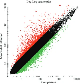
The results of the microarray in this study were ana-lyzed using SAM withP £ 0.05 and |logFC| > 1 as the screening criteria. In total, 559 RNA fragments showed dif-ferential expression between the AMI and control groups, of which 271 genes were down- regulated and 288 were up-regulated (Figure 1). Red dots represent the up-regu-lated genes, green dots represent the down-reguup-regu-lated genes, and black dots represent the unaltered genes. The top differ-entially expressed genes are shown in Table 4. The details of the gene chips have been uploaded to the Gene Expres-sion Omnibus (GEO) database for consultation: GEO se-ries accession number GSE97320 (https://www.ncbi.nlm.nih.gov/geo/query/acc.cgi?acc=GS E97320).
Table 1- Baseline information of the study subjects for microarray assay.
Parameters AMI group
(n=3)
Control group (n=3)
P
Age(years) 53.0±13.1 53.7±4.7 0.938
Gender(M/F) 2/1 3/0 1.000
HDL-C(mmol/l) 1.46±0.95 1.41±0.16 0.940
LDL-C(mmol/l) 2.18±0.84 2.31±0.74 0.846
Smoking history(yes/no) 2/1 1/2 1.000
Drinking history(yes/no) 0/3 1/2 1.000
History of Statins adminis-tration(yes/no)
0/3 0/3 1.000
Family history(yes/no) 0/3 0/3 1.000
Table 2- Primer sequences for fluorescence quantitation.
Gene Primer sequence (5’- > 3’)
CYP4F3 F ATTGGTTCTTGGGTCACCTG
R GATGTAGGTGGGGTGGAAGA
TBL1XR1 F CACCCGCTGCATTGATTTCTA
R TACGGCATCTATCAGGGACAG
GBGT1 F TGGGTGTATCTTGAGAACTGGC
R GTACTGTGACCATACCACGGG
USP25 F GATGAAAGGTGTCACAACATAATGAAA
R CCACTCCTCATATTCCTCCAAGTTT
FDFT1 F ACTTCCCAACGATCTCCCTTG
R CCCATTCTCCGGCAAATGTC
RORA F ACTCCTGTCCTCGTCAGAAGA
R CATCCCTACGGCAAGGCATTT
IL13RA1 F TCCAATTCCTGATCCTGGCAAGATT
R TTCTATCAGCACTACAGAGTCGGTT
GAPDH F CTCCTGGAAGAT GGTGATGG
R ACGGATTTGGTC GTATTGGGCG
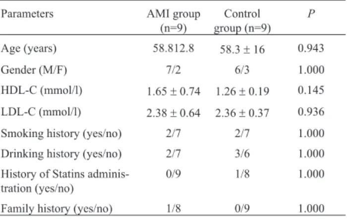
Table 3- Baseline information of the study subjects for real-time fluores-cence quantitative PCR verification.
Parameters AMI group
(n=9)
Control group (n=9)
P
Age (years) 58.812.8 58.3±16 0.943
Gender (M/F) 7/2 6/3 1.000
HDL-C (mmol/l) 1.65±0.74 1.26±0.19 0.145
LDL-C (mmol/l) 2.38±0.64 2.36±0.37 0.936
Smoking history (yes/no) 2/7 2/7 1.000
Drinking history (yes/no) 2/7 3/6 1.000
History of Statins adminis-tration (yes/no)
0/9 1/8 1.000
These differentially expressed genes were then sub-jected to GO analysis, and the results revealed that these genes belong to 128 categories of cellular localization, are involved in 521 biological processes, and are suggested to have 151 molecular functions (details are shown in supple-mentary material Tables S1-S3).
The Kyoto Encyclopedia of Genes and Genomes (KEGG) pathway analysis of the559 differentially expres-sed genes revealed that these genes participate in 107 gene pathways, and the primary pathways, as shown in Table S4.
RT-qPCR analysis
In this study, the dissolution curves and amplification curves of the candidate genes and reference genes met the quantitative requirements.
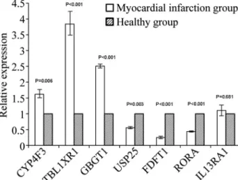
The RT-qPCR results showed that transcripts of the
CYP4F3/ TBL1XR1/GBGT1genes in the peripheral blood of the AMI group were up-regulated compared to that in the control group, whereasUSP25/RORA/FDFT1were down-regulated compared to the control group;IL13RA1 expres-sion showed no significant difference between the two groups(Table 5 and Figure 2). These results showed the same trend as the results of the microarray, indicating that the microarray was accurate and could be used for screen-ing differentially expressed genes.
Discussion
This study analyzed the differentially expressed genes in peripheral blood of patients with AMI and found 559 differentially expressed genes in the above patients,
among which 288 were up-regulated and 271 were down-regulated.
Among the target genes,CYP4F3encodes a member of the large cytochrome P450 family, belonging to the CYP4F subfamily. P450 enzymes are wide-spectrum bio-logical catalysts in nature and can act on a variety of substances. During the reperfusion phase, injection of non-specific inhibitors of CYP, such as chloramphenicol, cime-tidine, and sulfaphenazole (selective inhibitor of CYP2C9), could significantly reduce ROS generation in the rat heart and reduce the infarct size (Hunteret al., 2005). Further-more, the myocardial protective effects of the medication during the reperfusion phase showed that medication to pa-tients with AMI after ischemia might still be effective, and this finding is of important clinical significance (Nithi-patikomet al., 2004). Moreover, 20-HETE, a metabolite of CYP4F3, is a strong contraction agent acting on small
arte-Figure 1- Scatterplot of differentially expressed genes between AMI and healthy people.
Table 4- The top differentially expressed genes.
Gene Symbol logFC P
FRG1JP -2.40024 0.00023
LOC202181 -1.83279 0.001522
PTGS2 2.901423 0.001881
TNFRSF10C 2.295785 0.002082
LIN7A 1.664351 0.003145
KRT23 3.612449 0.003452
PRKCI -1.56355 0.003518
ABCC4 1.822611 0.003814
DZIP3 -1.985 0.004079
VNN3 1.724204 0.004178
LOC105377200 -1.85184 0.004275
NFE4 2.009371 0.004284
ENOSF1 -1.29599 0.004697
SLAIN2 -1.43069 0.004855
GBGT1 1.642487 0.004933
MMP25 2.366192 0.005301
THBD 2.157065 0.005327
CXCL5 2.92028 0.005388
G0S2 2.437448 0.005758
NMT2 -2.18653 0.00612
SMIM14 -1.2562 0.006733
GVINP1 -1.23053 0.00683
ADM 2.490298 0.007169
PI3 3.355852 0.007782
MBOAT7 2.153989 0.007905
LINC00152 1.528425 0.007979
C4A 1.890805 0.007996
FGFR1OP2 -1.31358 0.008
FPR1 1.912927 0.008116
rioles. By inhibiting the KCa channel, activating the L-type Ca2+ channel, and activating PKC, it could increase the intracellular Ca2+ content and cause shrinkage in small ar-teries. The injury might be associated with blockage of the heart sarcKATP pathway. In this study, CYP4F3 was upregulated in the AMI group, suggesting that CYP4F3 might participate in the myocardial injury and repair pro-cesses of AMI.
USP25 is a member of the USPS family and partici-pates in all processes of tumor occurrence. It is known that USP25 is involved in the metastasis of non-small cell lung cancer cells, which is promoted by inducingmiR-200C(Li
et al., 2014). Studies also showed that USP25 could nega-tively regulate the IL17-mediated inflammatory response (Zhonget al., 2012). In this study, USP25 in the AMI group was down-regulated compared to the control group. AMI is associated with inflammation, but the detailed mechanisms by which USP25 affects the occurrence of AMI still need to be explored.
The protein encoded by theRORAgene is a member of the family of NR1 subunit hormone receptors and plays an important role in regulating the metabolism of lipids and glucose, as well as insulin expression (Vu-Dacet al., 1997). TheRORAgene is considered a predisposing gene for dia-betes in Mexican Americans (Hayeset al., 2007). Further-more, mutation of an individual base in this gene is posi-tively correlated with incidence of diabetes in the Chinese population (Zhanget al., 2016). The protein encoded by the
FDFT1gene is the first enzyme in cholesterol biosynthesis, and studies have found that this gene can affect blood lipids, blood sugar, and inflammation, thus participating in obesity-related coronary heart disease, diabetes, and coro-nary artery calcification (Dinget al., 2015).
Other than the target genes, genes showing high dif-ferential expression includedPTGS2,encoding a cyclooxy-genase that is a key enzyme in prostaglandin biosynthesis and is thought to be involved in the occurrence of myocar-dial infarction, hypertension, and diabetes. Studies have shown that polymorphism in thePTGS2gene reduces the risk of myocardial infarction and stroke by affecting COX-2 activity and reducing the formation of atherosclerotic plaques (Cipollone et al., 2004). Relevant experiments have shown that the rs20417 mutant of thePTGS2gene can significantly reduce the risk of cardiovascular events (Ross
et al., 2014). Animal experiments have demonstrated that inhibitingPTGS2expression can increase susceptibility to salt-sensitive hypertension (Zhang MZet al., 2015). The protein encoded by thePRKCIgene is one of the members of the protein kinase C family, which is known to affect glucose degradation by participating in insulin-mediated glucose transport (Bandyopadhyay et al., 2004). In the platelet activation pathway, the activated PRKCI protein activates the Ras-associated protein, RAP-1a, thereby indi-rectly promoting platelet aggregation. Among the screened genes, a large part has been found to be associated with tu-mor occurrence or participating in biological processes such as RNA degradation; yet not all genes were found to
Table 5- RT-PCR results of the candidate genes.
DCq
Gene DDCq 2-DDCq
P
AMI group Control group
CYP4F3 12.3±0.11 12.98±0.22 -0.68±0.19 1.62±0.27 0.006
TBL1XR1 2.47±0.36 4.38±0.07 -1.91±0.29 3.84±0.78 < 0.001
GBGT1 8.45±0.20 9.78±0.24 -1.32±0.07 2.51±0.13 < 0.001
USP25 7.07±0.04 6.24±0.17 0.83±0.15 0.56±0.07 0.003
FDFT1 5,24±0.35 3.16±0.73 2.08±0.47 0.250.08 < 0.001
RORA 6.49±0.48 5.29±0.39 1.21±0.14 0.44±0.04 < 0.001
IL13RA1 5.97±0.27 6.07±0.27 -0.10±0.27 1.11±0.35 0.681
Cq: quantification cycle;DCq =Cq of the target gene - Cq of the endogenous control gene;DDCq=DCq of the AMI group -DCq of the control group;2-DDCq represents relative expression.
be related to the formation of myocardial infarction, and the roles of some genes are not yet clear. The impact of these differentially expressed genes and their expression changes on the formation of myocardial infarction still needs further verification.
Compared with genes, pathways may play a more im-portant role in the onset of AMI. The KEGG pathway anal-ysis showed that the 559 differentially expressed genes par-ticipated in 107 pathways, including the systemic lupus erythematosus pathway, apoptosis, mitogen-activated pro-tein kinase (MAPK) signaling, and insulin signaling. Sys-temic lupus erythematosus (SLE) is an autoimmune disease involving multiple systems and multiple organs, and the ex-pression of a variety of autoantibodies. The autoantibodies deposited in renal glomeruli and autoantigen immune com-plexes mediate systemic inflammatory responses by acti-vating complement proteins or neutrophils and macro-phages via FcgR. SLE was found to have the same risk factors as MI, including hypertension, hyperlipidemia, smoking, or diabetes (Petriet al., 1992). Patients with SLE were also at higher risk of cardiovascular diseases (Tazi Mezaleket al., 2014; Schuettet al., 2015). A study in Tai-wan revealed that the risk ratio of AMI combined with SLE was 5.11, which was 6.28 in females, and MI patients with SLE exhibited a higher mortality rate (Linet al., 2014). The existence of SLE could cause blood vessel inflammation, thus inducing vascular remodeling and platelet aggrega-tion, as well as atherosclerosis (Quinlanet al., 2016). The pathway analysis revealed the most differentially expressed genes in SLE (17 genes), which is consistent with related reports indicating that these differentially expressed genes lead to disorders of related inflammation adjustment due to expression changes, thus affecting the occurrence of MI.
MAPK is a group of serine/threonine protein kinases that can be activated by different extracellular stimuli, such as cytokines, neurotransmitters, hormones, cell stress, or cell adhesion. The MAPK pathway included MAP kinase kinase kinase (MKKK), MAP kinase kinase (MKK), and MAPK; these three kinases can be activated consecutively and co-regulate a variety of important cellular physiologi-cal/ pathological processes, such as cell growth, differentia-tion, adaptation to environmental stress, and inflammation. P38-MAPK is an important apoptotic mediator, and after ischemia, P38 can be rapidly activated through phosphoryl-ation, and its concentration could be increased by reper-fusion (Sanchezet al., 2012). In addition, p38-MAPK is involved in a variety of inflammatory reactions, including the reaction process of myocardial injury (Gaoet al., 2015; Guo et al., 2015). In this study, eight differentially ex-pressed genes were involved in the normal transduction of this signaling pathway, and abnormal expression of these genes was involved in the occurrence of MI and the forma-tion of reperfusion injury.
The toll-like receptor signaling pathway includes re-ceptors using different modes that can detect bacteria,
vi-ruses, fungi, and parasites, using pathogen-associated mo-lecular modes. Each receptor binds to a specific ligand, ini-tiates an innate immune response towards a particular pathogen, and then activates the acquired immune re-sponse. Studies have shown that TLR4-mediated signals may induce myocardial dysfunction during myocardial ischemia/reperfusion (Liet al., 2015). Toll-like receptor 9 plays an important role in the development of stress-induced inflammation and heart failure (Omiya et al., 2016).
It is well known that diabetes is an independent risk factor for MI. The insulin signaling pathway can regulate the glucose content of the body by over-regulating the deg-radation and transformation of glucose, and indirectly in-fluence lipid synthesis, thus affecting the occurrence of myocardial infarction. Glucose is the main energy substrate supporting myocardial contraction, which can increase myocardial contraction, and insulin can regulate this by regulating the blood sugar balance. Moreover, insulin can directly act on the myocardium by mediating the Akt sig-naling pathway, stimulating the production of vascular en-dothelial growth factors and vessels, inhibiting apoptosis, promoting cell survival, and ultimately improving myocar-dial microcirculation and coronary artery resistance by in-creasing the synthesis of ribosomes and proteins, thereby increasing myocardial blood flow (Iliadiset al., 2011). It was found that strictly controlling blood glucose after myo-cardial infarction can reduce the senescent myocyte precur-sor cells, thus increasing the possibility of recovery of the ischemic myocardium (Marfellaet al., 2012).
Owing to funding constraints, the gene chips in this study were limited, and the number and size of samples used for verification was relatively small. However, the verification results were consistent with the microarray re-sults, indicating that the microarray results were relatively accurate, and could be used for analyzing AMI-related pathways and screening the target genes. This study used clinically obtained venous blood as samples, which may fa-cilitate clinical diagnosis and treatment using the discov-ered target genes in the future.
Conclusions
Gene expression differences were observed in the pe-ripheral blood of patients with AMI and healthy individu-als. The microarray revealed 559 differentially expressed genes in the peripheral blood of Northeast Chinese Han pa-tients with AMI. RT-qPCR verified that the results of the microarray were relatively accurate and could be used for screening differentially expressed genes. Abnormal regula-tion of SLE and MAPK metabolic pathways might have a significant impact on the occurrence of MI.
being used as clinical targets for the treatment of AMI pa-tients.
Acknowledgments
This study was funded by the Excellent Talent jects in New Century of Ministry of Education (2008), Pro-jects of Jilin Provincial Science and Technology Department in 2009 (20090734), and Projects of Jilin Pro-vincial Department of Finance in 2012 (2012009), and spe-cial Acknowledgments should be given to Professor Zhihui Zhao and his research team for their technical guidance.
References
Arvind P, Jayashree S, Jambunathan S, Nair J and Kakkar VV (2015) Understanding gene expression in coronary artery disease through global profiling, network analysis and inde-pendent validation of key candidate genes. J Genet 94:601-610.
Bandyopadhyay G, Standaert ML, Sajan MP, Kanoh Y, Miura A, Braun U, Kruse F, Leitges M and Farese RV (2004) Protein kinase C-l knockout in embryonic stem cells and adipocytes impairs insulin-stimulated glucose transport. Mol Endocri-nol 18:373-383.
Barth AS and Tomaselli GF (2015) Gene scanning and heart at-tack risk. Trends Cardiovasc Med 26:260-265.
Cipollone F, Toniato E, Martinotti S, Fazia M, Iezzi A, Cuc-curullo C, Pini B, Ursi S, Vitullo G, Averna M,et al.(2004) A polymorphism in the cyclooxygenase 2 gene as an inher-ited protective factor against myocardial infarction and stro-ke. JAMA 291:2221-2228.
den Hoed M, Strawbridge RJ, Almgren P, Gustafsson S, Axelsson T, Engström G, de Faire U, Hedblad B, Humphries SE, Lindgren CM,et al.(2015) GWAS-identified loci for coro-nary heart disease are associated with intima-media thick-ness and plaque presence at the carotid artery bulb. Athero-sclerosis 239:304-310.
Ding J, Reynolds LM, Zeller T, Müller C, Lohman K, Nicklas BJ, Kritchevsky SB, Huang Z, de la Fuente A, Soranzo N,et al.
(2015) Alterations of a cellular cholesterol metabolism net-work are a molecular feature of obesity-related type 2 diabe-tes and cardiovascular disease. Diabediabe-tes 64:3464-3474. Do R, Stitziel NO, Won HH, Jørgensen AB, Duga S, Angelica
Merlini P, Kiezun A, Farrall M, Goel A, Zuk O,et al.(2014) Exome sequencing identifies rare LDLR and APOA5 alleles conferring risk for myocardial infarction. Nature 518:102-106.
Erdal C, Karakülah G, Fermanci E, Kunter I, Silistreli E, Canda T, Erdal E and Hepaguslar H (2012) Early biventricular molec-ular responses to an acute myocardial infarction. Int J Med Sci 9:74-82.
Gao XF, Zhou Y, Wang DY, Lew KS, Richards AM and Wang P (2015) Urocortin-2 suppression of p38-MAPK signaling as an additional mechanism for ischemic cardioprotection. Mol Cell Biochem 398:135-146.
Guo F, He H, Fu ZC, Huang S, Chen T, Papasian CJ, Morse LR, Xu Y, Battaglino RA, Yang XF,et al.(2015) Adipocyte-derived PAMM suppresses macrophage inflammation by in-hibiting MAPK signalling. Biochem J 472:309-318.
Hayes MG, Pluzhnikov A, Miyake K, Sun Y, Ng MCY, Roe CA, Below JE, Nicolae RI, Konkashbaev A and Bell GI (2007) Identification of type 2 diabetes genes in Mexican Ameri-cans through genome-wide association studies. Diabetes 56:3033-3044.
Hunter AL, Bai N, Laher I and Granville DJ (2005) Cytochrome p450 2C inhibition reduces post-ischemic vascular dysfunc-tion. Vascul Pharmacol 43:213-219.
Iliadis F, Kadoglou N and Didangelos T (2011) Insulin and the heart. Diabetes Res Clin Pract 93(Suppl 1):S86-S91. Isordia-Salas I, Mendoza-Valdez AL, Almeida-Gutiérrez E and
Borrayo-Sánchez G (2010) Genetic factors of the hemostatic system in young patients with myocardial infarction. Cir Cir 78:93-97.
Karahan Z, Ugurlu M, Uçaman B, Ulug AV, Kaya I, Cevik K, Öztürk Ö and Iyem H (2015) Relation between apolipo-protein E gene polymorphism and severity of coronary ar-tery disease in acute myocardial infarction. Cardiol Res Pract 2015:363458.
Kathiresan S, Melander O, Anevski D, Guiducci C, Burtt NP, Roos C, Hirschhorn JN, Berglund G, Hedblad B, Groop L,et al.(2008) Polymorphisms associated with cholesterol and risk of cardiovascular events. N Engl J Med 358:1240-1249. Li J, Tan Q, Yan M, Liu L, Lin H, Zhao F, Bao G, Kong H, Ge C,
Zhang F,et al.(2014) miRNA-200c inhibits invasion and metastasis of human non-small cell lung cancer by directly targeting ubiquitin specific peptidase 25. Mol Cancer 13:166.
Li J, Xie C, Zhuang J, Li H, Yao Y, Shao C and Wang H (2015) Resveratrol attenuates inflammation in the rat heart sub-jected to ischemia-reperfusion: Role of the TLR4/NF-kB signaling pathway. Mol Med Rep11:1120-1126.
Lin CY, Shih CC, Yeh CC, Chou WH, Chen TL and Liao CC (2014) Increased risk of acute myocardial infarction and mortality in patients with systemic lupus erythematosus: Two nationwide retrospective cohort studies. Int J Cardiol 176:847-851.
Maciejak A, Kiliszek M, Michalak M, Tulacz D, Opolski G, Matlak K, Dobrzycki S, Segiet A, Gora M and Burzynska B (2015) Gene expression profiling reveals potential prognos-tic biomarkers associated with the progression of heart fail-ure. Genome Med 7:26.
Marfella R, Sasso FC, Cacciapuoti F, Portoghese M, Rizzo MR, Siniscalchi M, Carbonara O, Ferraraccio F, Torella M, Petrella A,et al.(2012) Tight glycemic control may increase regenerative potential of myocardium during acute infarc-tion. J Clin Endocrinol Metab 97:933-942.
Nithipatikom K, Gross ER, Endsley MP, Moore JM, Isbell MA, Falck JR, Campbell WB and Gross GJ (2004) Inhibition of cytochrome P450omega-hydroxylase: A novel endogenous cardioprotective pathway. Circ Res 95:e65.
O’Gara PT, Kushner FG, Ascheim DD, Casey Jr DE, Chung MK, de Lemos JA, Ettinger SM, Fang JC, Fesmire FM, Franklin BA,et al.(2013) 2013 ACCF/AHA guideline for the man-agement of ST-elevation myocardial infarction: A report of the American College of Cardiology Foundation/American Heart Association Task Force on Practice Guidelines. J Am Coll Cardiol 61:e78.
independently of inflammation. Am J Physiol Heart Circ Physiol 311:H1485-H1497.
Petri M, Spence D, Bone LR and Hochberg MC (1992) Coronary artery disease risk factors in the Johns Hopkins Lupus Co-hort: Prevalence, recognition by patients, and preventive practices. Medicine 71:291-302.
Qaseem A, Fihn SD, Williams S, Dallas P, Owens DK, Shekelle P and Clinical Guidelines Committee of the American College of Physicians (2012) Diagnosis of stable ischemic heart dis-ease: Summary of a clinical practice guideline from the American College of Physicians/American College of Car-diology Foundation/American Heart Association/American Association for Thoracic Surgery/Preventive Cardiovascu-lar Nurses Association/Society of Thoracic Surgeons. Ann Intern Med 157:729-734.
Quinlan C, Marks SD and Tullus K (2016) Why are kids with lupus at an increased risk of cardiovascular disease? Pediatr Nephrol 31:861-883.
Roberts R and Stewart AF (2012) Genes and coronary artery dis-ease: Where are we? J Am Coll Cardiol 60:1715-1721. Ross S, Eikelboom J, Anand SS, Eriksson N, Gerstein HC and
Mehta S (2014) Association of cyclooxygenase-2 genetic variant with cardiovascular disease. Eur Heart J 35:2242-2248.
Salo PP, Vaara S, Kettunen J, Pirinen M, Sarin AP, Huikuri H, Karhunen PJ, Eskola M, Nikus K, Lokki ML,et al.(2015) Genetic variants on chromosome 1p13.3 are associated with non-ST elevation myocardial infarction and the expression of DRAM2 in the Finnish population. PLoS One 10:e0140576.
Sanchez A, Tripathy D, Yin X, Desobry K, Martinez J, Riley J, Gay D, Luo J and Grammas P (2012) p38 MAPK: A media-tor of hypoxia-induced cerebrovascular inflammation. J Alzheimers Dis 32:587-597.
Schuett KA, Lehrke M, Marx N and Burgmaier M (2015) High-risk cardiovascular patients: Clinical features, comorbidi-ties, and interconnecting mechanisms. Front Immunol 6:591.
Silbiger VN, Luchessi AD, Hirata RD, Lima-Neto LG, Cavichioli D, Carracedo A, Brión M, Dopazo J, García-García F, dos Santos ES, et al. (2013) Novel genes detected by trans-criptional profiling from whole-blood cells in patients with early onset of acute coronary syndrome. Clin Chim Acta 421:184-190.
Tazi MZ, Harmouche H, Ammouri W, Maamar M, Adnaoui M and Cacoub P (2014) Atherosclerosis in systemic lupus erythematosus. Presse Med 43:1034-1047.
The CARDIoGRAMplusC4D Consortium, Deloukas P, Kanoni S, Willenborg C, Farrall M, Assimes TL, Thompson JR,
Ingelsson E, Saleheen D, Erdmann J,et al.(2013) Large-scale association analysis identifies new risk loci for coro-nary artery disease. Nat Genet 45:25-33.
Tokat B, Kurt O, Bugra Z, Ozturk O and Yilmaz-Aydogan H (2013) Investigation of the monocyte diapedesis-related LFA-1 and JAM-A gene variants in Turkish coronary heart disease patients. Meta Gene 2:1-10.
Vu-Dac N, Gervois P, Grotzinger T, De Vos P, Schoonjans K, Fruchart JC, Auwerx J, Mariani J, Tedgui A and Staels B (1997) Transcriptional regulation of apolipoprotein A-I gene expression by the nuclear receptor RORA. J Biol Chem 272:22401-22404.
Wang M, Luo J, Wan L, Hu T, Li S and Zhan C (2015) Screening genes associated with myocardial infarction and transverse aortic constriction using a combined analysis of miRNA and mRNA microarray. Gene 571:245-248.
Welter D, MacArthur J, Morales J, Burdett T, Hall P, Junkins H, Klemm A, Flicek P, Manolio T, Hindorff L,et al.(2014) The NHGRI GWAS Catalog, a curated resource of SNP-trait associations. Nucleic Acids Res 42:D1001-D1006. Zhang MZ, Yao B, Wang Y, Yang S, Wang S, Fan X and Harris
RC (2015) Inhibition of cyclooxygenase-2 in hematopoietic cells results in salt-sensitive hypertension. J Clin Invest 125:4281-4294.
Zhang Y, Lin P, Jiang H, Xu J, Luo S, Mo J, Li Y and Chen X (2015) Extensive serum biomarker analysis in patients with ST segment elevation myocardial infarction (STEMI). Cytokine 76:356-362.
Zhang Y, Liu Y, Liu Y, Zhang Y and Su Z (2016) Genetic variants of Retinoic Acid Receptor-Related Orphan Receptor Alpha determine susceptibility to type 2 diabetes mellitus in Han Chinese. Genes (Basel) 7:54.
Zhong B, Liu X, Wang X, Chang SH, Liu X, Wang A, Reynolds JM and Dong C (2012) Negative regulation of IL-17-media-ted signaling and inflammation by the ubiquitin- specific protease USP25. Nat Immunol 13:1110-1117.
Supplementary material
The following online material is available for this article: Table S1 - GO analysis – Cellular localization.
Table S2 - GO analysis – Biological process. Table S3 - GO analysis – Molecular function. Table S4 -Main KEGG pathways.
Associate Editor: Houtan Noushmehr