Dissertation presented to obtain a Master degree in Biotechnology at the Universidade Nova de Lisboa, Faculdade de Ciências e Tecnologia
Supervisor: Dr. Paula Alves,
Animal Cell Technology Unit, IBET/ITQB-UNL
Faculdade de Ciências e Tecnologia
Departamento de Química
Microencapsulation technology: a powerful tool for
human embryonic stem cells expansion and
cryopreservation
Cláudia Susana Pedreira Correia
Acknowledgements
I would like to acknowledge all the people directly or indirectly involved in this thesis. To Dr. Paula Alves, for giving me the opportunity to do my master thesis at the Animal Cell Technology Unit at ITQB/IBET, for the good working conditions offered and for being a strong example of leadership. Also, I would like to thank the chance of attending the international symposium “Stem cells in biology and disease” as well as the iPS Cells Technology course that positively contributed to my scientific formation.
To Prof. Susana Barreiros for always being available to help during the entire Master degree. To Prof. António Laires for accepting to be jury of my thesis.
To Dr. Heiko Zimmermann and his group at Fraunhoffer-IBMT for kindly providing the cGMP grade ultra high viscous alginate.
To Margarida Serra for having taught me the majority of what I learnt this year and for always being there to help me. A deep thanks for your confidence, support, constant encouragement and friendship. It is a pleasure to work with you and to be your friend. To all the TCA colleagues for the good working environment. A special acknowledgment to: Eng. Marcos Sousa and Dr. Catarina Brito, for always being available to help in anything; Rui Tostões for the help with confocal images; Rita Malpique for helpful discussions on cryopreservation studies; Carina Silva for the initial formation.
A todos os amigos que fiz durante os meus cinco anos académicos na FCT, pelos bons momentos passados dentro e fora desta instituição. Um agradecimento especial à Joana, à Cris, à Lúcia, ao Hélio, ao André e à Vera pela amizade e por me fazerem sempre rir.
Aos amigos de longa data, dos quais nunca me esqueço.
À minha cara-metade por estar sempre presente, por todo o apoio durante estes últimos anos e por ter partilhado comigo alguns dos momentos mais felizes da minha vida.
Preface
This work was performed in the scope of the project - Integrated strategy for expansion, neuronal differentiation and cryopreservation of human embryonic stem cells (PTDC/BIO/72755/2006) funded by FCT (Fundação para a Ciência e Tecnologia). The main aims of this project are:
i) Scale-up of undifferentiated hESCs production, with the goal of obtaining large expansion of healthy undifferentiated hESCs.
ii)Optimize scalable protocols to direct and control differentiation of hESCs into neuronal lineages.
iii)Improvement of cryopreservation protocols to successful storage and transport of viable and genetically stable stocks of both undifferentiated and differentiated hESCs. This thesis contributed to achieve the objectives i) and iii).
This work led to the following article:
Abstract
Human embryonic stem cells (hESCs) are known by their ability to either self-renewal and differentiate into any adult cell type. These properties confer to hESCs a huge applicability for cell therapy, tissue engineering and drug screening. However, successful implementation of hESCs-based technologies requires the production of large numbers of well characterized cells and their efficient long-term storage. In this study, alginate microencapsulation technology was used in order to develop an efficient, scalable and integrated 3D culture system for expansion and cryopreservation of pluripotent hESCs. Three strategies were outlined: microencapsulation of hESCs as single cells, cell aggregates and cells immobilized on microcarriers.
Encapsulation of hESCs immobilized on microcarriers was the best strategy to expand and cryopreserve pluripotent hESCs. The culture of encapsulated hESCs-microcarriers in spinner vessels assured an approximately 20-fold increase in cell concentration. Moreover, this strategy improved twice cell survival after cryopreservation by a slow-freezing rate procedure, comparatively with non-encapsulated culture. Microencapsulation also protected hESC aggregates from damage caused by stirring, allowed the control of aggregates size and the maintenance of cells pluripotency for two weeks.
This work demonstrates that microencapsulation technology is a powerful tool to enhance growth and post-thawing recovery of pluripotent hESCs. The 3D culture systems developed herein represent a promising vehicle to assist the transition of hESCs to the clinical and industrial fields.
Resumo
As células estaminais embrionárias humanas (hESCs) são conhecidas pelas capacidades de proliferar indefinidamente em cultura e de diferenciar em qualquer tipo de célula adulta. Estas propriedades conferem às hESCs uma enorme aplicabilidade em terapia celular, engenharia de tecidos e no desenvolvimento de novas drogas. No entanto, para que a implementação de tecnologias baseadas em hESCs seja bem-sucedida, é necessário assegurar a produção de um elevado número de células bem caracterizadas e o seu armazenamento adequado a longo prazo. Neste estudo, a tecnologia de microencapsulação em alginato foi utilizada para desenvolver um sistema de cultura 3D eficiente, escalonável e integrado para a expansão e criopreservação de hESCs pluripotentes. Três estratégias foram delineadas: encapsulação de hESCs como células individuais, encapsulação de agregados de hESCs e encapsulação de hESCs imobilizadas em microsuportes.
A encapsulação de hESCs imobilizadas em microsuportes revelou ser a melhor estratégia para expandir e criopreservar hESCs pluripotentes. A cultura de hESCs-microsuportes encapsulados em vasos agitados permitiu um aumento de aproximadamente 20 vezes na concentração celular. Além disso, com esta estratégia de cultura 3D, conseguiu-se duplicar a percentagem de sobrevivência das células, imediatamente após criopreservação por um processo de congelamento lento, em relação à cultura não-encapsulada. É importante realçar que a microencapsulação permitiu proteger os agregados de hESCs dos danos causados pela agitação, controlar o tamanho dos mesmos e manter o estado pluripotente das células por duas semanas.
Em suma, este trabalho demonstra que aplicando a tecnologia de microencapsulação é possível melhorar o crescimento de hESCs pluripotentes e a recuperação das células após os processos de congelamento/descongelamento. Desta forma, os sistemas de cultura 3D aqui desenvolvidos constituem uma estratégia promissora para acelerar a transição de hESCs para a área clínica e industrial.
Abbreviations list
ASC adult stem cells
ASMA α‐smooth muscle actin bFGF basic fibroblast growth factor DAPI 4,6‐diamidino‐2‐phenylindole
DMEM Dulbecco’s minimum essential medium DMSO dimethyl sulfoxide
CPA cryoprotective agent EB embryoid bodies ECM extracellular matrix EG ethylene glycol FBS fetal bovine serum FDA fluoresceíne diacetate FOXA2 forkheadbox A2 HSA human serum albumin
HEPES 4‐(2‐hydroxyethyl)‐1‐piperazineethanesulfonic acid hESCs human embryonic stem cells
ESCs embryonic stem cells hFFs human foreskin fibroblasts ICM inner cell mass
IgG immunoglobulin G IgM immunoglobulin M IVF in vitro fertilization
KO-SR knockout serum replacement LN2 liquid nitrogen
mEFs mouse embryonic fibroblasts mEFs-CM conditioned media in mEFs mESCs mouse embryonic stem cells NEAA non-essential aminoacids non-CM non-conditioned media PBS phosphate buffered saline Pen/Strep Penicillin/Streptomycin PI propidium iodide
PFA paraformaldehyde
Oct‐4 transcription factor octamer‐4 OPS open pulled straw
ROCKi Rho-associated kinase (ROCK) inhibitor, Y-27632 SCED single cell enzymatic dissociation
SD standard deviation
SSEA‐1 stage‐specific embryonic antigen‐1
SSEA‐4 stage‐specific embryonic antigen‐4
TRA‐1‐60 tumour rejection antigen‐1‐60
Contents
Acknowledgements……….……….…..i
Preface……….………..……….………..ii
Abstract…….………..………....iii
Resumo…….……….………...iv
Abbreviations list………...v
1. Introduction ... 1
1.1. Human embryonic stem cells (hESCs) ... 1
1.1.1. Sources of pluripotent stem cells ... 1
1.1.2. Advantages of using hESCs instead ASCs or primary cells ... 4
1.1.3. hESCs applications ... 4
1.1.4. Using hESCs in the clinic: critical issues ... 5
1.2. 3D models for hESCs culture ... 8
1.2.1. Culture of hESCs as aggregates ... 9
1.2.2. Culture of hESCs on microcarriers ... 10
1.3. hESCs cryopreservation ... 10
1.3.1. Cryopreservation methodology ... 11
1.3.2. Techniques to cryopreserve hESCs ... 12
1.4. Cells microencapsulation ... 14
1.4.1. History and the concept ... 14
1.4.2. Using cell microencapsulation in the clinic: critical issues ... 16
1.4.3. Microcapsule materials ... 17
1.4.4. Microcapsule formation ... 19
1.4.5. Microencapsulation of stem cells for clinical application ... 19
1.5. Microencapsulation in stem cell bioprocessing ... 19
1.5.1. Expansion of encapsulated hESCs ... 20
2. Aim of the thesis ... 24
3. Material and Methods ... 25
3.1. hESCs culture on feeder layer... 25
3.2. Preparation of mEFs conditioned medium... 25
3.3. Encapsulation of human embryonic stem cells ... 25
3.4. Three-dimensional hESC cultures ... 26
3.4.1. Culture of hESCs in Erlenmeyer ... 26
3.4.2. Culture of hESCs in spinner vessels ... 27
3.5. Cell cryopreservation ... 29
3.5.2. Vitrification ... 29
3.5.3. Assessment of hESCs survival after thawing ... 30
3.6. Evaluation of cellular viability ... 30
3.7. Evaluation of the culture metabolic activity ... 31
3.8. Evaluation of cell growth ... 31
3.9. Cell characterization ... 31
3.10. In vitro pluripotency test via embryoid bodies (EBs) formation ... 32
4. Results ... 33
4.1. Evaluation of two different UHVNT alginate concentrations ... 33
4.2. Strategy I – Encapsulation of hESCs as single-cells ... 33
4.2.1. Culture of encapsulated hESCs as single cells in Erlenmeyer ... 33
4.3. Strategy II – Encapsulation of hESCs as aggregates ... 35
4.3.1. Culture of encapsulated hESCs as aggregates in Erlenmeyer ... 35
4.3.2. Expansion of encapsulated hESC aggregates in spinner vessels ... 36
4.3.3. Effect of two alginate types in expansion of hESC aggregates in spinner vessels ... 37
4.3.4. Characterization of expanded hESC aggregates... 38
4.3.5. Cryopreservation of encapsulated hESCs aggregates ... 40
4.4. Strategy III – Encapsulation of hESCs immobilized on microcarriers ... 41
4.4.1. Culture of hESCs immobilized on microcarriers in Erlenmeyer ... 41
4.4.2. Expansion of encapsulated hESCs immobilized on microcarriers in spinner vessels ... 44
4.4.3. Characterization of expanded hESCs ... 47
4.4.4. Cryopreservation of encapsulated hESCs immobilized on microcarriers ... 49
5. Discussion ... 53
6. Conclusion ... 61
1. Introduction
1.1.Human embryonic stem cells (hESCs)
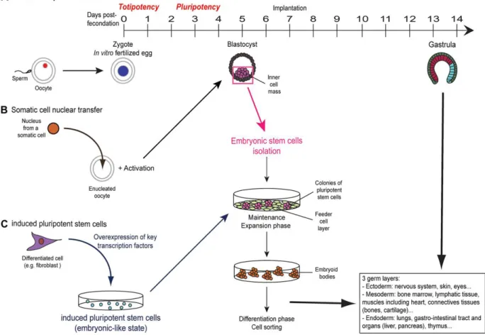
Human embryonic stem cells (hESCs) have an enormous potential for cell-based therapy, due to their ability to self-renew indefinitely and to differentiate into all mature cell types of the human body (pluripotent nature). The first reports describing hESCs potential were published in 19841 and 19942, but it was only in 1998 that Thomson and co-workers described the isolation of hESCs from blastocysts and the creation of the first permanent and characterized hESC lines for research3.
hESCs are derived from the inner cell mass (ICM) of blastocysts3 (Fig. 1.1). A blastocyst is a pre-implantation embryo that develops 5 days after the fertilization. It contains all the material necessary for the development of a complete human being. In normal development, the blastocyst would implant in the wall of the uterus to become the embryo and continue developing into a mature organism. The outer cells (trophoblast) would begin to form the placenta and the cells from the ICM would begin to differentiate into the progressively more specialized cell types of the body.
Today, more than 1000 hESC lines are described in the literature4. Some of these cell lines are well characterized and organized in international stem cell banks, for example, the hESCreg (www.hescreg.eu), the UK stem cell bank (www.ukstemcellbank.org.uk), and the National stem cell bank (www.nationalstemcellbank.org)5.
hESC lines can be identify by the presence of surface marker antigens (Tra series, SSEA series, GCT series, HLA, and CD markers) and transcriptional factors (Oct4, Nanog), by the ability to differentiate into tissues originating from all three germ layers in vivo (via
teratoma formation) and in vitro (via embryoid body differentiation), by the chromossomal
stability with serial culture, alkaline phosphatase positiveness and high telomerase activity6, 7.
1.1.1.Sources of pluripotent stem cells
Fig. 1.1 - hESC line derivation. Main steps of the protocol for establishment of hESCs lines9. The main suppliers of blastocysts for stem cell
research are in vitro fertilization (IVF) clinics. The process of
IVF requires the retrieval of a woman’s eggs via a surgical procedure after undergoing an intensive regimen of
“fertility drugs”. When IVF is used for reproductive purposes, all of the donated eggs are fertilized in order to
maximize their chance of producing a viable blastocyst that
can be implanted. Because not all the fertilized eggs are
implanted, this has resulted in a large bank of excess
blastocysts that are currently stored for use in medical
research. Importantly, the unused blastocysts can only be utilized for research purposes with the written informed consent of the donors.
The creation of stem cells specifically for research using IVF rises ethical issues because it involves the destruction of human embryos. Accordingly, other
exhibit similar features to embryonic stem cells, including cell morphology, cell-surface markers, growth properties, telomerase activity expression, and epigenetic marks of pluripotent cell–specific genes13, 14 and can give rise to cells derived from all three germ layers in vitro and in vivo. This technology can be used to generate patient-specific cell
types11, 15, opening the door to “personalized” cell-based therapy. However, generation of iPSCs still suffers from low efficiency and high cost10. Furthermore the viral expression vectors used to obtain iPSCs16, the potential for insertional mutagenis11 and the recent knowledge that hiPSCs expresses cancer hallmarks17 have raised additional concerns regarding the safety of these cells for clinical applications.
Fig. 1.2 – Isolation/generation, culture and differentiation of pluripotent stem cells. The development of pluripotent stem cells by A) in vitro fertilization, B) somatic cell nuclear transfer and C) generation of induced pluripotent stem cells is illustrated. Typically pluripotent cells are expanded in culture on feeder cell layers.
Fig. 1.3 - Human embryonic stem cells applications 20 1.1.2.Advantages of using hESCs instead ASCs or primary cells
Another type of stem cells is also currently used in research, particularly adult stem cells (ASCs) found in specialized tissues of the body, including the brain, bone marrow, liver, skin and gastrointestinal tract10. However these cells are less flexible than hESCs, because their differentiation potential is limited. ASCs form only a restricted number of cell types, usually only the cell types of the lineage of their origins, thus they are classified as multipotent stem cells. Moreover, hESCs are also generally more easier to isolate, purify and maintain in vitro than ASCs (with the relative exception of hematopoietic stem cells)18 (Tab.
1.1). Therefore, ASCs have a limited usefulness for tissue engineering or regenerative
medicine compared with hESCs. In addition primary cells suffer from having an even lower
expansion and differentiation potential (Tab. 1.1). Nevertheless, hESCs are difficult to control in what concerns their stem cell fate, requiring more complex and robust bioprocesses (Tab. 1.1).
Tab. 1.1 - Comparison between human embryonic stem cells (ESCs), adult stem cells (ASCs) and primary cells types. Legend: + low, ++ medium, +++ high. (Adapted from Polak et al. 19).
1.1.3.hESCs applications
The unlimited self-renewal
ability and pluripotency of hESCs
gives them limitless applications
(Fig. 1.3). Indeed hESCs are
excellent candidates to cure
diseases by repairing or replacing
damaged cells and tissues (cell
therapy or regenerative
medicine), since they could be
used to generate an infinite quantity of cells with a clinical interest, including retina cells21,
dopaminergic neurons22, motor neurons23, OPCs (oligodendrocyte progenitor cells)24, Cell type Expansion
potential
Differentiation potential
Cell availability
Ease of regulation
Bioprocess complexity
ESCs +++ +++ +++ + +++
ASCs + ++ ++ ++ ++
Fig. 1.4 – Criteria that have to be addressed before hESCs approval for clinical use.
cardiomyocytes25, pancreatic β-cells26 and hepatocytes27. Moreover, the US Food and Drug
Administration recently approved the world’s first phase I clinical trial using hESCs derivatives (oligodendrocyte progenitor cells) in patients with spinal cord injury10.
Another interesting application of hESCs is to serve as pharmacological and
cytotoxicity screening platforms, supporting the discovery of new drugs for therapeutic use28.
Given the high costs spending to bring a new drug to market, there are great advantages of
having access to large numbers of biologically relevant human cells for early testing and
screening. For example, pure cultures of hepatocytes, cardiomyocytes and neuronal cells
derived from hESCs would provide robust cell-based in vitro assays for toxicity
measurements and for drugs being development for cardiovascular or neurodegenerative
disorders, respectively5.
ESCs are also valuable models for scientific research. They can lead to a better
understanding of the basic biology of the human body, embryonic development,
pathogenesis of congenital defects and cancer formation6. In fact, it is possible to derive disease-specific hESCs from embryos with diagnosed mutations by preimplantation genetic diagnosis29. For instance hESC lines derived from embryos with Fanconi anemia-A mutation and fragile X mutation have already been established29. These hESC lines will provide in vitro
models for study the phenotype of these mutations, allowing the identification of new treatments for these diseases.
1.1.4.Using hESCs in the clinic: critical issues
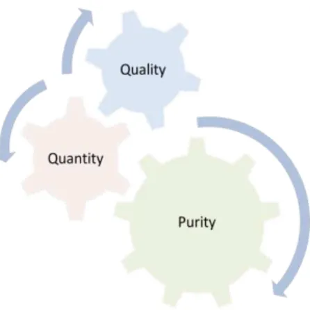
Even though hESCs hold great promise for the cure of various human disorders, there are three significant issues that need to be addressed prior these cells being approved for clinical and industrial applications: the right quality, quantity and purity of hESCs and/or their derivatives have to be achieved (Fig. 1.4).
1.1.4.1. Quality
Initial methods to culture hESCs were based on techniques originally developed to
culture mouse ESCs3, that involved the culture on a layer of mitotically inactivated mouse
embryonic fibroblasts (mEFs) and medium supplemented with 20% fetal bovine serum (FBS).
pluripotent properties3. However, it was realized that the continuous use of mEFs and animal-derived components in hESCs cultures would hinder the development of clinical applications due to: the presence of non-human sialo proteins in culture, which are immunogenic for humans; the risk of transmitting animal virus or prion material; and difficulty with quality control of these undefined components30-32. Subsequently, improvements to these procedures have largely focused on removing the undefined and non-human components.
Serum can be replaced by Knock-Out Serum Replacement (KO-SR), commercially available serum substitute with less batch-to-batch variability than serum. However, KO-SR is still not yet a fully defined product33 and has animal-derived components, namely AlbuMAX, a lipid-rich albumin fraction of bovine serum and bovine transferrin32, but in spite of that it has been validated for culture of clinical grade cells34.
Regarding the use of mEFs, it is not entirely understood the purpose of growing hESCs on feeder cell layer. However it has been suggested that feeder cells provide a suitable attachment substrate and important soluble factors for the maintenance of undifferentiated hESCs31. Although, irradiated or treated with mitomycin C, feeder cells are still capable of stimulate ESC growth and also inhibit their differentiation through the secretion of specific growth factors and cytokines35. Since the exposure to animal components presents a serious risk to the patients, mEFs have been replaced by human fibroblasts. These feeders equally support hESCs growth and maintenance36-38. Nevertheless,
due to the drawbacks associated to the use of co-culture systems, several attempts to
establish feeder-free systems for hESCs propagation have been performed. A successful feeder-free hESC culture system was developed in which undifferentiated cells were maintained long term on a Matrigel layer in mEFs-conditioned medium39. Matrigel is a soluble basement membrane extract from mouse sarcoma which contains extracellular molecules, such as laminin, collagen IV, growth factors and other unknown components31. Moreover, a single extracellular matrix (ECM) component such as fibronectin, has also been successfully used to support undifferentiated growth of hESCs in non-conditioned medium supplemented with KO-SR and various growth factors40, 41. Basic fibroblast growth factor (bFGF) is one example, it plays a key role in sustaining hESC self-renewal42, and is included in nearly all the reported medium formulations for hESC propagation30.
culture matrices, culture media, hESCs passaging and cryopreservation) have to minutely follow the FDA (Food and Drug Administration) regulations43. Importantly, cell phenotype and function should be characterized and evaluated during culture. Undifferentiated hESCs have to maintain their pluripotency and karyotype stability after expansion while hESCs derivatives must express markers of the specific cell lineage and be fully functional after differentiation.
1.1.4.2. Quantity
Another important challenge is to achieve sufficient numbers of stem cell for an effective therapy. For example, 1x10⁹ to 2x10⁹ cardiomyocytes are required to replace damaged cardiac tissue after myocardial infarction44 and 1.3x10⁹ insulin producing β-cells per 70-kg patient45 are needed for insulin independence after islet transplantation. To achieve these high cell numbers scalable bioprocesses need to be developed.
The requirement of two-dimensional (2D) surfaces, such as well-plates and tissue-culture flasks for cell attachment is the first hurdle to limit large stem cell production and clinical applications19. Indeed there are several drawbacks related with the 2D culture systems namely: i) low reproducibility due to the uncontrolled culture conditions, ii) achievement of low cell yields, iii) limitations in scaling-up, iv) labor-intensive procedures, v) inability to support complex cellular growth configurations19. Therefore, the establishment of novel three-dimensional (3D) culture systems with a high available surface area for cellular attachment and growth that resemble the in vivo conditions by accounting for the
cell–cell, cell–matrix and cell–growth factor interactions (see section 1.2) became a priority. In addition, in order to improve cell density or the expansion rate of the 3D systems cultures, bioreactors or stirred vessels that can accommodate dynamic culture conditions have been used46, 47. Stirred suspension bioreactors makes possible to overcome the mass transport limitations of static cultures due to the constant agitation rate promoted for example by an impeller. However, they require careful impeller design and the delineation of the optimum stirring speed for each culture. Distinct cell types have different sensitivities/necessities in terms of the shear stress (force exerted over the cells due to the flow of the media)46. Consequently, an inappropriate agitation could cause unsuitable shear stress that can damage the cells.
low46, 48. So far, fully controlled stirred tank bioreactors appear as promising candidates for the expansion of hESCs since at least a 10-fold increase in cell density is achieved when compared with the traditional 2D culture systems47.
Another worth mentioning aspect is that clonal efficiency of hESC is extremely low. These cells are very sensitive to single-cell dissociation and recover poorly when plated at clonal density after a passage30, which hinders cell passage from 2D to 3D culture system. In fact, cell-cell interactions seem critical for efficient hESCs propagation, since it was demonstrated that the loss of gap junctions between hESCs can increase cell apoptosis and inhibit colony growth49. For these reasons, hESCs have been routinely passaged in cell clumps. In order to facilitate up-scaling of hESCs culture, it was developed a new single-cell enzymatic dissociation (SCED) culture system using recombinant protein-based TrypLE Select50. Nevertheless, clonal survival of hESCs can also be enhanced by culturing in the presence of Rho-associated kinase inhibitor (ROCKi)51.
1.1.4.3. Purity
The tumorigenic potential of pluripotent cells is other important hurdle in the safety utilization of these cells. At present, protocols for the differentiation of hESCs are generally inefficient, resulting in low differentiated cell yields and contamination by other cell types. Of greater concern is the persistence of undifferentiated hESCs and the possibility of these cells form malignant tumors when transplanted in the host52. Therefore the use of robust methods for i) differentiation, ii) selection of pure populations of specialized cells and iii) demonstration of their genetic and epigenetic stability will be essential before these cells being used clinically10.
1.2.3D models for hESCs culture
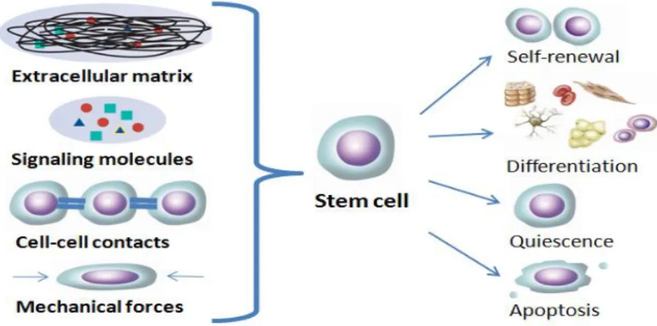
expression, cytoskeletal organization and cell morphology47. Besides that, cells constantly remodel local ECM by degrading or synthesizing new ECM elements58.
Fig. 1.5 – Components of the cellular in vivo microenvironment. The synergy of biochemical (signaling molecules), biophysical (ECM or substrates) and mechanical (hemodynamic forces or shear forces) factors with cell-cell interactions determine the fate of pluripotent stem cells. (Adapted from Abraham et al. 55)
Thus, culturing stem cells in 3D microstructures that closely mimic stem cells native microenvironment is imperative to enhance cells performance and fully exploit cells potential.
hESCs are highly anchorage dependent and need a surface to attach and proliferate. Therefore, growth of these cells in a 3D configuration (Fig. 1.6)59 requires normally either the use of a support system, such as
microcarriers, or the formation of small aggregates. Moreover, polymeric matrices like alginate hydrogels can also be a good option for hESCs cultivation since this type of matrices better represents the geometry, chemistry and
signaling of in vivo environment than
2D cultures60 (see section 1.5.).
1.2.1.Culture of hESCs as aggregates
The cells grew 70-fold, reaching 3.6x10⁷ cell/mL after 28 days and gave rise to cells of the three germ layers.
hESCs differentiate extensively when cultured as aggregates61, therefore is difficult to control the expansion of undifferentiated hESCs and their further directed differentiation into specific cell types. Indeed, there are only a reduced number of studies that reported the expansion of undifferentiated hESCs as aggregates (see Tab. 1.2; Page 22).
1.2.2.Culture of hESCs on microcarriers
Microcarriers are spherical particles, composed of various materials including cellulose, glass, plastic, and polyester. They have a typical diameter of 100-250 μm and can be compact or porous63. Currently many types of microcarriers are commercial available and they are used for the growth of several adherent cell types. Indeed, the attributes of microcarriers make them also attractive for culture and directed differentiation of hESCs. First, they allow the translation of anchorage-dependent hESCs from 2D culture into suspension culture by providing a high attachment surface. Also, microcarrier cultures are characterized by high surface-to-volume ratio, which allows higher cell densities than 2D cultures. More important, the available area for cell growth can be adjusted easily by changing the amount, porosity and size of microcarriers61, 63.
Normally microcarriers have to be customized, for example, by attaching synthetic peptides or extracellular matrix molecules (collagen, Matrigel, fibronectin…) in order to improve the adhesion of hESCs. In fact, different types of microcarriers were already tested to culture hESCs and some distinct results were obtained. For example, Nie et al. reported that Cytodex 3 beads appeared to promote better attachment and viability of hESCs63, whereas others observed that hESCs on these beads exhibited the poorest growth with little or no recovery of viable cells after 48–72 hours64. Such discrepancies may be due to the use of different hESC lines. Nevertheless, the adhesion efficiency of hESCs increased when the beads were coated with Matrigel or seeded with mouse embryonic fibroblasts (mEFs)63. For instance, our group recently demonstrates a 12-fold in yield of hESCs cultured in Cytodex3 microcarriers coated with Matrigel in 300mL perfused bioreactors fully controlled, operating with pO2 at 30% air saturation65. See Tab. 1.2 (Page 22) for more examples.
1.3.hESCs cryopreservation
To fully exploit the potential of hESCs in medicine and research, freezing, cryostorage
Cryopreservation is traditionally defined as the maintenance of biologics at temperatures typically bellow the glass transition of pure water (‐132°C), at which biological metabolism is dramatically diminished66.
An effective method for hESCs cryopreservation is critical for their use in clinical and
research applications. A suitable cryopreservation enables a good storage and transportation
of the cells between the sites of collection, processing and clinical administration67. This
makes possible the exchange of cells between research centers, which promotes scientific collaboration and facilitates widespread use of hESCs. Also, the ability to preserve cells
permits the banking of stem cells until later use. An inefficient hESCs cryopreservation
increases time between cell storage and use in experimental or clinical settings, because of
an extended lag in establishing a viable highly populated culture following thawing. Even
though hESCs are self-renewable, aging cultures can acquire chromosomal abnormalities and
lose their differentiation potential, thus efficient procedures to preserve hESCs in low
passage numbers are indispensable.
1.3.1.Cryopreservation methodology
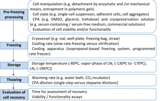
Typically, cryopreservation procedure involves the steps described in Fig. 1.7. First cells are subjected to a pre‐freezing treatment in order to leave the cells in the state that
they will be frozen (e.g. single‐cell suspension, cell aggregates, adherent cell monolayers)
and the cell viability is evaluated. Second cells are transferred into the cryovessel on which they will be frozen (e.g. vial, well‐plate, straw). Before freezing, samples are loaded with a cryoprotective agent (CPA) like DMSO or glycerol to help minimizing the injury to cells during freezing and thawing. The cells are then frozen at the desired cooling rate to the storage temperature at which they will be stored. After thawing CPAs are removed from the sample by dilution. At this time post-thaw cell recovery is evaluated.
CPAs are usually classified based on their ability to diffuse across the plasm
membrane of cell in penetrating (e.g. DMSO, glycerol and 1,2‐propanediol) or non -penetrating CPAs (hydroxyethyl starch, sucrose and polyssaccharides).
Non‐penetrating CPAs are generally relatively high molecular weight, long chain
polymers that are soluble in water and can only be taken up by cells through endocytosis or induced processes. They are thought to act by dehydrating the cells before freezing, thereby reducing the amount of water that the cell needs to lose to remain close to the osmotic equilibrium during freezing70. On the other hand, trehalose and other polysaccharides protect membranes and proteins against the destructive effects of dehydration by serving as a substitute for structural water associated with their surface71.
Fig. 1.7 – Main steps composing of a cryopreservation procedure for mammalian cells. For each step the most relevant parameters are listed
1.3.2.Techniques to cryopreserve hESCs
Two different methods have generally been used to preserve hESCs, namely slow freezing-rapid thawing (for larger numbers of cells) and vitrification (for smaller number of cells)72. These procedures are based on well-established protocols developed for mESCs and embryos, respectively73.
1.3.2.1.Slow freezing rate
The conventional slow‐freezing rate consists in freezing the samples in the presence
of a CPA at a slower cooling rate to minimize the probability of intracellular ice formation, which is likely to damage the cells. Slow cooling leads to a better cell dehydration during the cryopreservation process74. Briefly, hESCs colonies or single cell suspension are exposed to the CPA solution (normally 5-20% dimethyl sulfoxide (DMSO) in culture medium or serum)
Pre-freezing processing
Cell manipulation (e.g. detachment by enzymatic and /or mechanical means, entrapment in polymeric gels)
Cell state (e.g. single-cell suspension, adherent cells, cell aggregates) CPA (e.g. DMSO, glycerol, trehalose) and cryopreservation solution (e.g. serum-containing / serum-free medium, commercial solutions) Evaluation of cell viability and/or functionality
Cryovessel (e.g. vial, well-plate, freezing bag, straw) Cooling rate (slow-rate freezing versus vitrification)
Cooling apparatus (isopropanol-based freezing system, programmed rate freezer)
Freezing
Storage temperature (-80ºC, vapor-phase of LN2 (-130ºC to -170ºC),
LN2 (-196ºC))
Storage
Warming rate (e.g. water bath, CO2 incubator)
CPA dilution (single-step versus stepwise dilutions) Thawing
Evaluation of cell recovery
and placed in a programmed rate freezer or in an isopropanol bath intended to cool the sample at 1°C/min. Slow cooling protocols are effective for the preservation of mESCs, however, applying these to hESCs, has met serious difficulties. Normally these procedures
result in low post‐thaw survival, low plating efficiencies, high differentiation rates and loss of
pluripotency, presumably due to ice crystal formation that disrupts cell‐cell adhesion75-77. For instance, in early studies colony recovery after thawing were very low: 16%77, 23%78 and 30%79. Trying to improve post thaw recovery it has been tested the additions of other components to the cooling solution. For instance, extracellular matrix molecules (human type IV collagen or laminin) and trehalose were found to improve post thaw recovery and reduce differentiation of the colonies80, 81.
In all referred studies, hESCs were cryopreserved in small clumps in order to prevent cell loss from apoptosis after single cell dissociation. However, recently it was reported the cryopreservation of hESCs as single cells suspension by slow freezing rate using ROCKi. Cell survival increased from 30.5±5.2% (absence of ROCKi) to 56.4±7.2% (presence of ROCKi)82.
1.3.2.2.Vitrification
Vitrification procedures aim at preventing ice formation throughout the sample by applying extremely high cooling rates (>104 °C/min) together with high concentrations of
CPAs (6 ‐ 8M)83
. Increasing the concentration of non-penetrating CPAs that interact strongly with water prevents water molecules from interacting to form ice. Moreover high cooling rates through the temperature region of potential crystallisation allows reaching the amorphous glassy state before ice crystals have the opportunity to form84. Therefore, samples pass directly from a liquid phase to a glassy state without suffering any nucleation. Briefly, the method consist in a brief stepwise exposure of hESCs colonies (100–400 cells) to two vitrification solutions of increasing CPA concentrations, in which the common components are DMSO, EG and sucrose. The colonies are then loaded into open pulled straws and directly plunged into liquid nitrogen. To avoid ice crystallisation during thawing, colonies are rewarmed as rapidly as possible by direct immersion into pre-warmed culture medium solutions containing decreasing concentration of sucrose73. High recovery rates (>75%) are described for vitrified undifferentiated colonies75, 77, 78.
1.3.2.3. Slow freezing rate vs. vitrification of hESCs
limitations. There is an increased risk of microbiological contamination and transmission of infection associated with the use of open/non sterile straws and the direct exposure to liquid nitrogen85. Furthermore, the process uses high concentrations of a CPA that is toxic to cells at room temperature and is difficult to apply to bulk quantities of hESCs. Typically, each straw will hold only 8–12 colony fragments which must be prepared, transferred through the vitrification solutions, loaded into a straw and plunged into liquid nitrogen. The process is extremely labor-intensive and operator-dependent, thus is difficult to reproduce73. All this makes current vitrification protocols unsuitable for development of a scalable process and enable its use in hESC banks67.
Using the slow freezing method, large cell numbers can be frozen in one vial, making easy handling of bulk quantities of hESCs. Therefore, an efficient slow freezing of hESCs in suspension (single cells or hESCs growing in 3D), which can easily and efficiently be stored in
cryovials seems to be the optimal scalable system to preserve hESCs. Recently, Nie et al.
developed a scalable method to cryopreserve hESCs in cryovials. They reported that the
cryopreservation of hESCs adherent on mEFs coated microcarriers using slow freezing
method, improved post thaw recovery when compared to standard cryopreservation of
hESCs colonies63.
1.4.Cells microencapsulation
1.4.1.History and the concept
The concept of enclosing transplantable cells within a semi-permeable polymeric capsule to protect them from immune rejection was proposed by Chang in 196486. Twenty years later encapsulation was successfully used to immobilize xenograft islet to aid in glucose control for diabetes in rats. Diabetic state was corrected for several weeks87.
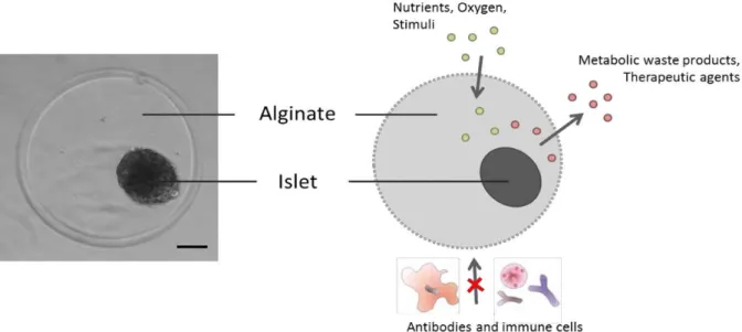
The polymer microcapsule modulates the bidirectional diffusion of molecules (Fig. 1.8). Nutrients, oxygen, waste products and biotherapeutic agents freely diffuse across the microcapsule, whereas high molecular weight molecules, such as antibodies, immunocytes and other immunologic moieties, are excluded90. This barrier can thus isolate the
transplanted cells from the host’s immune response making possible the graft long-term function91, without the administration of immunosuppressive drugs. Furthermore, this technology allows the controlled and continuous ‘de novo’ delivery of therapeutic products to the host92. Indeed, cell microencapsulation ensures a higher success than the encapsulation of therapeutic protein. The therapeutic products are produced and secreted
de novo at a constant rate by the encapsulated cells, giving rise to a more physiological and
effective concentration of the drug, over time. This approach corresponds more to the natural behavior of the cells, and can minimize unintentional side effects93. In case of capsule damage, the fast release of high protein concentrations that could be toxic for the patient is avoided92, 93.
Other advantage of this approach is to make possible the transplantation of allogenic (non-patient) or even xenogenic cells (non-human), which could be a mean of overcoming the obstacle of limited supply of donor cells. Cells can also be genetically modified prior to their encapsulation, to produce or secrete a desired protein in vivo.
Fig. 1.8 – Concept of cell microencapsulation. This technique consists of enclosing biologically active material within a polymeric matrix (e.g. alginate) that is designed to circumvent immune rejection. The microcapsule membrane allows the bi-directional diffusion of nutrients, oxygen and waste, and the secretion of the therapeutic agents, but prevents immune cells and antibodies from entering the capsule. Scale bar: 200 µm.
environment wherein cells can grow to mimic the in vivo process, this makes the
microencapsulation technique also very attractive in regenerative medicine19.
1.4.2.Using cell microencapsulation in the clinic: critical issues
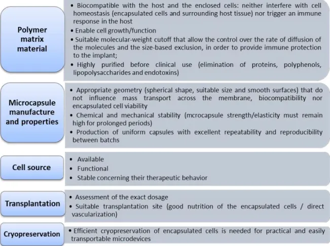
Nowadays an increasing number of biotechnology and pharmaceutical industries have focused their interest on cell microencapsulation for the treatment of endocrine diseases, such as anemia94, pituitary95, dwarfism96, Hemophilia B97, kidney98 and liver99 failure, central nervous system insufficiencies100 and diabetes mellitus87. However, the first clinical trials using cell microencapsulation have demonstrated limited reproducibility. The duration of immunoprotection and/or function of the transplants were subjected to a large variability89. The main causes of cell microencapsulation technology failure in vivo include hypoxia (due to the great distance between the encapsulated islets and the blood supply), biocompatibility of the encapsulating material and insufficient immune-protective properties of the microcapsules101, 102. Indeed, the clinical implementation of cell microencapsulation in humans requires higher levels of quality, efficacy and biosafety. Strict requirements
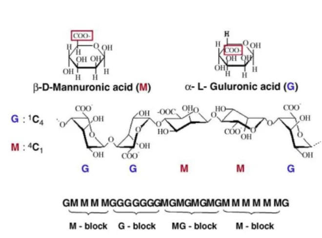
Fig. 1.10 - Structure of alginate. Alginate molecules are linear block copolymers of β-D-mannuronic (M) and α-L-guluronic acids (G) with a variation in composition and sequential arrangements. (Adapted from de Vos et al. 91).
concerning the exact selection and purity of matrix materials, microcapsule manufacture and final properties, cell source and site of transplantation must be thoroughly fulfill90, 92, 101,
103-106. Fig. 1.9 describes the main requirements for this technology succeeds.
1.4.3.Microcapsule materials
Microcapsules are almost exclusively produced from hydrogels since they provide a highly hydrated microenvironment for embedded cells that guide cellular processes such as differentiation, proliferation and migration107. Moreover, their properties can be designed to achieve ideal biocompatibility, degradation and physical characteristics depending on the application108. Some materials used for microencapsulation are alginate87, chitosan109, agarose110, poly(hydroxyethylmetacrylate-methyl methacrylate) (HEMA-MMA)111, copolymers of acrylonitrile (AN69)112, and polyethylene glycol (PEG)113.
1.4.3.1.Alginate
Alginate is the most common used polymer for cell microencapsulation, due to the following characteristics: i) has easy gelling properties; ii) allows the processing of the capsules at physiological conditions (room temperature, physiological pH and isotonic solutions)91; iii) has an excellent availability, biocompatibility (after purification) and biodegradability; iv) does not interfere with cellular function of the islets89, 93. Indeed, it was already reported
that alginate-based microcapsules have great potential for transplantation of Langerhans’ islets and other factor-secreting cells and tissues114.
Alginates are a family of unbranched anionic polysaccharides extracted from brown algae (Phaeophyta). They occur extracellularly and intracellularly at approximately 20% to 40%
The proportion and distribution of these two monomers is of paramount importance since they have a significant impact on some of the alginate gel properties (viscosity, permeability, biocompatibility, stability, mechanical resistance, biodegradability)107, 115. For instance, stiffness of cross-linked alginates increases as follows: MG blocks < MM blocks < GG blocks, whereas flexibility or elasticity increases in the opposite way114. Therefore, the M:G ratio, length of polymeric chains, and the ratio of homologous to heterologous chains must be carefully selected to optimize microcapsules properties. Nevertheless, it should be referred that these properties depend on the source from which the polymer is isolated and also on the harvesting and extraction processes60.
1.4.3.1.1.UHV Alginate
Ultrahigh viscosity (UHV) alginate (>30 mPa s; 0,1% w/v solution115) is a type of alginate designed taking into account the proportion of M/G monomers. It is composed by a 1:1 mixture of alginates from Lessonia nigrescens and Lessonia trabeculata algae species.
Both species grow from central Peru to southern Chile. L. nigrescens stipes are very elastic and flexible because of high M alginates (~60%). By contrast, L. trabeculata stipes are very stiff due to a high content of G (~90%)115. Therefore, UHVNT alginate meet the demands of
high stability and flexibility (they are composed by 35% M and 65% G blocks) and has mechanical and elastic properties required for medical, pharmaceutical and biotechnologic applications114. It should be noted that this alginate is subject to a detailed purification process, thus the final product fulfills the standards for routine clinical application115.
1.4.3.1.2.Commercial Alginate
However, some companies have focused on the purification of alginate allowing to guarantee clinical-grade qualities. For instance it is now possible to buy ultrapure alginates with endotoxin levels of less than 100EU/g91 and lacking immunogenic effects. Pronova UP MVG alginate (Pronova Biomedical) is an example. It is isolated from Laminaria hyperborea
stipe and has a high G content, a high viscosity (316 mPa s; 1% w/v solution) and a high molecular weight (231 000 g/mole)117.
1.4.4.Microcapsule formation
Microcapsules are normally produced by forcing the alginate-cells suspension (by using a syringe or a motor-driven prison) through a nozzle with a coaxial air jet to facilitate break-off (Fig. 2.1). The resulting droplet is transformed into a rigid bead by gelification in an oppositely charged, di- or trivalent ion solution. The most used cross-linkers of the polymeric chains are Ba2+ and Ca2+ 88. Microcapsule size is controlled via the air and alginate flows.
Air-jet generators however can create “tails” during break-off that can elicit immunologic reactions, and in some cases small air bubbles can be trapped in the alginate, limiting diffusion and leading to poor long-term stability114. Therefore, tails and air bubbles should be avoided during the process.
1.4.5.Microencapsulation of stem cells for clinical application
Although the availability of allogenic or xenogenic mature cells is not a major problem, cell therapy based on encapsulated mature cells still has some drawbacks. The time of secretion of therapeutic proteins is often limited93. Still, mature differentiated cells cannot be expanded easily as stem cells. So the combination of stem cells and encapsulation technology has the potential to expand the current application range of this approach.
Although most of the published works about stem cells microencapsulation are only in vitro
approaches, some in vivo studies have been developed such as the implantation of
encapsulated bone marrow mesenchymal stem cells to improve the formation of the
osseous and cartilaginous architecture118-120. Moreover, the encapsulation of these cells with
hepatocytes improved hepatocyte-specific functions in vivo121.
1.5.Microencapsulation in stem cell bioprocessing
Cell microencapsulation is an attractive tool to achieve stem cell bioprocesses issues
like control, reproducibility, validation and safety. In addition, it could be a valuable strategy
1.5.1.Expansion of encapsulated hESCs
Cell encapsulation has been shown to be a good strategy to culture cells since it allows higher cell survival during long-term suspension culture91. Cell encapsulation provides a growth support for the islets, prevents excessive aggregation of free cells which can interfere with availability of nutrients and oxygen for the cells in the core of the aggregates91 and protects cells from shear stress122. In fact, 3D cultures are normally highly dependent of the agitation rate. Low stirring speeds result in a few oversized aggregates and/or excessive agglomeration of microcarriers which may cause the formation of necrotic centers. More intense agitation rates induce high shear, compromising cell viability, morphology, gene expression and differentiation potential123. Cell encapsulation overcomes this hurdle by protecting the cells from the damage caused by stirring. Moreover, the scaffold environment can be customized by the incorporation of primordial tissue124, growth factors125, and small functional groups126, to design suitable microenvironments for hESCs attachment, growth and/or differentiation. The main studies of culture hESCs encapsulated in alginate are described in Tab. 1.2 (Page 23).
1.5.2.Cryopreservation of encapsulated cells
The numerous applications of cell microencapsulation make imperative the development of effective cryopreservation protocols for microcapsules that would allow the good storage of encapsulated cells until their use.
The relatively large size (500-600 μm) and fragile semipermeable membrane of the microcapsules makes them particularly prone to cryodamage by ice crystallisation127. Nevertheless, same studies have been reported in which encapsulated cells were successfully cryopreserved. Stensvaag et al. cryopreserve recombinant human embryonic kidney cells (HEK 293 cells) secreting endostatin, encapsulated in alginate beads, using a slow freezing procedure. Cellular viability, alginate structure, and protein secretion were maintained128. Wu et al. vitrified hepatocytes encapsulated in 2 types of engineered collagen
matrices and achieved post-thaw viabilities of 86%129. Heng et al. developed an efficient
cryopreservation medium for microencapsulated cells (2.8M (20%) DMSO and 0.25M ssucrose) that could give high post-thaw cell viability (>95%)127. Malpique et al.
2. Aim of the thesis
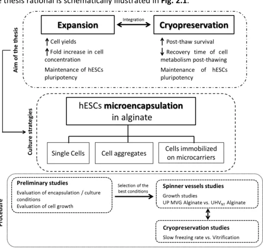
The main aim of this thesis was the development of an efficient and scalable methodology to integrate expansion and cryopreservation of human embryonic stem cells. This approach should allow the achievement of high cell yields of pluripotent human embryonic stem cells and, at the same time, high recovery rates of undifferentiated cells after thawing. To achieve this goal, cell microencapsulation in alginate was used and three strategies were outlined: encapsulation of single cells, encapsulation of hESC aggregates and encapsulation of hESCs immobilized on microcarriers.
On a first approach preliminary studies were performed in small scale suspension systems (Erlenmeyer) in order to optimize specific culture parameters. The best conditions were selected and reproduced on a larger scale culture system (spinner vessels), using two different alginate matrices (UHVNT and UP MVG alginates). In parallel, different methods for
cryopreservation of 3D hESC cultures were investigated including slow freezing rate and vitrification.
The thesis rational is schematically illustrated in Fig. 2.1.
3. Material and Methods
3.1.hESCs culture on feeder layer
hESCs cells (SCEDTM461, Cellartis AB) were routinely propagated as colonies in static conditions (6 well-plates) on a feeder layer of human foreskin fibroblasts (ATCC collection,
Cat. No. CRL‐2429) inactivated with mitomycin C (ihFF) in standard culture medium (DMEM -KO) (DMEM-KO supplemented with 20% (v/v) KO-SR, 1% (v/v) MEM-NEAA, 0.1mM 2-mercaptoethanol, 2mM Glutamax, 1% (v/v) Pen/Strep, 0.5% (v/v) Gentamycin (all from Invitrogen) and 10 ng/mL bFGF (Perprotech), as previously reported50. Every 10-12 days, i.e. when the hESC colonies covered approximately 75-85% of the surface area of the culture well, the colonies were digested with TrypLETM Select (Invitrogen) for 6‐8 min, and the resulting single cell suspension was transferred to fresh ihFF feeders (at splitting ratios between 1:4 and 1:24). Culture medium was replaced with fresh medium every 1–3 days.
3.2.Preparation of mEFs conditioned medium
For the production of the conditioned medium (mEFs-CM) for use in 3D hESC cultures, mouse embryonic fibroblast (mEFs, Millipore) were mitotically inactivated and replated on gelatin-coated T-flasks at 5.5x10⁴ cell/cm2 in DMEM-KO medium without bFGF (0.5mL/cm2). mEFs were cultured at 37ºC with 5% (v/v) CO2 and conditionated media were collected daily
for a total of 10 days per batch. Before feeding hESCs cultures, mEFs-CM was filtered and supplemented with 10 ng/ml bFGF and 0.1 nM Rapamycin (Sigma).
3.3.Encapsulation of human embryonic stem cells
Alginates: A 1:1 mixture of purified UHV alginates from L. nigrescens (UHVN) and L.
trabeculata (UHVT) - UHVNT alginate -was used at 0.4% or 0.7% (w/v) in 0.9% NaCl solution115, 130, 141
. UP MVG alginate (Pronova UP MVG NovaMatrix) was used at 1.1% (w/v) in 0.9% NaCl solution139.
Microcapsules formation: Microcapsules were formed by passing the alginate-cells mixture using 1 mL syringe through an air‐jet generator (Fig. 3.1; Page 28), at an air flow rate of 2-3.5 L/min and an air pressure of 1 bar. These encapsulation parameters yielded microcapsules with a diameter of approximately 500-700 µm. For cross‐linkage of the UHVNT alginate,
microcapsules were dropped directly from the droplet generator’s nozzle into a 20 mM BaCl2
solution, adjusted to 290 mOsm using NaCl buffered at pH 7 with 5 mM histidine. For
7.4 was used. Both alginates microcapsules were washed twice with 0.9% NaCl solution and one time with DMEM-KO medium before being transferred to culture systems.
Alginate dissociation: Ba2+-UHVNT alginate was dissolved by incubating the microcapsules in a
20 mM Na2SO4 solution for 20 min at 37°C, in a humidified atmosphere of 5% CO2 in air130.
Ca2+-UP MVG alginate was dissolved by incubating the microcapsules with a chelating solution (50 mM EDTA and 10 mM HEPES in PBS) for 5 min at 37ºC139.
3.4.Three-dimensional hESC cultures
The 3D hESC strategies and culture parameters tested are summarized in Tab. 3.1 and 3.2. 3.4.1.Culture of hESCs in Erlenmeyer
Single Cells: Before dissociation from 2D static cultures, hESCs colonies were pre-treated for 1 hour with 5 µM Y-27632, a ROCK inhibitor (ROCKi, Calbiochem). The single cell suspension, resulted from colonies dissociation using Tryple Select, was immediately encapsulated at different concentrations (0.75, 2 and 3 x10⁶ cell/mL alginate) using two alginate types (Tab. 3.1). In all cultures the final hESCs concentration was 1.5 x10⁶ cell/mL. hESCs-microcapsules were then inoculated into 125 mL Erlenmeyer (Corning), cultured in 15 mL mEFs-CM supplemented with 10 µM ROCKi, at 37ºC and 5% CO2 stirred at 70 rpm using an orbital
agitation.
hESC aggregates: hESCs were dissociated from the 2D static cultures and inoculated as single cells at 1.5 105 cell/mL into 125 mL Erlenmeyer. Cells were cultured in 10 mL mEFs-CM supplemented with 10 µM ROCKi, at 37ºC and 5% CO2, using an orbital agitation of 70 rpm.
Encapsulation was performed at day 2 or 5 (Tab. 3.1); aggregates were pre-treated with 5 µM ROCKi for 1 hour and then transferred to 15mL falcon tubes to allow their deposition and culture medium removal. The encapsulation procedure was done after ressuspension of the aggregates with equal volume of alginate solution. Microcapsules were cultured in Erlenmeyers, with 15 mL of mEFs-CM supplemented with 10 µM ROCKi.
hESCs immobilized on microcarriers: hESCs were inoculated at 4.5 105 cell/mL into 125 mL
Erlenmeyer containing 3 g/L (dry weight) Cytodex3 microcarriers (GE Healthcare) coated with Matrigel (BD Biosciences) or human plasma fibronectin (Millipore). The microcarriers
Erlenmeyers were placed inside an incubator (37ºC, 5%CO2) under intermittent stirring in
order to obtain a homogeneous cell distribution on the supports. After 6h, 2.5 mL of fresh medium was added to cultures and agitation rate set to 70 rpm. By day 3, the volume was adjusted to 15 mL. The encapsulation was performed at different time points (Tab. 3.2) using the same procedure described for aggregate cultures.
In all strategies, non-encapsulated cells culture was performed and run in parallel using the same culture conditions. The media were only partially (50%) replaced when necessary.
Tab. 3.1 – Culture parameters of single cells and hESCs aggregates 3D culture strategies
3.4.2.Culture of hESCs in spinner vessels
hESC aggregates: hESCs were inoculated at 1.5 105 cell/mL into 300 mL Erlenmeyer (Corning) in 50 mL mEFs-CM supplemented with 10 µM ROCKi. Cells were cultured at 37ºC, 5%CO2, using an orbital agitation of 70 rpm. Encapsulation was performed at day 2 and then
microcapsules were transferred to 125 mL spinner vessels (Wheaton) equipped with paddle impellers and cultured in 100 mL of mEFs-CM at 45 rpm for additional 16 days (Fig. 3.1). Culture medium was partially replaced three times a week. This was done by stopping agitation (to induce capsules sedimentation), removing 50% of the medium and feeding with 50% of fresh medium. Non-encapsulated aggregate cultures were also performed and run in parallel. Both cultures were monitored in terms of cell viability, metabolic activity, aggregate size and composition during time. For flow cytometry analysis, aggregates were transferred to gelatin coated surfaces, in mEFs-CM, where cells were able to migrate. After 2-3 days cells were dissociated using TrypLE Select.
hESCs immobilized on microcarriers: hESCs were inoculated at 4.5 105
cell/mL into 125 mL spinner vessels with paddle impellers containing Cytodex3 microcarriers (2 or 3g/L, Tab. 3.2)
Culture Strategy
Scale of culture
Type of culture Culture medium
Encapsulation time point
Cell concentration per alginate
Single cells Erlenmeyer
Encapsulated in UHVNT alginate
mEFs-CM day 0
0.75x10⁶ cell/mL Alg. 2 x10⁶ cell/mL Alg. 3 x10⁶ cell/mL Alg. Encapsulated in UP
MVG alginate
mEFs-CM day 0 3 x10⁶ cell/mL Alg.
hESC aggregates
Erlenmeyer
Non-encapsulated mEFs-CM -
--- Encapsulated in
UHVNT alginate
mEFs-CM day 2 day 5
Spinner vessels
Non-encapsulated
mEFs-CM day 2 Encapsulated in
UHVNT alginate Encapsulated in UP
coated with Matrigel. Cells were cultured in 25mL of mEFs-CM and the spinner vessels were placed at 37ºC, 5% CO2 under intermittent stirring. After 6h, fresh mEFs-CM was added to
cultures and agitation rate set to 24 rpm. By day 3, the volume was completed to 100 mL. The encapsulation was performed at day 6. In some experiments, fresh coated microcarriers (1 or 2 g/L) were added to the cultures 1 hour before encapsulation (Tab. 3.2). After encapsulation, hESCs were cultured in the same conditions for additional 13 days (Fig. 3.1). Medium was partially (50%) replaced daily. Non-encapsulated cell-microcarrier cultures were also performed and run in parallel. Cultures were monitored in terms of cell viability, cell concentration, metabolic activity and cell characterization during time. For flow cytometry analysis, hESCs were dissociated from the supports after 5 min of incubation with Tryple Select at 37ºC.
After expansion of both cell aggregates and hESC-microcarriers cultures, hESCs were dissociated and plated on a top of a monolayer of ihFFs for further characterization studies. Tab. 3.2 – Culture parameters of hESCs immobilized on microcarriers culture strategy
Scale of culture
Type of culture Coating of microcarriers Culture medium Encapsulation time point Concentration of microcarriers added initially Concentration of microcarriers added at day 6
Erlenmeyer
Non-encapsulated Matrigel mEFs-CM
- 3 g/L -
Fibronectin DMEM-KO Encapsulated in
UHVNT alginate Matrigel
mEFs-CM
day 0 (after 8h)
3 g/L -
day 1 day 3 day 6 Spinner vessels Non-encapsulated
Matrigel mEFs-CM day 6
3 g/L -
2 g/L 1 g/L Encapsulated in
UHVNT alginate Matrigel mEFs-CM day 6
3 g/L -
2 g/L 1 g/L Encapsulated in UP
MVG alginate Matrigel mEFs-CM day 6
2 g/L 1 g/L 2 g/L 2 g/L
3.5.Cell cryopreservation
Cultures of non-encapsulated and encapsulated hESCs (cell aggregates and cell immobilized on microcarriers) were harvested from the spinner vessels at days 14 and 13, respectively, and cryopreserved using two different procedures: slow freezing rate and vitrification. Before cryopreservation, hESCs were pre-treated with 5 µM ROCKi for one hour.
3.5.1.Slow Freezing Rate
Freezing: At the moment of freezing, after deposition of the microcapsules, culture medium was removed and cryopreservation medium (90% KO-SR and 10% (v/v) DMSO (Sigma) supplemented with ROCKi (5µM)) was added. Cell suspension obtained was then transferred to cryovials (1 mL/vial). The cells were allowed to equilibrate in the cryopreservation medium for 20 min at 4ºC. Samples were frozen to -80ºC in an isopropanol-based freezing
system, (“Mr. Frosty”, Nalgene) at a rate of cooling very close to 1ºC per minute, and stored in the gas phase of a liquid nitrogen (LN2) reservoir until their thawing.
Thawing: Following storage, cells were quickly thawed by placing the cryovials in a 37ºC water bath, a stepwise dilution (1:1, 1:2, 1:4) in mEFs-CM was performed immediately after thawing in order to dilute the DMSO while reducing the osmotic shock. Cells were further cultured for post-thaw studies (cell viability, metabolic activity and differentiation state) in petri-dishes in mEFs-CM suppletemented with 5 M of ROCKi. Changes of media were performed daily. Whenever possible microcapsules were dissolved, hESCs were dissociated with TrypLE Select, transferred to ihFFs monolayers and maintained in culture for several passages for post-thaw studies of growth and pluripotency.
3.5.2.Vitrification
Freezing: Two serum‐free vitrification solutions (VS) were used in the freezing process: VS1 included 10% DMSO and 10% ethyleneglycol (EG); VS2 contained 0.5 M sucrose, 30% DMSO and 30% EG. Cells/capsules were incubated in VS1 for 1 min followed by VS2 for 5 seconds and by 10 seconds in LN2. Cell strainers of 70 µm (BD) were used to transfer the
microcapsules between solutions. As these were preliminary studies the cells were thawed
immediately after the cooling process instead of being stored in the vapor‐phase of LN2.
incubation for 1 min in 2x diluted WS1, followed by 5 min incubation in WS2. Cells were then cultured in mEFs-CM supplemented with 5 M of ROCKi.
3.5.3.Assessment of hESCs survival after thawing
The percentage of hESCs survival/recovery after thawing was determined by calculating the ratio between the number of viable hESCs after cryopreservation and the number of initially frozen viable hESCs, counted using a Fuchs-Rosenthal haemocytometer chamber (Invitrogen) and the Trypan Blue (Invitrogen) exclusion method.
3.6.Evaluation of cell viability
Cell membrane integrity assay: The qualitative assessment of the cell plasma membrane integrity during culture was done using the enzyme substrate fluorescein diacetate (FDA; Sigma-Aldrich) and propidium iodide (PI; Sigma-Aldrich). Cells were incubated with 20 µg/mL FDA and 10 µg/mL PI in PBS for 2-5 min and then visualized using fluorescence microscopy (Leica Microsystems GmbH). FDA is a non-polar, non-fluorescent fluorescein analogue which enters all cells freely. In viable cells, FDA is converted by intracellular esterases to highly fluorescent fluorescein. Being highly polar, fluorescein becomes trapped within cells that possess membrane integrity (viable cells) and confers to them green fluorescence. PI is a polar, highly fluorescent (red color) compound which can only enter in cells that lack membrane integrity. It intercalates into the major groove of the DNA, therefore nucleus dying/dead cells stain red142.
Trypan Blue exclusion method: The total number of viable cells was determined by counting the colorless cells in a Fuchs-Rosenthal haemocytometer chamber after incubation with Trypan Blue dye (0.1% (v/v) in PBS). Dead cells with damaged membranes stain blue in the presence of this compound.
Lactate dehydrogenase (LDH) activity: LDH activity from the culture supernatant was dermined spectrophotometrically (at 340 nm) following the rate of oxidation of NADH to NAD+ coupled with the reduction of pyruvate to lactate. The specific rate of LDH release (qLDH) was calculated for each time interval using the following equaton: , where is the change in LDH activity over the time period , and