Relationship of Electromechanical Dyssynchrony in Patients
Submitted to CRT With LV Lead Implantation Guided by Gated
Myocardial Perfusion Spect
Erivelton Alessandro do Nascimento,
1,2Christiane Cigagna Wiefels Reis,
2Fernanda Baptista Ribeiro,
3Christiane
Rodrigues Alves,
2Eduardo Nani Silva,
3Mario Luiz Ribeiro,
3Claudio Tinoco Mesquita
2Instituto Estadual de Cardiologia Aloysio de Castro,1 Rio de Janeiro, RJ - Brazil
Programa de Pós-graduação em Ciências Cardiovasculares da Universidade Federal Fluminense (UFF),2 Niterói, RJ - Brazil
Hospital Universitário Antônio Pedro - Universidade Federal Fluminense,3 Niterói, RJ – Brazil
Mailing Address: Erivelton Alessandro do Nascimento • Rua Gil Ferreira, 182. Postal Code 27283-570, Jardim Primavera, Volta Redonda, RJ - Brazil
E-mail: [email protected], [email protected]
Manuscript received March 19, 2017, revised manuscript February 05, 2018, accepted May 09, 2018
DOI: 10.5935/abc.20180159
Abstract
Background: Heart failure (HF) affects more than 5 million individuals in the United States, with more than 1 million hospital admissions per year. Cardiac resynchronization therapy (CRT) can benefit patients with advanced HF and prolonged QRS. A significant percentage of patients, however, does not respond to CRT. Electrical dyssynchrony isolated might not be a good predictor of response, and the last left ventricular (LV) segment to contract can influence the response.
Objectives: To assess electromechanical dyssynchrony in CRT with LV lead implantation guided by GATED SPECT.
Methods:This study included 15 patients with functional class II-IV HF and clinically optimized, ejection fraction of 35%, sinus
rhythm, left bundle‑branch block, and QRS ≥ 120 ms. The patients underwent electrocardiography, answered the Minnesota
Living with Heart Failure Questionnaire (MLHFQ), and underwent gated myocardial perfusion SPECT up to 4 weeks before CRT, being reassessed 6 months later. The primary analysis aimed at determining the proportion of patients with a reduction in QRS duration and favorable response to CRT, depending on concordance of the LV lead position, using chi-square test. The pre- and post-CRT variables were analyzed by use of Student t test, adopting the significance level of 5%.
Results: We implanted 15 CRT devices, and 2 patients died during follow‑up. The durations of the QRS (212 ms vs 136 ms)
and the PR interval (179 ms vs 126 ms) were significantly reduced (p < 0.001). In 54% of the patients, the lead position was
concordant with the maximal delay site. In the responder group, the lateral position was prevalent. The MLHFQ showed a
significant improvement in quality of life (p < 0.0002).
Conclusion: CRT determines improvement in the quality of life and in electrical synchronism. Electromechanical synchronism relates to response to CRT. Positioning the LV lead in the maximal delay site has limitations. (Arq Bras
Cardiol. 2018; 111(4):607‑615)
Keywords: Heart Failure; Cardiac Resynchronization Therapy; Eletrodes,Implanted;, Stroke Volume; Radionuclide Imaging.
Introduction
Heart failure (HF) affects more than 5 million individuals in the United States. Approximately 550,000 new cases are diagnosed annually, and decompensated HF accounts for over 1 million hospital admissions per year.1 Projections show that HF prevalence will increase by 46% from 2012 to 2030, resulting in more than 8 million individuals with HF aged 18 years and older.2 As a consequence of this epidemiological transition, of the advances in healthcare and of population aging, the prevalence of coronary artery disease, systemic
arterial hypertension, obesity and diabetes mellitus is increasing and will have a significant impact on HF incidence in developing countries.3
Cardiac resynchronization therapy (CRT) has become an option to treat advanced symptomatic HF with: (A) left ventricular dysfunction; (B) electrical dyssynchrony; and (C) optimized clinical therapy. The technique has shown a significant improvement in
New York Heart Association functional class (NYHA FC) and in left ventricular ejection fraction (LVEF) of individuals with severe ventricular dysfunction and left bundle-branch block (LBBB).4 However, a significant group of patients does not respond favorably to CRT.5-7 Patients with coronary artery disease and previous acute myocardial infarction are less likely to respond, because of the presence of fibrosis. The selection criteria for CRT currently used do not seem ideal, because previous studies on CRT using those criteria have found a significant percentage of patients (20% to 40%) who did not benefit from the therapy.6,7
of mechanical dyssynchrony. Thus, it is worth studying the ventricular synchronism before CRT to estimate the patient’s response, because the procedure involves high costs. Phase analysis to assess left ventricular (LV) dyssynchrony has been incorporated into gated myocardial perfusion SPECT (GATED SPECT).8 In addition to the synchronism parameters and in a highly reproducible way, phase analysis provides the assessment of the last ventricular segment to contract. Patients with LBBB tend to begin LV mechanical contraction earlier in the cardiac cycle in the septal wall, and later in other myocardial regions because of the deceleration of the nervous impulse propagation along the conduction system, causing late activation, the most common last site of contraction being located in the posterolateral wall.9
The present study aimed at assessing the ability to analyze LV synchronism by use of GATED SPECT to predict the response to CRT and guide LV lead implantation.
Methods
The present study contains national data that are part of the international multicenter project VISION CRT, which assesses the value of phase analysis by use of GATED SPECT in patients who will be submitted to CRT, coordinated in multiple countries by the International Atomic Energy Agency. It is a non-blind clinical trial that included consecutive patients, who underwent 12-lead electrocardiogram at rest immediately before undergoing GATED SPECT and speckle-tracking echocardiography. In addition, the patients answered the Minnesota Living with Heart Failure Questionnaire (MLHFQ) within 4 weeks before the implantation of the CRT device and 6 ± 1 months after that implantation for comparison. Thus, the scintigraphic parameters of ventricular function [LVEF, end-diastolic volume (EDV), end-systolic volume (ESV), LV mass] were assessed, as were the parameters to assess dyssynchrony by use of phase analysis. The analysis of GATED SPECT with the software ECT Synctool, version 3.0, used the following parameters for mechanical dyssynchrony: standard deviation (SD) > 43° and histogram bandwidth (HBW) > 135°.
The inclusion criteria were as follows: patients stable and
older than 18 years, in NYHA FC ≥ II, with LVEF ≤ 35% of
ischemic or non-ischemic cause, sinus rhythm, QRS duration
≥ 120 ms, LBBB morphology, being followed up at or
referred to two tertiary institutions of the Brazilian Unified Health System. Patients with cardioverter-defibrillator implanted for primary or secondary prevention of sudden cardiac death were included.
Patients with any of the following characteristics were excluded: severe disease and life expectancy shorter than 1 year; right bundle-branch block; pregnancy or breastfeeding; acute coronary syndromes; coronary artery bypass grafting or percutaneous coronary intervention within 3 months from study entrance and up to 6 months after CRT.
The definition of ‘responder to CRT’ considered the presence of two of the following findings: 1. improvement of at least one NYHA FC; 2. improvement of at least 5 points
in the MLHFQ; 3. improvement of LVEF ≥ 5%; 4. reduction in ESV ≥ 15%; 5. reduction in HBW < 51°. The categorical
variables were presented in nominal and ordinal forms.
The LV pacing lead position was classified as follows: 1. concordant, when positioned in the maximally delayed segment; 2. adjacent, when located in up to one segment away from the maximal delay site; and 3. remote, when located more than one segment away from the maximal delay site.
This project was approved by the Ethics Committee in Research of the Antônio Pedro University-affiliated Hospital/ Fluminense Federal University (No 884844).
Statistical analysis
Statistical analysis was performed with EXCEL (2010, Microsoft Corporation) and SPSS software, version 21.0 (2012, IBM Corporation), and data were shown as means and standard deviations. The categorical variables were compared by use of Fisher exact and chi-square tests, while paired Student t test was used for numerical variables. The Kolmogorov-Smirnov test showed the normal distribution of the continuous variables. Pearson’s linear correlation coefficient was calculated for the continuous variables. The significance level adopted in the statistical analysis was 5%.
Results
From July 2014 to August 2016, 15 patients were included in the study and 2 patients were lost to follow-up because of death (Table 1). Mean follow-up was 193 ± 16 days.
All QRS intervals had a duration longer than 150ms and LBBB morphology. After CRT, a significant reduction was observed in the duration of QRS intervals (212 ms vs 136 ms;
p < 0.001) and of PR intervals (179 ms vs 126 ms; p < 0.001).
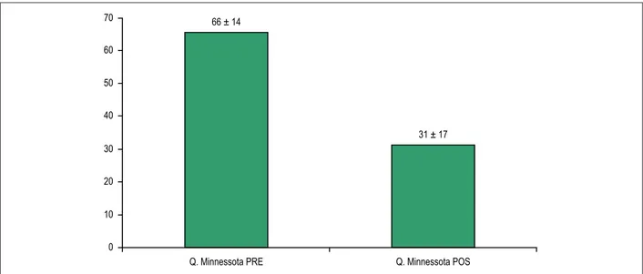
No change was observed in the QT interval after CRT. The impact of CRT on the quality of life was recorded by use of the MLHFQ, with significant response (p=0.0002) when comparing the mean score before and after CRT (Figure 1).
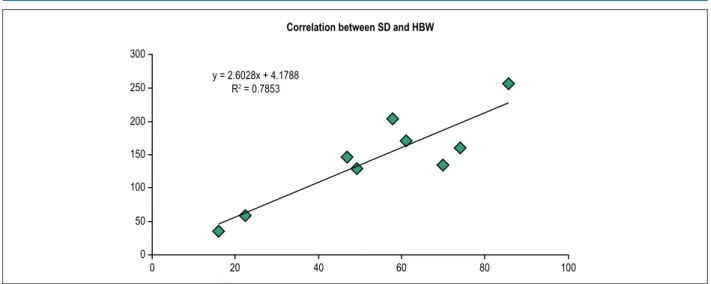
Analysis with HBW (Figure 2) showed that the longer the QRS duration, the higher the HBW value, showing that HBW and SD also have a direct relationship, because their linear correlation coefficient is good (Figure 3).
The group of patients with a significant improvement in LVEF 6 months after CRT (6 patients) had a lower pre-CRT LVEF than that of non-responders (7 patients) (Figure 4).
When assessing the electrocardiographic parameters associated with clinical response to the CRT device, the responder group showed a significant reduction in the PR
interval in ms (p < 0.0001), which did not reach significance
in the non-responder group (p = 0.09). This is influenced by the need for constant ventricular stimulation in CRT, which normally leads to more reduced PR intervals.
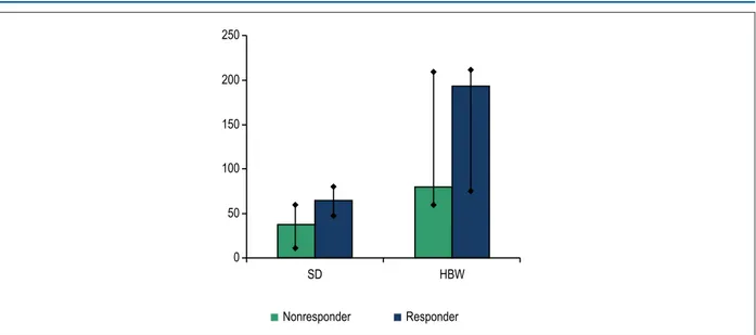
When classifying the patients as responders and non-responders, the SD and HBW values were higher in responders than in non-responders. The HBW difference between the groups showed statistical significance by use of Student t test (Figure 5).
Figure 1 – Comparison of the mean score of the Minnesota Living with Heart Failure Questionnaire before and after cardiac resynchronization therapy, using Student t test. 70
60
50
40
30
20
10
0
66 ± 14
31 ± 17
Q. Minnessota PRE Q. Minnessota POS
dyssynchrony do not respond to CRT on GATED SPECT because they have no change suggestive of mechanical dyssynchrony on baseline tests. Likewise, patients with severe mechanical dyssynchrony and changes on baseline tests show a marked improvement in the GATED SPECT parameters after CRT, mainly HBW.
Of the group of responders, 77.7% had the pacing lead implanted in the lateral region, 11.1% in the posterolateral region, and 11.1% in the posteroseptal region (Figure 6).
When assessing synchronism by use of myocardial perfusion imaging, concordant LV lead positioning was achieved in 54% of the cases (Figure 7 illustrates a concordant implantation), the major reason for not reaching concordance being the anatomical variability of the veins related to coronary sinus, as well as the absence of tributaries reaching the site determined by myocardial perfusion imaging. One of the patients had an aneurysmal coronary sinus, which prevented the lead from being anchored. Thus, the LV lead implantation was converted to the epicardial pathway, in the maximally delayed site.
Discussion
In our study, we observed that CRT led to patients’ clinical improvement and to a reduction in electrical and mechanical dyssynchrony. Although CRT is associated with the improvement of several clinical parameters, not every patient benefited from that, and longer QRS duration on the electrocardiogram and increased SD and HBW on GATED SPECT were markers of higher likelihood of clinical response. In addition, we observed that GATED SPECT could identify the last myocardial segment to contract, the ideal LV lead implantation site on CRT. However, because of anatomical limitations, that identification led to concordant implantation in only 54% of the cases. Table 1 – General baseline characteristics of the patients submitted
to cardiac resynchronization device implantation
Demographic characteristics N or mean ± SD
Total of patients 15
Age (years) 63.21 ± 7.7
Body mass index (kg/m2) 26.92 ± 5.4
Male sex 4
Diabetes mellitus 6
Hypertension 9
Dyslipidemia 8
Smoking 0
Previous coronary artery disease 7
Previous infarction 7
Coronary artery bypass grafting 2
Percutaneous coronary intervention 0
NYHA functional class II 1
NYHA functional class III 7
NYHA functional class IV 5
Beta-blocker 13
Angiotensin-converting-enzyme inhibitor 8
Angiotensin-receptor blocker 7
Acetylsalicylic acid 2
Diuretics 8
Statin 5
Aldosterone antagonist 8
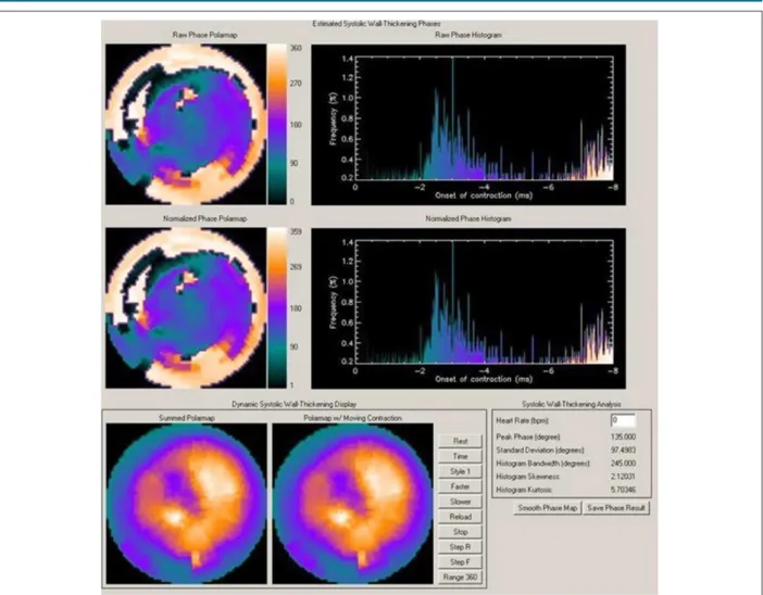
Figure 2 – Gated myocardial perfusion SPECT with analysis of ventricular synchronism in a patient with dilated cardiomyopathy and left bundle-branch block, showing marked dyssynchrony with HBW of 245° and SD of 97°.
The population of the present study reflects the profile of patients normally treaded at high-complexity hospitals, and most of them had coronary artery disease. As shown in previous studies, most of our patients had NYHA FC III or IV at the time of CRT.10 The clinical-functional assessments of the present study, NYHA FC and MLHFQ, confirmed the benefit of CRT reported previously.10-13 In this study, 77.8% of the patients had a reduction of at least one NYHA FC and a significant improvement in their quality of life, as shown on the MLHFQ after CRT. Although the MLHFQ assesses subjective data, it refers to the patients’ perception of their symptoms, which is aligned with the results of a previous study.4
In most patients, CRT is associated with clinical benefits. Some electrocardiographic parameters are considered predictors of a higher chance of response to treatment, such as the longer QRS duration, and the benefit increases even more when QRS duration is > 150 ms, as reported by Poole et al.14 In our study, all patients had QRS duration > 150 ms (mean QRS duration, 212 ms), which increased the likelihood of response. Supporting such data, the COMPANION study has shown no benefit of CRT for patients with QRS duration
< 147 ms12 when assessing the primary outcome of death or hospitalization due to any cause. However, the RAFT study,13 assessing the primary outcome of death or hospitalization due to HF, has found a higher benefit of CRT in individuals with QRS duration > 150 ms.
Our sample did not include patients with non-LBBB morphology, nonspecific intraventricular conduction disorders and/or right bundle-branch block, which might have led to the clinical benefit observed. Those findings have also been reported in several recent studies,7,15 which have shown a reduction or even absence of CRT benefit in that group of patients. It is worth noting that our study, even recruiting
all patients with LVEF < 35%, QRS duration > 150 ms and
LBBB morphology, identified 27% of them as non-responders (clinical criteria/death). Those figures are aligned with those reported in the literature.6,15
Figure 3 – Correlation between SD and HBW before cardiac resynchronization therapy (R2: 0.78) 300
200
100
100 250
150
50
20 40 60 80
0 0
y = 2.6028x + 4.1788 R2 = 0.7853
Correlation between SD and HBW
Figure 4 – Distribution of the mean pre- and post-cardiac resynchronization therapy left ventricular ejection fraction according to clinical response to implantation. Pre-CRT LVEF PRE: pre-cardiac resynchronization therapy left ventricular ejection fraction; Post-CRT LVEF: post-cardiac resynchronization therapy left ventricular ejection fraction; RESPOND: responder group; NONRESPOND: non-responder group. (Student t test).
60%
50%
40%
30%
20%
10%
Pre-CRT LVEF Post-CRT LVEF
RESPOND NONRESPOND
On GATED SPECT, SD and HBW could assess mechanical dyssynchrony before CRT. On the assessment 6 months after CRT, those variables showed no significance, probably because not all patients had the LV lead positioned in the maximally delayed segment. On GATED SPECT, the significant cardiac function data were LV ESV and LV mass, probably due to reverse remodeling determined by CRT.
In the search for a relationship between responders to CRT and the presence of mechanical dyssynchrony on myocardial perfusion imaging, responders had higher SD and HWB values as compared to non-responders (HBW of 177° vs 76°, and SD of 62° vs 36°, respectively). Such findings are aligned with those reported by Henneman et al.,16 whose study showed significantly higher values of dyssynchrony parameters in responders as compared to non-responders (HBW of 175° vs
117°, and SD of 56° vs 37°, respectively). In addition, responders
had longer QRS duration than non-responders, supported by the finding of the direct relationship between them.
When assessing the maximal delay site of LV activation, the presence of fibrosis in the site and adjacent to it can be determined, which can influence the response to CRT. Daoulah et al.17 have shown that the presence of transmural fibrosis in the posterolateral region before CRT is associated with a 75% lower chance of echocardiographic or clinical response to that therapy. In our study, 11.1% of the patients had the lead implanted in the posterolateral region; however, no fibrosis was reported in that region in our sample, and 7 patients had history of previous myocardial infarction.
Figure 5 – Distribution of the mean pre-cardiac resynchronization therapy SD and HBW according to clinical response. SD: standard deviation; HBW: histogram bandwidth (Student t test).
250
150
50
0 200
100
SD HBW
Nonresponder Responder
Figure 6 – Response to cardiac resynchronization therapy according to the left ventricular lead implantation site. Post Lat - posterolateral region; Post Sep - posteroseptal region. 8
7
6
5
4
3
2
1
0
Lateral Post Lat Post Step
Responder Non-responder
Despite the limitation of LV lead positioning in the last activation site, the vascular technique has some benefits: 1. less invasive procedure with smaller peri- and postoperative complications; 2. lower chronic stimulation thresholds; and 3. shorter hospital length of stay. One strategy to overcome the limitation of stimulating the maximal delay site in LV contraction is the possibility of using multipoint pacing. This stimulation has been made possible with the development of technologies of multipolar leads that
stimulate the left ventricle in several sites, generating several possibilities of stimulating vectors.20 In addition, GATED SPECT can identify the last LV activation site at the same time it identifies if the area has fibrosis, contributing, thus, to select patients for CRT.
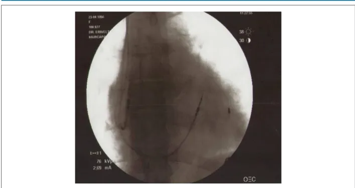
Figure 7 – Fluoroscopy during implantation of the cardiac resynchronization device with left ventricular lead implanted in the maximal delay site determined on GATED SPECT.
Considering the present study’s findings, it seems that the use of electrocardiography in isolation, based on QRS duration and its morphology to select patients for CRT, is not a good isolated preditor; however, the association of the duration and morphology criteria with the imaging criteria of mechanical dyssynchrony can provide a better response to CRT.
The benefit of using imaging techniques, especially GATED SPECT with phase analysis, to detect mechanical dyssynchrony and to guide lead positioning in the site of maximal conduction delay should increase the number of responders, but larger studies with LV lead positioning guided by imaging techniques are required to draw definite conclusions.
Conclusion
1) Patients submitted to CRT have a good clinical response, with a reduction in electrical dyssynchrony assessed on electrocardiography and a reduction in mechanical dyssynchrony assessed on GATED SPECT.
2) Responders to CRT have longer QRS duration before the implantation of the CRT device as compared to non-responders. In addition, responders had a significant reduction in the PR interval duration as compared to non-responders.
3) Electrical dyssynchrony is not necessarily associated with mechanical dyssynchrony, as shown on GATED SPECT. 4) The phase analysis of GATED SPECT showed that the parameters SD and HBW are associated with higher likelihood of responding to CRT.
5) Although GATED SPECT indicates the last myocardial segment to contract for the LV lead positioning in this site, that is not always possible because of anatomical variability (tributaries) and caliber of the coronary sinus.
Author contributions
Conception and design of the research and Statistical analysis: Nascimento EA, Reis CCW, Mesquita CT; Acquisition of data and Critical revision of the manuscript for intellectual content: Nascimento EA, Reis CCW, Ribeiro FB, Alves CR, Silva EN, Ribeiro ML, Mesquita CT; Analysis and interpretation of the data and Writing of the manuscript: Nascimento EA, Reis CCW, Silva EN, Mesquita CT; Obtaining financing: Mesquita CT.
Potential Conflict of Interest
No potential conflict of interest relevant to this article was reported.
Sources of Funding
This study was funded by Agência Internacional de Energia Atômica.
Study Association
This article is part of the thesis of master submitted by Erivelton Alessandro do Nascimento, from Universidade Federal Fluminense.
Ethics approval and consent to participate
1. Hunt AS; American College of Cardiology; American Heart Association Task Force on Practice Guidelines (WritingCommittee to Update the 2001 Guidelines for the Evaluation and Management of Heart Failure). ACC/AHA 2005 guideline update for the diagnosis and management of chronic heart failure in the adult: a report from the American College of Cardiology/American Heart Association Task Force on Practice Guidelines (Writing Committee to Update the 2001 Guidelines for the Evaluation and Management of Heart Failure), J Am Coll Cardiol. 2005; 46(6):e1-82. Erratum in: J Am Coll Cardiol. 2006;47(7):1503-5.
2. Heidenreich PA, Albert NM, Allen LA, Bluemke DA, Butler J, Fonarow GC, et al; American Heart Association Advocacy Coordinating Committee; Council on Arteriosclerosis, Thrombosis and Vascular Biology; Council on Cardiovascular Radiology and Intervention; Council on Clinical Cardiology; Council on Epidemiology and Prevention; Stroke Council. Forecasting the impact of heart failure in the United States: a policy statement from the American Heart Association. Circ Heart Fail. 2013;6(3):606-19.
3. Cubillos-Garzon L, Casas J, Morillo C, Bautista L. Congestive heart failure in Latin America: The next epidemic. Am Heart J. 2004;147(3):412-7.
4. Abraham WT, Fisher WG, Smith AL, Delurgio DB, Leon AR, Loh E, et al; MIRACLE Study Group. Multicenter InSync Randomized Clinical Evaluation. Cardiac resynchronization in chronic heart failure. N Engl J Med. 2002;346(24):1845-53.
5. Mozaffarian D, Benjamin EJ, Go AS, Arnett DK, Blaha MJ, Cushman M, et al. American Heart Association Statistics Committee and Stroke Statistics Subcommittee. Heart disease and stroke statistics--2015 update: a report from the American Heart Association. Circulation. 2015;131(4):e29-322. Erratum in: Circulation. 2016;133(8):e417. Circulation. 2015;131(24):e535.
6. Bakker PF, Meijburg H, Dejonge N, Mechelen RV, Wittkampf F, Mower M, et al. Beneficial effects of biventricular pacing in congestive heart failure. [absctract]. Pacing Clin Electrophysiol. 1994;17:820.
7. Daubert C, Gold MR, Abraham WT, Ghio S, Hassager C, Goode G, et al; REVERSE Study Group. Prevention of disease progression by cardiac resynchronization therapy in patients with asymptomatic or mildly symptomatic left ventricular dysfunction: insights from the European cohort of the REVERSE (Resynchronization Reverses Remodeling in Systolic Left Ventricular Dysfunction) trial. J Am Coll Cardiol. 2009;54(20):1837-46.
8. Zhou Y, Faber TL, Patel Z, Folks RD, Cheung AA, Garcia EV, et al. An automatic alignment tool to improve repeatability of left ventricular function and dyssynchrony parameters in serial gated myocardial perfusion SPECT studies. Nucl Med Commun. 2013;34(2):124-9.
9. Almeida AL, Gjesdal O, Mewton N, Choi EY, Tura GT, Yoneyama K, et al. Speckle Tracking pela ecocardiografia bidimensional: aplicações clínicas. Rev bras ecocardiogr imagem cardiovasc. 2013;26(1):38-49.
10. Gervais R, Leclerq C, Shankar A, Jacobs S, Eiskjaer H, Johannessen A, et al; CARE-HF investigators. Surface electrocardiogram to predict outcome
in candidates for cardiac resynchronization therapy: a sub analysis of the CARE-HF trial. Eur J Heart Fail. 2009;11(7):699-705.
11. Cazeau S, Leclercq C, Lavergne T, Walker S, Varma C, Linde C, et al; Multisite Stimulation in Cardiomyopathies (MUSTIC) Study Investigators. Effects of multisite biventricular pacing in patients with heart failure and intraventricular conduction delay. N Engl J Med. 2001;344(12):873-80.
12. Bristow MR, Saxon LA, Boehmer J, Krueger S, Kass DA, De Marco T, et al; Comparison of Medical Therapy, Pacing, and Defibrillation in Heart Failure (COMPANION) Investigators. Cardiac-resynchronization therapy with or without an implantable defibrillator in advanced chronic heart failure. N Engl J Med. 2004;350(21):2140-50.
13. Tang ASI, Wells GA, Talajic M, Arnold MO, Sheldon R, Connolly S, et al; Resynchronization-Defibrillation for Ambulatory Heart Failure Trial (RAFT) Investigators. Cardiac resynchronization therapy for mild-to-moderate heart failure. N Engl J Med. 2010;363(25):2385-95.
14. Poole JE, Singh JP, Birgersdotter-Green U. QRS duration or qrs morphology: what really matters in cardiac resynchronization therapy? J Am Coll Cardiol. 2016;67(9):1104-17.
15. Moss AJ, Hall WJ, Cannom DS, Klein H, Brown MW, Daubert JP, et al; MADIT-CRT Trial Investigators. Cardiac-resynchronization therapy for the prevention of heart-failure events. N Engl J Med. 2009;361(14):1329-38.
16. Henneman MM, Chen J, Ypenburg C, Dibbets P, Bleeker GB, Boersma E, et al. Phase analysis of gated myocardial perfusion single-photon emission computed tomography compared with tissue Doppler imaging for the assessment of left ventricular dyssynchrony. J Am Coll Cardiol. 2007;49(16):1708-14.
17. Daoulah A, Alsheikh-Ali AA, Al-Faifi SM, Ocheltree SR, Haq E, Asrar FM, et al. Cardiac resynchronization therapy in patients with postero-lateral scar by cardiac magnetic resonance: a systematic review and meta-analysis. J Electrocardiol. 2015;48(5):783-90.
18. Singh JP, Fan D, Heist EK, Alabiad CR, Taub C, Reddy V, et al. Left ventricular lead electrical delay predicts response to cardiac resynchronization therapy. Heart Rhythm. 2006;3(11):1285-92. Erratum in: Heart Rhythm. 2006 Dec;3(12):1515.
19. Ellenbogen KA, Gold MR, Meyer TE, Fernndez Lozano I, Mittal S, Waggoner AD, et al. Primary results from the SmartDelay determined AV optimization: a comparison to other AV delay methods used in cardiac resynchronization therapy (SMART-AV) trial: a randomized trial comparing empirical, echocardiography-guided, and algorithmic atrioventricular delay programming incardiac resynchronization therapy. Circulation. 2010;122(25):2660-8.
20. Pappone C, Ćalović Ž, Vicedomini G, Cuko A, McSpadden LC, Ryu K, et al. Multipoint left ventricular pacing improves acute hemodynamic response assessed with pressure-volume loops in cardiac resynchronization therapy patients. Heart Rhythm. 2014;11(3):394-401.