I – Introduction
All cells are variations on a basic structural theme: a package of highly structured cytoplasm contained within a lipid barrier. The cytoplasm is subdivided by internal membrane system into several compartments specialized for particular function. The mechanical strength and capacity to change shape of a cell, its own movement and internal action are largely provided by an intracellular network of specialized polymers, the cytoskeleton. The actin cytoskeleton involves a multitude of proteins and regulatory mechanisms implicated in assembly, disassembly and organization of actin filaments, making this system vulnerable to genetic alterations that may cause diseases, such as cancer. In cancer cells, structural and functional perturbations of the actin cytoskeleton correlate with higher proliferation rates and uncontrolled movement (Giganti and
Friederish, 2003). Therefore, the study of the actin dynamics during tissue morphogenesis is important for cancer therapeutics.
1 – Drosophila as a model system
Drosophila melanogaster is one of the most used model organisms and is
commonly known as the “fruit fly”. Among many advantages of using flies as a research model are their short life cycle, high fecundity, small genome, easy and low cost culturing and their powerful genetic tools (reviewed in St. Johnston, 2002). Furthermore, 197 out of 287 human genes, known to cause disease when misregulated have
Drosophila homologues that can produce very similar symptoms when misregulated in
flies (reviewed in St. Johnston, 2002).
1.1 – Advantage of using Drosophila genetics
Genetic tools commonly used in Drosophila allow the identification and characterization of genes that are involved in a wide range of biological processes
(reviewed in St. Johnston, 2002). Some of such genetic tools available are the balancers chromosomes, the phenotypic markers and non-recombination in males. The balancer chromosomes are chromosomes with one or more inverted segments that suppress recombination and allow lethal mutations or transgenes to be maintained without selection. A large number of phenotypic markers, both recessive and dominant, are available and allow the selection of flies carrying the chromosome of interest, by eye, body, bristle, larval and wing phenotype. By using Mendel’s law and following a dominant phenotypic marker, it’s easy to successfully follow a multi-generation cross. Also, there is another powerful technique available which is the P-elements transformation. Using this technique it is possible to introduce any gene of interest within the Drosophila genome as soon as the construct of interest has been placed between P-elements. The P-elements are transposable pieces of DNA that randomly insert into genomic DNA with the help of the transposase enzyme. Transgenic flies are selected using a variety of phenotypic markers such as eye color, body color or resistance to antibiotics (Socolish, 2003).
Moreover, there is another useful genetic tool used to analyze gene function known as genetic mosaics that will be frequently used throughout this work. By genetic mosaic technique, genetic changes can be induced in a subset of cells or tissues in an individual organism and examine genetic changes that would be otherwise lethal if applied to the entire organism (see Materials and Methods) (Blair, 2003).
1.2 – The Drosophila life cycle
The developmental period of Drosophila depends at which temperature the flies are grown. Drosophila undergoes a process of metamorphosis which proceeds as follows: embryo, larvae, pupae and ends in the adult fly. The shortest developmental time (egg to adult) takes 7 days and it is achieved at 28ºC. The developmental time increase at higher temperatures due to heat stress. Under ideal conditions, the development time at 25°C is 8.5 days, as it is shown in Fig. 1.1. At room temperature the whole cycle takes 12 days. Females lay some 400 eggs (embryos) per day into their food (Ashburner et al., 2005).
Figure 1.1 – Schematic representation of Drosophila melanogaster life cycle at 25ºC (adapted from
Griffiths et al., 2000).
1.3 – Drosophila imaginal discs are typical epithelial structures
1.3.1 – Epithelial structure of the wing imaginal disc
Each Drosophila external adult structure (such as wings, leg, eyes and antennas) derive from internal structures called imaginal discs (as shown in Fig.1.5). Imaginal discs form during embryogenesis from small clusters of cells (about 40 cells for the wing disc). That proliferate during the whole larval development to form folded, single layer, epithelial sacs (about 10000-50000 cells for the wing disc) (reviewed in St. Jonhston, 2002).
Figure 1.5 – Location and identification of Drosophila imaginal discs (adapted from Carballo
and Hwang, 2000).
Each of these sacs consists of a columnar epithelium covered by a peripodial membrane (Fig. 1.7) (Schock and Perrimon, 2002). The columnar cells are a laterally coherent sheet polarized along the apico-basal axis. These epithelial cells contain adherens junctions (AJ) composed mainly of E-cadherin and α- and β-catenin (in
Drosophila encoded by armadillo). The AJ localize apically and links the actin cytoskeleton (also polarized) of neighboring cells (Fig.1.6). Apical to the AJs, Drosophila has two complexes that are involved in regulating the establishment and maintenance of epithelial polarity. These complexes are the Baz/Par-6/aPKC complex and Crb/Stardust/Patj complex (Fig.1.6). Present basal to AJ, exists the septate junctions that are formed by Dlg/Scribble/Lgl complex (Gibson and Perrimon, 2003) (Fig.1.6).
Figure 1.6 – Schematic representation of a Drosophila epithelial cell (modified from Schock and Perrimon,
2002). The major junctional complexes are represented: the apically localized subapical region, the adherens junction and the septate junction.
Many tissues are composed of epithelial sheets that function as a protection from the external environment and separate two different chemical milieus (Schock and Perrimon, 2002). Imaginal discs are therefore considered an interesting model to study epithelial morphogenesis, because they are true epithelial tissue, which are differentiated and easy to dissect, since they are floating in the larvae. In addition, their advantages rely mostly on the great success of Drosophila as a genetic model organism (referred in Section 1).
1.3.2 – Patterning of the wing imaginal disc
Growth and patterning of wing imaginal discs occurs during larval development. The late third instar disc is extensively patterned by complex expression patterns of genes, dividing the disc into territories called “compartments” (Whittle, 1990). The first compartment established is the anterior-posterior (A/P) boundary during embryonic stage (Cohen, 1993). This boundary requires the engrailed gene product in the posterior compartment for its maintenance (Lawrence and Morata, 1976). The other boundary that
occurs during wing development is the dorsal-ventral (D/V) boundary and it is formed during the second larval instar. This happens by the relationship between the epidermal growth factor (EGF) and Wingless (Wg) signaling (Klein, 2001). The two boundaries observed in the wing imaginal disc are represented in Fig. 1.7. Along these axis we can observe 3 main morphological regions: the wing blade, hinge and notum. The blade (green), which will give rise to the adult wing, the hinge (yellow) that will give rise to the structures that attaches the wing to the thorax of the fly and finally the notum that will give rise to the thorax of the adult fly (Butler et al., 2003).
Figure 1.7 – Structure of the wing imaginal disc (adapted from Butler et al., 2003). Third instar wing
imaginal disc showing the anterior-posterior and dorsal-ventral compartments boundaries and major regions in the disc. Blue indicates the notum, yellow indicates the hinge and green indicates the blade.
Another system subdivides the disc along the proximal-distal axis early in development. In the proximal-distal patterning, the wing disc forms both distal (wing) and proximal (notum) structures. It is known that Decapentaplegic (dpp) gene is required for proximal-distal patterning of all adult appendages (Posakony et al., 1990), while
apterous (ap), scalloped (sd), vestigial (vg) and wingless (wg) genes are specifically
required for formation of the wing region of the disc (Lindsley and Zimm, 1992). This last four genes can be subdivided in two classes that reflect either an early or late requirement in disc development. Therefore, vg and sd are required later in development (during the third larval instar). In contrast, ap is required before wing specification to established the proper domains of sd and vg expression. Interestingly, wg has the earliest requirement and influences both the notum/wing and dorsal/ventral boundaries. wg plays
is on the top of genetic regulatory hierarchy guiding wing formation (Williams et al, 1993). Wg was one of the first members of Wnt family to be identified (Baker, 1987), and its function is required throughout development in a wide range of patterning events at different times and in different tissues (Cadigan and Nusse, 1996). In the case of the wing imaginal disc, Wg is involved in the specification of the wing margin, which is required for the promotion of cell proliferation and patterning of wing cells (Neumann and Cohen, 1997). Wg is expressed in the two concentric rings of the hinge (Neumann and Cohen, 1996). Thus, this secreted signaling molecule is a developmental regulator of epithelial tissue growth in flies.
Figure 1.8 – Schematic representation of a wing imaginal disc showing the major patterning genes
involved in the proximo-distal axis and the different regions of the disc (adapted from Gómez-Skarmeta et al, 2003).
1.3.3 – Growth control of the wing imaginal disc
The development of a functional organ requires not only patterningmechanisms that confer proper identities for its constituent cells, but also growth-regulatory mechanisms that specify the final size of the organ (Barolo and Posakony 2002). In
Drosophila, the same extracellular signaling pathways responsible for pattern formation
also directly regulate cell proliferation. This naturally links growth and patterning during development (Baker, 2007). There are several developmental regulators of epithelial
tissue growth in flies, the already mencioned EGF, Wg and Decapentaplegic (DPP), and also Notch. These growth regulators interact with pathways and control networks, such as the Hippo signal transduction pathway, which is required to restrict cell and tissue growth in Drosophila. The Hippo signaling pathway is important during the development and it is a vulnerable target for misregulation in cancer. The Hippo pathway consists of several negativegrowth regulators acting in a kinase cascade that ultimatelyphosphorylates and inactivates Yorkie (Yki), a transcriptional co-activator that positively regulates cell growth, survival,and proliferation. The upstream components Fat, Merlin and Expanded transduce extracellular signals that promote the phosphorilation and activation of Warts by Hippo. In turn, Salvador and Mats activate the kinase activity of Hippo and Warts, respectively. Activated Warts phosphorilates and inactivates the transcriptional co-activator Yorkie, promoting tissue growth (reviewed in Saucedo and Edgar, 2007).
Recent studies have shown that inactivation of Hippo signaling allows cells in the developing fly epithelium to outcompete between them (reviewed in Saucedo and Edgar, 2007). This mechanism, called cell competition, compares cells within a growing population and eliminates the weaker ones by JNK-mediated apoptosis (Moreno et al., 2002). The winner cells not only survive but also proliferate to fill the space left by the disappearing cells. One required property of cell competition is that the loser cells can survive if they are surrounded by cells of the same genotype. So, the elimination of a cell by this mechanism is a non-autonomous process and is caused by the presence of wild-type cells (Moreno et al., 2002).
2 – The actin cytoskeleton
Tissue patterning must be translated into morphogenesis through cell shape changes mediated by remodeling of the actin cytoskeleton (Janody and Treisman, 2006). The actin cytoskeleton plays an important role in numerous cellular processes in all eukaryotic organisms. It is responsible for the generation and maintenance of cell morphology and polarity, in endocytosis and intracellular trafficking, in contractility,
intracellular protein in eukaryotes, with only a few amino acids sequence differences between distant evolutionarily species, like humans and slime molds (Wagner et al., 2002). Actin is found in two main states: globular actin (G-actin) a monomeric form, with about 42kDa, and filamentous actin (F-actin) a helical polymer (Reisler and Egelman, 2007). G-actin has the ability to polymerize into F-actin and F-actin to depolymerize into G-actin. Actin filaments are polarized structures (Yamasaki, 2005) with a fast-growing end or barbed end, where actin monomers are preferentially incorporated, and a slow-growing end or pointed end, where actin filaments are disassembled by loss of monomers (Cooper, 2000). This is an ATP dependent process, called treadmilling (schematized in Fig. 1.9). F-Actin G-Actin Barbed end (+) Pointed end (-) F-Actin G-Actin Barbed end (+) Pointed end (-)
Figure 1.9 – Process of treadmilling. Actin monomers are incorporated at the barbed end, while
disassemble from the pointed end.
The polarized actin cytoskeleton is a key feature of epithelial cells. As it has already been mentioned, a continuous band of actin filaments (localized apically) links epithelial cells to each other, forming the zonula adherens (ZA). This structure, called actin belt, is extremely important for the maintenance of epithelia, since it provides adhesive strength between epithelial cells (Baum and Perrimon, 2001). This structure is formed from actin bundle arrangements and is composed of cortical actin filaments that encircles the cell together with the adherens junctions (Abe and Takeichi, 2007). In addition, non-muscle cells contain stress fibres localized at the basal sites. Stress fibers are bundles of actomyosin that are the major mediators ofcell contraction and allow cells
2007).
The actin cytoskeleton forms and maintains morphological features in differentiated cells that correlate with their specialized functions. These features include membrane protrusions such as microvilli, bristles, filopodia and lamellipodia. Microvilli structures are observed in the thorax of Drosophila and are involved in mechanosensing. In Drosophila, bristles are actin based structures that have sensory function. Lamellipodia and filopodia are membrane extensions at the leading edge of cells which formation is involved in either motility or interactions with other cell types (Revenu, 2004). The most common F-actin arrangements in these different membrane structures occur in different forms: as cortical actin found in all cells and as bundled or branched assemblies (Fig. 1.10) (Röper et al., 2005). Functionally, bundles and networks (branched actin) have identical roles in a cell: both provide a framework that supports the plasma membrane and, therefore, determines a cell’s shape. Structurally, bundles differ from networks
mainly in the organization of the actin filaments. In bundles the actin filaments are closely packed in parallel arrays, whereas in a network the actin filaments criss-cross, often at right angles, and are loosely packed (Winder and Ayscough, 2005). The actin
framework formed, whether in bundles or networks, depends on what actin binding proteins (ABPs) are associated to the F-actin.
Figure 1.10 – Schematic organization of actin bundles and networks. Actin filaments in bundles are
crosslinked into parallel arrays by small proteins that align the filaments closely with one another. In contrast, networks are formed by large flexible proteins that crosslink orthogonal filaments (Cooper, 2000).
since they may mediate intercellular communication (Marzesco et al., 2005). Some protrusions have been described also in the wing imaginal discs. The apical side of imaginal discs is decorated with microvilli (Ursprung, 1972), while filopodia are present at their basal side (Eaton et al., 1995). Also, cells at the periphery of the columnar cell sheet have long, planar filopodia-like protrusions, called cytonemes (Ramirez-Weber and Kornberg, 1999).
2.1 – Like in Vertebrates, Drosophila contains six actin genes
There are many fundamental similarities in the biology of Drosophila and vertebrates. Most of higher organisms contain multiple actin genes. For instance, humans, mice and flies have six actin genes (Röper et al., 2005). In Drosophila, Actin protein is encoded by: actin5C, actin42A, actin57B, actin79B, actin87E and actin88F genes.
Drosophila actin genes can be subdivided into 3 groups, based on the developmental
stage where they are expressed (Fyrberg et al., 1983). Two actin genes encode cytoplasmic actins (actin 5C and 42A), while four encode muscle-specific actin isoforms. The four muscle actins can be further subdivided into flight muscle actins (actin 79B and
actin 88F) and larval muscle actins (actin 57B and actin 87E) (Fig. 1.11) (Fyrberg et al.,
1983). All six Drosophila actins are more closely related to vertebrate cytoplasmic actins than to any of the muscle-specific vertebrate actins, with the two fly cytoplasmic actins being the most similar to the two vertebrate cytoplasmic actins (Fig. 1.11).
Throughout this work and accordingly to the available literature the term actin genes and actin isoforms will be used to design the same concept. However, it is important to notice that by using actin isoform we do not refer to protein isoforms that come from a unique gene but rather the 6 actin proteins which derive from 6 independent genes.
Figure 1.11 – Phylogenetic relationship between all fly and human actin genes (Röper et al., 2005). All fly
actins are more closely related to vertebrate cytoplasmic actins than to vertebrate muscle actins.
Within species and even between distant species like fly and human, the actin proteins differ from each other in only a few aminoacids (Fig. 1.12) (Röper et al., 2005). In Drosophila melanogaster, the six actin genes encode proteins whose aminoacids sequences share greater than 85% homology (Tobin et al., 1980; Fyrberg et al., 1981; Sanchez et al., 1983). This high conservation observed at the protein level raises an interesting question: what is the reason for the existence of multiple actin genes and why they have been retained through evolution.
Figure 1.12 – Sequence alignment of the six Drosophila and two human actin proteins (Röper et al., 2005). Colors indicate residues conserved between
groups of actins in flies or human: blue indicates cytoplasmic actin-specific residues; red indicates residues specific to fly cytoplasmic actins; yellow, human cytoplasmic actins; green, fly muscle-actins-specific residues; pink and purple, residues specific for actin 79B and 88F, respectively; orange, human muscle-specific residues; grey, all other non-conserved residues.
2.2 – Actin “isoforms” are highly regulated
Although actin protein sequences are very similar, each actin gene exhibits a unique profile of transcript accumulation throughout development (Fyrberg et al., 1983; Sanchez et al., 1983), suggesting that they have evolved independent regulatory programs (Vigoreaux and Tobin, 1987). In fact, actin genes have several levels of regulation (Fyrberg et al., 1983; Röper et al., 2005).
2.2.1 – Actin genes are regulated at a transcription level
The first level of regulation of actin genes is at the transcription level. For instance, when fragments of cloned actin genes were used to examine the levels of individual mRNAs in different developmental stage organisms and dissected body parts, it was found that each actin gene is transcribed to form functional mRNA, which accumulates with a distinct pattern (Fyrberg et al., 1983). The timing of mRNA expression of actin genes during development of Drosophila shows that for the larval muscle actins, the actin 57B mRNA is expressed during embryogenesis and larval stages and actin 87E mRNA is expressed throughout life. The flight muscle actins 79B mRNA and 88F mRNA are mostly expressed during pupal and adult stages. For the two cytoplasmic actin, although actin 5C mRNA is expressed throughout life, actin 42A mRNA is mostly expressed during embryonic and pupal stage (Fyrberg et al., 1983). These observations suggest a temporal expression restriction. In addition, within specific tissues each actin gene mRNA seems to be spatially restricted. For instance, actin 57B mRNA and 87E mRNA are transcribed in the body wall muscle; actin 79B mRNA in the wing, thorax and leg muscle, while, actin 88F mRNA is transcribed in the indirect flight muscle (Fyrberg et al., 1983). So, at the transcriptional level, mRNA expression is restricted at both, temporal and spatial expression patterns (Fig. 1.13A) (Fyrberg et al., 1983). In wing imaginal discs, the actin 57B mRNA is expressed in the wing notum at third instar larvae but not in the wing hinge or blade (Fig. 1.13B), whereas nothing is known about expression pattern of the rest of the actin genes.
The second level of actin genes regulation is the alternative splicing (FlyBase). In addition, actin production at specific locations within a cell can also be regulated, by actin mRNAs subcellular localization. For instance, the lethal mutation of cytoplasmic actin 5C gene can be rescued by reintroducing the protein-coding region of the other cytoplasmic actin 42A gene, under the control of the regulatory regions of the actin 5C (Wagner et al., 2002). This indicates that the amino acids differences between these two cytoplasmic actin isoforms are not important for function, suggesting that it is the regulated expression of actin 5C gene that is essential for the fly (Wagner et al., 2002). However, another study showed that flightless flies, which resulted from null mutations in a flight muscle actin gene, the actin 88F gene, could not be rescued by other types of actin genes, cytoplasmic or larval (Fyrberg et al., 1998).
A B
Butler et al., 2003 Butler et al., 2003 Butler et al., 2003 Figure 1.13 – Restriction of temporal and spatial expression patterns of Drosophila actin mRNAs. (A)
Diagram of the timing of expression of the six actin mRNAs during Drosophila development using northern blot analysis (Fyrberg et al., 1983). White boxes equal no expression, and the darker the boxes are shaded, the higher the expression level is (Röper et al., 2005). (B) Expression pattern of actin 57B mRNA in third instar wing imaginal disc (Butler et al., 2003). Note that actin 57B might be more strongly expressed in the wing notum region of the disc.
2.2.2 – Actin genes might be regulated at a post-translational level
The different actin genes might also be regulated at the protein level (Röper et al., 2005). When actin-GFP fusion proteins are overexpressed in Drosophila follicular
instance, actin 5C and 42A incorporate strongly in stress fibers while actin 57B is more cortical, being weaker in that cellular structure (Fig. 1.14) (Röper et al., 2005). In addition, some ABPs interact more efficiently with specific actin filament structures, like the mammal β-cap73 that prevents polymerization of the β-filamentous actin but not the
α-filamentous actin (Welch and Herman, 2002). Another ABP, Ezrin in vertebrates, also interacts specifically with β-actin but not with the α-actin filaments (Shuster and Herman, 1995). These experiments strongly support the idea that the different actin isoforms might have specialized functions.
Figure 1.14 – Actin genes incorporate into different actin filaments structures in the Drosophila follicle
epithelium (Röper et al., 2005). (A) Diagram illustrating the follicle epithelium. (B-D) Confocal sections through the epithelium, overexpressing fusion proteins between GFP and actin 5C (act 5C-GFP) (B), 42A (act 42A-GFP) (C) or 57B (act 57B-GFP) (D). Egg chambers are stained with Phalloidin in red, which stain actin filaments and with an anti-GFP antibody. Note that Act 42A-GFP and Act 5C-GFP incorporate more strongly in stress fibers (white arrow), while Act 57B-GFP incorporate strongly in cortical filaments.
There are several possible reasons why organisms have multiple, highly similar actin genes. One explanation is, the organisms need a large quantity of actin, and the best
way to supply enough actin may be to have multiple genes. In this case, the amino acid sequence differences between the actin genes would have no functional consequences. Another reason could be, some cells need more actin than others and by having multiple genes provides a mechanism for differential regulation of actin expression. In this case, the amino acid differences would not be functionally significant either, but the regulation of the expression of genes would be critical. Finally, a strong reason could be, the small number of amino acid sequence differences may be functionally important. These differences might allow different genes to have different roles within the same cell (Wagner et al., 2002). For instance, it was demonstrated recently, that each actin gene, in
Paramecium tetraurelia, plays a role in a different cellular mechanism (Sehring et al.,
2006). Moreover, some actin genes act in cell division, while others act in vitality, cell shape or swimming behavior.
3 – The dynamic of the actin cycle is regulated by Actin
Binding Proteins
Most cells control the length, number and distribution of actin filaments under
conditions where total actinconcentration remains approximately constant (reviewed in
Dos Remedios et al., 2003). Actin filament growth, assembly and disassembly, and also, their organization into functional higher-order networks are known to be controlled by many actin binding proteins (ABPs) (Winder and Ayscough, 2005). Actin binds approximately 162 distinct and separate ABPs. At least 12 ABPs are membrane-associated proteins and another 9 are membrane receptors or ion transporters. About 13 ABPs cross-link actin filaments, while others enable filaments to interact with other elements of the cytoskeleton. In addition, actin binds 30 other ligands including drugs and toxins (reviewed in Dos Remedios et al., 2003).
3.1 – The Actin Binding Proteins
Dos Remedios classified the ABPs into seven groups, according to their function in the actin cycle. Monomer-binding proteins sequester G-actin and prevent its polymerization (e.g., thymosin β4 and DNaseI); Filament-depolymerizing proteins induce the conversion of F-actin to G-actin (e.g., CapZ and cofilin); Filament end-binding proteins cap the ends of the actin filament preventing the exchange ofmonomers at the pointed end (e.g., tropomodulin) and at the barbed end (e.g., CapZ); Filament severing proteins shorten the averagelength of filaments by binding to the side of F-actin and cuttingit into two pieces (e.g., gelsolin); Cross-linkingproteins contain at least two binding sites for F-actin, thus facilitate the formation of filament bundles, branching filaments,and three-dimensional networks (e.g., Arp2/3); Stabilizingproteins bind to the sides of actin filaments and prevent depolymerization(e.g., tropomyosin); Motor proteins use F-actin as a trackupon which to move (e.g., the myosin family of motors). However, it is important to note that, ABPs are not limited to one class. For instance, gelsolin is
capable of severing and capping the barbed end of actin filaments, and the Arp2/3
complex can nucleate filament formation, elongatefilaments, and establish branch points
in actin networks (reviewed in Dos Remedios et al., 2003). With such a broad and important function, ABPs are an appealing group of proteins to analyze to gain some insights on the final function of the actin cytoskeleton.
3.2 – Capping protein: a highly conserved heterodimer
One interesting ABP is capping protein (CP). In vitro studies showed that CP binds the barbed end of actin filaments through the C-terminal region of both units (Wear and Cooper, 2004), preventing the addition or loss of actin monomers (Schafer et al., 1995). The barbed end of F-actin has higher rates of association and dissociation with G-actin and a lower critical concentration for polymerization of G-actin than the pointed end.
formation of the short highly branched actin filaments required to generate protrusive force at the leading edge of migrating cells (Fig. 1.15) (Bear et al., 2002).
Figure 1.15 – CP together with Arp2/3 complex favors the formation of the short highly branched actin
filaments (adapted from Ayscough and Winder, 2004).
All together, the mechanism and regulation of CP have been studied, most of the available data results from in vitro biochemical studies and cells in culture. Since these analyses can be incomplete since in vitro analysis not exactly reproduce the in vivo complexities within a tissue (Hopmann and Miller, 2002), it is important to understand the role of CP and its regulation within a tissue.
In Drosophila, CP is a highly conserved and ubiquitous heterodimer of two protein subunits, each approximately of 30 kDa, encoded by the capping protein α and the capping protein β genes (Hart et al., 2000). Up to date, mutations in either of these genes give the same phenotype (Hopmann and Miller, 2003). Null mutations genes are lethal, but partial loss of function alleles are viable (Hopmann and Miller, 2003).
As previously proposed by in vitro studies, clonal analysis in the Drosophila wing imaginal disc showed that CP is required for the prevention of actin filaments polymerization. In this study, the cpa mutation increase actin filament polymerization (Janody and Treisman, 2006). Interestingly, CP doesn’t have the same developmental function in all epithelia. It was observed that, loss of cpa leads to accumulation of actin filaments in different regions of the wing disc. In the wing blade epithelium the actin filaments accumulate around the entire cell cortex, while in the notum and hinge, the actin filaments accumulate in the apical regions of cells (Fig. 1.16).
Also, in other study, it was demonstrated that loss of function of CP increases the amount of F-actin in Drosophila bristles (Hopmann and Miller, 2003). As already mentioned, Drosophila bristles are actin based structures that serve for the sensory function. Interestingly, CP seems to prevent polymerization of only a subpopulation of actin filaments within the bristles (Frank et al., 2006). CP does not primarily act directly
on actin bundles, but rather on a dynamicpopulation of actin filaments that are not part of
Figure 1.16 – Loss of cpa leads to accumulation of actin filaments in different regions of the wing disc
(adapated from Janody and Treisman, 2006). Optical cross sections through Drosophila third instar wing imaginal disc, comparing the notum and blade regions. The clones are negatively marked by GFP and discs are stained with Phalloidin to reveal F-actin (red).
the bundles, the actin snarls.
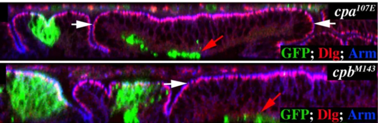
Clonal analysis in the Drosophila wing imaginal disc epithelium revealed a new feature of CP. They are required for the maintenance of cells within the wing blade epithelium (Janody and Treisman, 2006). This is because, mutant cells for either cpa or
cpb in these region suffer basal extrusion and apoptosis (Fig. 1.17). Interestingly, the
requirement for CP to maintain cells within the epithelia is restricted to the wing blade region, since cell extrusion and death caused by loss of CP occurred exclusively in the blade but not in the hinge and notum regions.
Surprisingly, it was found that broad depletion of cpb in the sd-Gal4 expression domain leads to tissue overgrowth and cell migration (Fig. 1.18) (Rebelo, unpublished data). The scalloped driver is express broadly in 1st instar imaginal wing imaginal discs
and this expression gets restricted mainly to the pouch region (Janody, unpublished data). In this experiment, although some cpb-depleted cells expressed activated Caspase 3, most of the cells were able to survive and overproliferate. However, this was not observed in
cpb mutant clones (Fig. 1.17). In a clonal situation, the mutant cells extruded basally and
died as already discussed. All together this data may suggest that cpb mutant clones dye probably due to a cell competition effect, since the cpb mutant cells behave differently, depending on the local environment, in small groups of cells or when the whole tissue is affected.
Figure 1.17 – Clones of mutant cells for cpa and cpb extrude from the wing blade epithelium (adapted from
Janody and Treisman, 2006). Optical cross sections through the wing disc epithelium. Mutant clones are marked positively by GFP (green) and discs are stained with anti-Dlg (red) and anti-Arm (blue) to outline apical membranes. The wing blade region is indicated with white arrows.
Figure 1.18 – Broad deplection of cpb, targeted to sd-expressing cells, leads to overgrowth (adapted from
Rebelo, unpublished). Standard optical section shows third instar wing imaginal disc marked with GFP (green) and stained with anti-Arm (blue) and anti-activated Caspase 3 (red). (A) cpb depletion with the scalloped driver, leads to overgrowth and cell migration.
II – Aims
Like in mammals, Drosophila has six actin genes, closely related to vertebrate cytoplasmic actins. Even though all fly actins are closely related, functional substitution tests have shown that some can compensate for loss of another actin while others cannot (Fyrberg et al., 1998; Wagner et al., 2002). This data suggests that actin genes are not functionally equivalent. For instance, the deleterious lack of cytoplasmic actin 5C can be rescued by reintroducing the coding sequence of the other cytoplasmic actin, actin 42A, under the regulatory control of actin 5C elements (Wagner et al., 2002). By contrast, lack of the indirect flight muscle-specific actin 88F cannot be rescued by expression of other types of actin genes, cytoplasmic or larval (Fyrberg et al., 1998). Also, all actins genes promote excessive actin filaments polymerization when overexpressed, but each actin seems to incorporate more efficiently in specific actin structures (Röper et al., 2005), suggesting that they promote different cytoskeleton structures. Despite this data, it is still not clear, the reason for the existence of multiple actin genes.
Some ABPs regulate specific actin filaments in mammals (Welch and Herman, 2002; Shuster and Herman, 1995), therefore, it may be possible that CP might prevent polymerization of specific actin isoforms to maintained cells within the epithelium.
The main goal of this work is to determine if the phenotype observed when the CP is depleted, which is the extrusion and death of wing blade cells, is a consequence of excessive actin filaments accumulation. To confirm this, it is necessary to test whether the overexpression of any specific actin isoform gives a similar phenotype to the loss of CP. To do so, I analyse here the developmental consequences of overexpressing each of the six actin genes during epithelial morphogenesis. As a model system I will use the epithelium of the wing imaginal discs of Drosophila’s third instar larvae.
In this work I will overexpress all the six Drosophila actin genes fused to GFP using the UAS-Gal4 system and the Gal80 system to drive expression in wing imaginal discs.
III – Materials and Methods
1 – Fly strains and genetics
1.1 – Methods of culturing flies
Fly stocks can be successfully cultured by periodic mass transfer of adults to fresh food in bottles or vials. Since temperature has a large effect on the rate of Drosophila development, flies were raised at 25ºC (unless otherwise indicated) and the crosses were cultured in small vials, under standard conditions. Virgin females are required for successful crosses; D. melanogaster adults do not mate for about 10h after eclosion, allowing virgins to be collected within 8-12h after the culture was cleared of adults (reviewed in Roberts, 1998).
1.2 – Genetics tools
1.2.1 – Fly stocks
Table 3.1 – Fly stocks used for the overexpression experiments of the different actin genes.
UAS lines References
UAS-actin5C-GFP (2-1) Verkhushaet al., 1999
UAS-actin42A-GFP (5-5) Röper et al., 2005 UAS-actin57B-GFP (6-1) Röper et al., 2005 UAS-actin79B-GFP (3-1) Röper et al., 2005 UAS-actin87E-GFP (7-6) Röper et al., 2005 UAS-actin88F-GFP (15-1) Röper et al., 2005
Drivers References
FRT40 or 42D Gal80 Lee and Luo, 2001 nubbin-Gal4 (nb-Gal4) Thompson and Cohen, 2006 daughterless-Gal4 (da-Gal4) Wodarz et al., 1995
engrailed-Gal4 (en-Gal4) Brand and Perrimon, 1993 scalloped-Gal4 (sd-Gal4) Willert et al, 1999
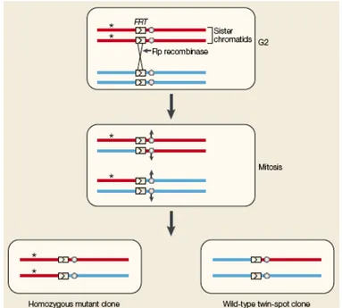
To generate mitotic clones usually the FLP/FRT system is used (Fig. 3.1). In a heterozygous parental cell, the site-specific recombinase FLP induces mitotic recombination between FRT sites on homologous chromosome arms. The FLP is expressed under the control of a heat shock promoter. Segregation of recombinant chromosomes at mitosis produces two daughter cells: a mutant cell bearing two copies of the mutant allele and a wild type cell containing only the wild type form of the gene (reviewed in St. Johnston, 2002).
1.2.3 – The use of the UAS-Gal4 System
The UAS-Gal4 system (Fig. 3.2) allows the selective expression of any cloned gene in a wide variety of cell and tissue specific patterns in Drosophila. A promoter directs expression of the transcriptional activator Gal4, and this in turn directs transcription of the Gal4 responsive UAS target gene. The Gal4 gene and the UAS target are initially separated into two distinct transgenic lines. In the Gal4 line, the activator protein is present, but has no target gene to activate. While in the UAS target gene line, the target gene is silent because the activator is absent. It is only when the Gal4 line is
Figure 3.1 – The FRT/FLP system (reviewed in St. Johnston, 2002).
crossed with the UAS target gene line that the target gene is turned on its progeny (reviewed in St. Johnston, 2002).
To drive the expression of each actin-GFP fusion gene in restricted expression domains in the wing imaginal disc, males carrying either one of the actin isoforms (see Table 3.1) were crossed with transgenic females bearing the following promotors: nb-Gal4, en-nb-Gal4, sd-Gal4 or da-Gal4.
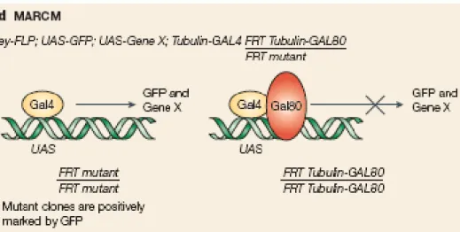
1.2.4 – The use of the MARCM system
The Gal80 system is based in a Gal4 inhibitor, the Gal80, which provides a way of controlling the UAS-Gal4 system (Lee and Luo, 1999). When Gal80 expression is driven with a tubulin promoter (tub-Gal80), it can inhibit the activity of a tubulin promoter-Gal4 construct. If Gal80 is removed, Gal4 is activated and drives the expression of a UAS construct. In the MARCM (Mosaic Analysis with a Repressible Cell Marker) technique (Fig. 3.3), the tub-Gal80 is removed using FRT mediated mitotic recombination (Lee and Luo, 1999). By this system the clones are marked positively
(Brumby and Richardson, 2005).
To generate clones marked positively by GFP in the wing imaginal disc, y-w-;
FRT42D, UAS-actin5C-GFP/CyO,y+ or y-w-; FRT40, actin42A-GFP or
UAS-actin57B-GFP or UAS-actin79B-GFP or UAS-actin87E-GFP or UAS-actin88F-GFP/CyO,y+ males were crossed with y-w-; hsFLP
122, UAS-GFP; FRT40 or 42D,
tub-Gal80; tub-Gal4/TM6B virgin females. To induce clones by using this system, the progeny was heat-shocked for 50min at 37ºC, 24 and 48hs after overnight collection, corresponding to the 1st and 2nd instar larvae (Fig. 3.4).
hs 2 Cross embryogenesis L1 L2 L3 L3 L3 pupae 24h hs 1 hs 2 Dissect Cross embryogenesis L1 L2 L3 L3 L3 pupae 24h hs 1 Dissect Cross embryogenesis L1 L2 L3 L3 L3 pupae 24h embryogenesis L1 L2 L3 L3 L3 pupae 24h hs 1 Dissect
Figure 3.4 – Time course of Drosophila development and the methodology used for clone induction. L1,
L2 and L3 correspond to 1st, 2nd, 3rd instar larvae, respectively. hs: heat shock.
2 – Western blot
2.1 – Embryos extract
To compare the level of overexpression of the six UAS-actin transgenes fused to GFP, y-w-; FRT42D, UAS-actin5C-GFP/CyO,y+ or y-w-; FRT40, UAS-actin42A-GFP or
UAS-actin57B-GFP or UAS-actin79B-GFP or UAS-actin87E-GFP or UAS-actin88F-GFP/CyO,y+ males were crossed with virgin females carrying the ubiquitous da-Gal4
driver. The six crosses, in addition to a control y-w- line, were left to lay overnight on
apple juice plates. Embryos were collected the following day under a GFP stereoscope to select embryos that expressed GFP. Embryos were then washed, immerged in bleach 100% for 5min to remove the chorion and washed thoroughly. Ten embryos were placed in a solution of 10μl 1:1 of SDS loading buffer/PBS. Extracts were kept at -20ºC before using.
2.2 – SDS-PAGE/Gel preparation
Before loading to a 12% polyacrylamide gel (30% acrylamide, Tris pH 8.8, 10% SDS, 10% APS and TEMED) covered by a stacking gel (30% acrylamide, Tris pH 6.8, 10% SDS, 10% APS and TEMED), samples were boiled for 5min, cooled down on ice and centrifuge for 1min at maximum speed. 5μl of embryonic extracts were used for loading. Electrophoresis was performed at 125V in running buffer (144g of glycine, 30g of Tris, 25ml of SDS 20% and H2O per liter).
2.3 – Proteins transfer to PVDF membrane
Following electrophoresis, the polyacrylamide gel was equilibrated in transfer buffer (Tris 480mM and glycine 390mM, 100% methanol, 10% SDS) for 20min. The PVDF membrane was treated with methanol for 10s, washed with water for 5min and equilibrated in transfer buffer for 10min. The transfer cassette was prepared as follows: 1 sponge, 2 Whatman papers, gel, membrane, 2 Whatman papers and 1 sponge. The
2.4 – Membrane staining
To make sure that total proteins were correctly transferred, the membrane was stained with a Ponceau S solution 1 % (w/v) in 5% acetic acid for 5min, then washed for 2min with water and for 20min with PBS, then blocked with 10% milk in PBST (PBS with 0,1% Tween 20) for 1h at room temperature (RT) with agitation to avoid unspecific binding of the antibody and washed again in PBST. The membrane was cut into two pieces, separating the actin-GFP bands from the loading control bands, and each of them were incubated for 1h at RT with shaking with the primary antibodies, diluted in 1% milk in PBST. The rabbit anti-GFP antibody (1:20000; Invitrogene), which recognizes each of the Actin-GFP fusion protein, was applied to the membrane containing proteins of high molecular weight, while the rabbit anti- β3-tubulin antibody (1:2000; Sigma), which reveals the quantity of protein for each extract, was applied to the membrane containing proteins of low molecular weight. Both membranes were then washed with PBST, and in PBST 5% milk and then incubated with the secondary antibody, anti-rabbit peroxidase conjugated with HRP (1:10000; Jackson Immunoresearch) in 1% milk in PBST. The incubation was performed in the dark and as described previously for the primary antibody. The membranes were then washed in PBST 3 times for 15min and washed in PBS for 15min. The kit ECL Plus Western Blotting Detection System (Amersham) was used to reveal the PVDF membrane. According to the kit instructions, a mix of solution A with solution B was added to the membrane for 5min. Storm equipment was used to scan the membrane. The intensity of the bands was quantified using the ImageJ programme.
3.1 – Antibody staining
Third instar larvae wing imaginal discs were dissected in 0,1M phosphate buffer pH 7,2 (72% Na2HPO4 0,5M and 28% NaH2PO4 1M) and fixed in 4% formaldehyde (FA)
in PEM 2x (0,1M PIPES pH 7,0; 2mM MgSO4; 1mM EGTA) for 30min on ice. Discs
were then washed in PBS 0,2% Triton (PBT) for 15min and incubated with the primary antibodies in PBT 10% Donkey serum overnight (ON) at 4ºC. The primary antibodies used in this work were a mouse anti-Armadillo (1:10; Hybridoma Bank) which marks adherens junctions and a rabbit anti-activated Caspase 3 (1:50; Cell Signaling Technology) which stains Caspase-dependant apoptotic cell death. Discs were then washed 3 times for 10min in PBT and incubated in the dark with the secondary antibodies in PBT 10% donkey serum for 2h at 4ºC. Secondary antibodies were an anti-mouse and an anti-rabbit conjugated to TRITC and Cy5 respectively (1/200; Jackson Immunoresearch). Imaginal discs were washed again 3 times for 10min with PBT before mounting between slides and coverslides in Vectashield medium (Vector Laboratories). Fluorescence images were obtained on a LSM 510 Zeiss Meta confocal microscope. In figures, the wing blade and hinge regions size were delimitated by measuring the distance between blade and hinge folding based on the Armadillo staining alone.
3.2 – Phalloidin staining
Third instar larvae wing imaginal discs were dissected and fixed as described in the previous section. This procedure was followed by incubation with Rhodamine-conjugated Phalloidin (Sigma), which stains F-actin, for 4min at 4ºC in the dark. Phalloidin was diluted in PBT 10% donkey serum and used at a concentration of 1:200. Mounting of the wing imaginal discs and image acquisition were performed as previously described.
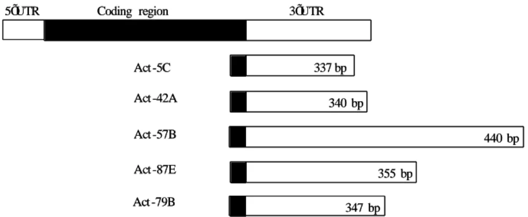
In order to determine the expression pattern of each actin isoform in the wing imaginal discs, I used common molecular biology techniques to create specific actin RNA probes. The cDNA sequences of the six actins are more or less 85% homologous to each other, while untranslated regions, comprising the 3’UTR are divergent (Fyrberg et
al., 1981). RNA probe, generated against the entire cDNA of one particular actin will
likely cross hybridize with all other actin RNA. Therefore, in order to generate RNA probes that recognizes specifically each actin isoform. I used the most divergent cDNA fragments for each gene, comprising the 3’UTR untranslated sequences, to subclone. The selected subcloned fragments and their sizes are represented in Fig. 3.5.
Coding region 5ÕUTR 3ÕUTR Act -5C Act -42A Act -57B Act -87E Act -79B 337 bp 340 bp 440 bp 355 bp 347 bp Coding region 5ÕUTR 3ÕUTR Act -5C Act -42A Act -57B Act -87E Act -79B 337 bp 340 bp 440 bp 355 bp 347 bp
Figure 3.5 – Representative diagram of transcribed region of each actin gene. In black is the coding region.
In white is represented the 3’UTR, the selected fragment used to subclone (adapted from Fyrberg et al.,1983).
The probe for the actin 88F gene was not made because there was not any cDNA clone available at the Drosophila Genomics Resource Center (DGRC).
4.1 – Classical techniques of Molecular Biology used in this study
4.1.1 – Preparation of competent cells
A pre-culture of DH5α cells was grown in LB medium, ON at 37ºC and inoculated the following day to a large volume of LB medium. The culture was incubated at 37ºC until the OD600 reached a density of 0,5. The culture was then placed on ice for
10min and centrifugated for 10min at 4ºC. The pellet was resuspended in a 100mM CaCl2
solution and incubated on ice for 20min, centrifugated again for 10min at 4ºC and resuspended in a 100mM CaCl2 solution supplemented with glycerol at 10%. Cells were
immediately frozen on a dry ice bath with ethanol and stored at -80ºC.
4.1.2 – DNA extraction (minilysats)
Transformed colonies were incubated in LB medium with 50μg/ml of Ampicillin or 2μl/ml of Chloranphenicol (see Table 3.2), ON at 37ºC. The culture was centrifugated for 2min at RT. Each pellet was ressuspended with 300µl of TS (50mM Tris pH 7,5 and 25% Sucrose) and mixed with 300µl of ELT (100mM EDTA, 2mg/ml Lysosyme and 0,1% TritonX100). The mixture was incubated 10min at RT and placed 10min at 70ºC before centrifugating for 15min at 4ºC. Pellets were discarded. Supernatants were then incubated with 550µl of 20% PEG6000, 1M NaCl for 30min at RT before centrifugation for 4min at high speed, to precipitate the plasmid. The pellets were then washed with 70% ethanol and ressuspended in TE 1x. Each minilysat was digested with the appropriate restriction enzyme and ran in an electroforese gel to confirm the identity of the plasmid amplified.
4.1.3 – DNA purification (phenol/chloroform method)
the mix was centrifugated for 1min to restore the two phases: Phenol (bottom phase), versus aqueous DNA solution, while contaminant proteins are left at the interphase (top phase). The DNA solution was then transferred to a new eppendorf tube, to which one volume of chloroform solution was added, forming two new phases. The mix was vortexed and centrifugated for 1min and the DNA solution was transferred to a new eppendorf tube. This step was repeated 3 times to remove any residual phenol. The purified DNA solution was then precipitated with three volumes of ethanol 100% supplemented with Ammonium Acetate to a final concentration of 2M. This mix was vortexed and centrifugated at 4ºC for 30min. The supernatant was discarded and the DNA pellet washed with 70% ethanol, was dried and ressuspended in TE 1x. The concentration of DNA was measured using the Nanodrop system.
4.2 – Cloning of the cDNA sequence used to generate a RNA
specific probe for each actin gene.
4.2.1 – Transformation of competent cells with clone containing the cDNA for each actin gene
Clones containing the entire cDNA for each actin gene were ordered to the DGRC (see Table 3.2).
Table 3.2 – List of cDNA clones used to subclone for each actin gene. Each cDNA clone has a specific
host vector and an antibiotic resistance gene.
Actin gene cDNA clone Host Vector Antibiotic resistance
Actin 5C GOLD RE 02927 pFLC-I Ampicillin
Actin 42A GOLD LD 18090 pBlueScript_SK(-) Ampicillin
Actin 57B EST LD 04994 pBlueScript_SK(-) Ampicillin
Actin 79B GOLD GH 04529 pOT2 Ampicillin
Actin 87E EST AT 14584 pOTB7 Chloranphenicol
with DH5α competent cells. Cells were heat shocked for 2min at 37ºC, transferred to 1ml of LB medium and incubated at 37ºC for 1h. Cells were then plated on LB supplemented with the appropriate antibiotic and incubated ON at 37ºC.
4.2.2 – PCR
In order to amplify the 3’ UTR of each actin gene, 3’ and 5’ primers that flank the region of interest were designed. PCR primers generally range in length from 15-30 bases and should contain 40-60% of G + C (Promega Technical Guide). Primers designed for each actin gene are described in Table 3.3.
Table 3.3 – List of 5’ and 3’ primers used to amplify each actin 3’UTR sequences.
Gene Primer Sequence (5’→ 3’)
Actin 5C Act5C (3’UTR5’)Act5C (3’UTR3’) GTAGTTCTTGTTATATTAAAGGGAAGGATCGCTTGTCTGG Actin 42A Act42A (3’UTR5’)Act42A (3’UTR3’) TTATTAAGAAGCGGTTACAAGGCAGTAGTCGGGCTGGG
Actin 57B Act57B (3’UTR5’) GCAGTTGCAGTTGCCTAG
Act57B (3’UTR3’) CAATCATGAGATTATTTTACAT
Actin 79B Act79B (3’UTR5’) GCATCCAAGCCACCCAAA
Act79B (3’UTR3’) CACATGGGTTATTGGTTTTTA
Actin 87E Act87E (3’UTR5’) GCGATCTAAACACCACAGA
Act87E (3’UTR3’) AGATTTAATTTATAGAGGATGC
To perform the PCR, GoTaq buffer 5x, MgCl2 solution 25mM, PCR nucleotide
mix 10mM, upstream primer, downstream primer, GoTaq polymerase 5u/µl, DNA template and nuclease free water were mixed on ice. The PCR was performed in a thermal cycler with the following parameters: an initial 2min denaturating cycle at 95ºC, 35 cycles that included 1min of denaturation at 95ºC, 1min of annealing at 58ºC and 1min of extension at 72ºC, a final extension cycle of 5min at 72ºC and 1 cycle of soak at 4ºC for indefinite time.
4.2.3 – PCR product purification
Purification of each PCR product was performed using E.Z.N.A Gel Extraction Kit (Omega bio-tek). According to the kit instructions, the PCR product was separated by electrophoresis on an agarose gel. The DNA fragment of interest was excised using a UV light box. The gel slice weight was measured and binding buffer was added in a ratio of 10µl buffer: 10mg agarose gel slice. The mixture was incubated at 55-60ºC for 7min or until the gel was completely melted and centrifuged briefly at RT. The dissolved gel slice was applied to HiBind DNA spin-column and centrifugated for 1min at 14000rpm. Binding buffer was added to the column and centrifuged for 1min. The column was washed twice with the SPW buffer and centrifugated at RT for 1min. The column was centrifugated for 1min to dry the column matrix. Elution buffer was applied directly to the centre of the column to elute the DNA. The eluted DNA was kept at 4ºC or -20ºC.
4.2.4 – Ligation within the pGEM-T easy vector
The pGEM-T easy vector (Fig. 3.6) is a convenient system for cloning PCR products. The vectors are prepared by cutting with EcoRV and adding a 3’ terminal thymidine to both ends. This, greatly improves the efficiency of the ligation of a PCR product into the plasmids by preventing recircularization of the vector and providing a compatible overhang for PCR products generated by certain thermostable polymerases (Promega technical manual).
Figure 3.6 – Schematic representation of pGEM-T easy vector with restriction sites (adapted from
Promega technical manual).
In order to ligate each PCR product corresponding to each actin isoforms 3’UTR sequence with the referred plasmid, purified PCR products were mixed with the vector in a 3 to 1 ratio, with 3 weiss/µl of T4 DNA ligase and 2x ligation buffer. The reaction was incubated for 1h at RT or ON at 4ºC.
4.2.5 – Transformation of competent cells with pGEM-T easy vector
One of the features of the pGEM-T easy vector (Promega) is the blue/white screening. The T7 and SP6 RNA polymerase promoters flank a multiple cloning region within the α- peptide coding region for β-galactosidase. Insertional inactivation of the α- peptide allows recombinant clones to be directly identified by colour screening on indicator plates (Promega technical manual). In order to amplify the DNA, the ligation reaction was mixed with DH5α competent cells and incubated on ice for 20min. Afterwards, a 90s incubation at 42ºC was done. The reaction was placed on ice for 2min. LB medium was added and incubated for 50min at 37ºC. XGal 20mg/ml and IPTG 0,8M were added to the LB Ampicillin (50µg/ml) plates. The culture was plated and incubated ON at 37ºC. The blue colonies (colonies with the insert) were picked and their DNA was extracted by minilysat method (described previously). To confirm the insertion, an
agarose gel electrophoresis was run. The resulted DNA solution will be used to perform specific probes.
5 – In situ hybridization
Since RNA probes are very sensitive to degradation by RNases, the water used for the whole in situ procedure was sterile and render RNase-free by a treatement with DEPC (Pyrokohlensaurediethylester 97%, diethyl pyrocarbonate 97%).
5.1 – Probe synthesis
DNA templates were either linearized by digestion with the SpeI enzyme or with the SacII enzyme and then purified via phenol/chloroform extraction (see Section 4.1.3). The DNA template linearized with SpeI enzyme was transcribed with the T7 RNA polymerase to produce the sense probe and the DNA template linearized with SacII enzyme was transcribed with the Sp6 RNA polymerase to produce the anti-sense probe. The DIG RNA Labeling Kit (SP6/T7) (Roche) was used to produce the RNA probes labelled with digoxigenin-UTP. RNA labelling reaction was performed with 1μg of purified DNA template. To perform the transcription reaction, 10x NTP labelling mixture, 10x transcription buffer, protector RNase inhibitor, RNA polymerase SP6 or T7 and water were added to the DNA template, on ice. The reaction was incubated for 2h at 37ºC and centrifugated briefly. To confirm that transcription of each RNA probe was efficient, 1µl of each transcription sample was run on a 2% agarose gel. Each probe was then broken in pieces with a solution of 2x carbonate buffer at pH 10,2 (120mM Na2CO3
and 80mM NaHCO3). The reaction was performed at 65ºC for 40min and stopped with a
solution of 3M NaAc at pH 6,0. Each probe was then precipitated ON at -20ºC by adding 4M of LiCl, 20mg/ml of E. coli tRNA in 100% ethanol and centrifugated at 4ºC for 45min. The pellet was washed in 70% ethanol, centrifugated at 4ºC for 30min,
resuspended in water and stored at -20ºC.
5.2 – Fixation of imaginal discs
Flies used for the in situ hybridization experiment were yw-. Third instar larvae
wing imaginal discs were dissected in PBS and fixed in PBS, 4% FA, 67mM EGTA for 30min. Discs were washed several times in methanol. Afterwards, discs were washed with ethanol 100%.
5.3 – Hybridization
Discs were washed for 60min with a solution of 50:50 of xylene/ethanol in a glass vial and then rinsed several times with ethanol and at the end with methanol. Discs were fixed for 5min with a 50:50 solution of 5% FA in methanol/PBT and fixed for 30min with 5% FA in PBT. After washing with PBT, the discs were incubated for 8-10min in 20mg/ml proteinase K. Discs were washed with PBT. Afterwards, discs were fixed with 5% FA in PBT for 30min at RT or ON at 4ºC and washed 10min with a solution of 50:50 of PBT/hybridization solution. Hybridization (hyb) solution is 50% formamide, 5x standard saline citrate (SSC), 100μg/ml heparin, 0,1% Tween 20 and 100μg/ml boiled salmon sperm DNA (ssDNA). Discs were incubated for 3hs in hyb solution at 55ºC. Discs were hybridized in 2μl of probe diluted in 60μl of hyb solution for 18-20hs at 55ºC. Rinsed with hyb solution and incubated in the same solution for 3hs at 55ºC.
5.4 – Pre-absortion of anti-digoxigenin (anti-DIG) antibody
y-w- embryos were collected after an ON collection, washed with water and
bleached for 5min. Then, the embryos were washed thoroughly. Embryos were fixed in a solution of 1:1 of PBS and heptane with 4% FA for 30min and washed in a 1:1 solution of methanol and heptane. Then, embryos were washed 3 times with methanol and 3 times
estimated. For 1 volume of embryos, a volume of anti-DIG antibody (Jackson ImmunoResearch) and 4 volumes of PBT were added. Embryos were incubated ON at 4ºC with shacking and centrifuged 5min at 4ºC. 1μl of azide 10% was added to 500μl of supernatant.
5.5 – Anti-DIG alkaline phosphatase reaction – Staining Reaction
Discs were successively washed at RT with decreasing series of hyb solution: 75% hyb in PBT, 50% hyb in PBT, 25% hyb in PBT and with PBT for 1h. Discs were incubated with the anti-DIG antibody, previously pre-absorbed ON at 4ºC at a 1:2000 final dilution. The following day, discs were washed several times for 1h in PBT before a final wash with the staining buffer (0,1M NaCl, 0,05M MgCl2, 0,1M Tris-HCl pH9,5 and
0,1% Tween 20). Revelation was performed in the dark with 20μl of NBT/BCIP solution (Roche) in the staining solution. To follow the staining reaction, discs were observed under a microscope from time to time. The reaction was stopped by 2 washes with the staining buffer and then rinsed with PBT and several times with ethanol 100%. Discs were then rehydrated in ethanol series in PBT and mounted in glycerol 80%.
IV – Results
It was shown by Janody and Treisman that mutant cells for CP induce excessive actin filaments polymerization. In this study, I want to investigate if the phenotype observed when the CP is lost is a consequence of excessive actin filaments accumulation. To do so, I used an overexpression approach, instead of a loss of function, to get more insights on whether each Drosophila actin genes have specific functions. In order to determine whether the overexpression of any specific actin isoform gives a similar phenotype to the loss of CP, which is the extrusion and death of wing blade cells, I analysed the developmental consequences of overexpressing each of the six actin genes during epithelial morphogenesis. To achieve this goal, I generated recombinant
Drosophila lines, between transgenes that encode for either of the six actin genes fused to
green fluorescent protein (GFP) and placed under the control of UAS-sites and FRT-sites. Afterwards, I expressed either of the six actin-GFP genes using the UAS-Gal4 (Brand and Perrimon, 1993) and the Gal80 (Lee and Luo, 2001) systems to drive expression in wing imaginal discs.
1 – All fly actin genes are overexpressed at a comparable
level.
Each actin cDNA fused to GFP was inserted into the Drosophila genome randomly by P-element transformation. Due to site specific chromatin organization, the accessibility to the UAS-sites, upstream of each actin-GFP target gene, can vary from one transgenic line to another, leading to different expression levels between each one. Since I want to determine whether any of the six actin isoforms, when overexpressed, gives similar phenotypes to the loss of CP and compare the behaviour of cells
Actin-GFP fusion protein.
To do so, I crossed females carrying the daugtherless-Gal4 (da-Gal4) driver to males bearing either one of the actin-GFP constructs. The da-Gal4 driver will express each target gene ubiquitously in the ectoderm. Embryos resulting from these crosses were collected to produce protein extracts that were analysed by Western Blot (see Materials and Methods). The membrane was blotted with an anti-GFP antibody that revealed a band of around 70KDa in each extract, while control y-w- embryonic extracts showed no
signal (Fig. 4.1). The intensity of the GFP signal for each Actin-GFP isoform, although showed small variations between each other, these variations did not appear to be significant (Fig. 4.1A).
To confirm that the quantity of the samples loaded into the gel were equal, I stained the same blot with an anti-β3 Tubulin antibody, revealing a band of around 55KDa for all embryonic extracts (Fig. 4.1 B). I compared the intensity of each band using the Threshold function of the ImageJ program. The ratio between the intensity of the signal given with the anti-β3 Tubulin antibody and the anti-GFP antibody for each genotype allowed me to compare the expression level for each Actin-GFP fusion protein (Chart 4.1).
Figure 4.1 – Western blot from Drosophila embryonic extracts, expressing each of the six Drosophila actin
genes fused to GFP under the control of da-Gal4 driver. The first lane indicates Drosophila embryonic extracts from y-w- embryos that do not express GFP. (A) Blot marked with the anti-GFP antibody that reveals each of the Actin-GFP fusion proteins. (B) Same blot marked with the Anti-β3 Tubulin antibody used as a reference to evaluate the quantity of loading extracts.
0.40 0.60 0.80 1.00 1.20 1.40 o ac tin -G F P BI/ β 3 tu b BI
Chart 4.1 also indicates that the intensity of the GFP staining for each Actin-GFP fusion protein has some variations and despite the fact that the major variation observed in Chart 4.1 is 0,74 fold, between the Actin 42A-GFP and Actin 79B-GFP overexpression, I assumed that those variations were not significant.
Therefore, the consequences of overexpressing each of the actin-GFP genes in disc epithelial tissues can be compared regardless of the different expression levels.
2 – Actin isoforms incorporate in filaments at various
subcellular locations within the wing disc when overexpressed.
The six Drosophila actin isoforms, when ectopically expressed, have the ability to associate with certain structures formed by endogenous actins, such as filopodia/lamellipodia, cortical actin, basal stress fibers, ring canals, actin struts, larval/adult muscles and Z-lines (Röper et al., 2005). Although the difference in the efficiency of incorporation into these structures is small, the different actin isoforms behave far from identically (Röper et al., 2005). In order to determine whether each actin-GFP isoform, when overexpressed in the wing imaginal disc, incorporates within actin filaments in specific subcellular locations, I overexpressed each of the 6 actin genes fused to GFP, by using the engrailed-Gal4 (en-Gal4) driver. Since en-Gal4 drives the
Chart 4.1 – Graph showing the ratio between the intensity of the signal obtained with the anti-β3 Tubulin antibody and the anti-GFP antibody for each genotype.Blue indicates cytoplasmic actins, yellow indicates larval muscle actins and pink indicates adult muscle actins.
expression of the UAS-target gene in the posterior compartment of the wing discs during all stages of larval development (Fig. 4.4 A), the anterior compartment can be used as a wild type control. Discs were stained with Phalloidin to reveal actin filaments (Fig. 4.2 A’-L’), and GFP revealed expression of each actin-GFP isoform in the posterior compartment (Fig. 4.2 A-L).
As expected, overexpressing either of the 6 actin-GFP isoforms promote excessive actin filaments formation in the posterior compartment of the wing imaginal disc (Fig. 4.2). Interestingly, actin filaments were observed to accumulate in different locations within the cell, depending of the Actin-GFP isoform overexpressed. For instance, Actin 5C-GFP (Fig. 4.2 G’) and Actin 57B-GFP (Fig. 4.2 I’) were mostly incorporated in filaments formed at the basal surface of wing disc epithelium. In contrast, Actin 42A-GFP (Fig. 4.2 B’), 79B-GFP (Fig. 4.2 D’), 87E-GFP (Fig. 4.2 E’) and 88F-GFP (Fig. 4.2 F’) were mostly incorporated in actin filaments at the apical surface of the wing disc epithelium. Further analysis of the F1 progeny of this set of experiment will be analysed in following sections.
This data shows that in the wing imaginal discs, each actin isoform, when overexpressed, might get incorporated in specific actin filaments structures within the cells. All together, this suggests that while the actin isoforms have very similar sequences, differing in only a few aminoacid residues, each one have specific cellular properties.
Figure 4.2 – Incorporation of the different actin isoforms in filaments in various locations of the wing disc. All panels
show standard confocal sections of third instar wing imaginal discs marked with GFP in green, which reveals each of the Actin-GFP fusion protein, and Phalloidin in red, which reveals F-actin. (A-F’) Apical sections. (G-L’) Basal sections. (A-L) Merged. (A’-L’) Phalloidin staining alone. The white arrows in G’, B’, I’, D’, E’, and F’ indicate strong actin