The role of Actin-Capping Protein and Src
signalling in tissue
growth and apoptosis during Drosophila wing
development
Barbara Zofia Jezowska
Dissertation presented to obtain a Ph.D degree in Developmental Biology
Instituto de Tecnologia Química e Biológica | Universidade Nova de Lisboa
Research work coordinated by:
This dissertation was sponsored by Fundação para
a Ciência e Tecnologia.
Apoio financeiro da FCT e do FSE no âmbito do
Quadro Comunitário de apoio, BD nº
D
ECLARAÇÃO
Esta dissertação é o resultado do meu próprio trabalho desenvolvido entre Outubro de 2007 e Outubro de 2011 no laboratório da Doutora Florence Janody, no Instituto Gulbenkian de Ciência, em Oeiras, Portugal, no âmbito do Programa Doutoral do Instituto Gulbenkian de Ciência PGD2007. Todas as colaborações estão indicadas em
cada capítulo, nas secções de ‘Material and Methods’ e ‘Acknowledgments’. Os
resultados do Capítulo 3 representam o artigo publicado:
Jezowska, B., Fernández, G.-F.B., Amândio, A.A., Duarte, D.P., Mendes, C., Brás-Pereira, C., Janody, F., 2011. A dual function of Drosophila capping protein on DE-cadherin maintains epithelial integrity and prevents JNK-mediated apoptosis. Developmental Biology.
Os resultados do Capítulo 4 estão a ser compilados num artigo, que será submetido brevemente.
A
CKNOWLEDGMENTS
/A
GRADECIMENTOS
First I would like to give a special thanks to my supervisor Florence Janody. She was always supporting, understanding when there were difficult moments in my live. She helped me to develop my ideas. She really showed me that she care about me and my scientific development. She is a fantastic Scientist and great person.
To Prof. António Coutinho and the directors of the IGC PhD program Henrique Teotónio and Thiago Carvalho for giving me an oprtunity to be a part of the program. Thank you!
To Beatriz – hope you know that I would “drawn in the spoon of water” if it
wouldn´t be you?! You are great friend that understand falls but helps to stand up and improve. Friend that is not afraid to tell you the truth. Beatriz you are amzing, strong and beautiful person. Keep on going and be a filer for the next generations of students in our lab! Gracias a mi amiga!
To Catarina that was helping me with the Portuguese parts of my Thesis and prepared
amazing “Jar” that that did not allow me to complain for few days!
To the all past end present members of the laboratory for help and great atmosphere!
To Sofia that is sweet and funny and let me into the gropu of her friends at IGC……which
was very important for me! To Mauricia that for being a good companion and for always caring about little things that most of us would miss! To Gaspar that is incredible smart, patient and happy to share his knowledge.
To my colleagues from PGD2007 –You are fantastic.
To Clara, fantastic friend and that understand me very well! I am so happy that I met you Clara!
To Roberto that helped me to deal with the Ilustrator and that become my friend! To Krzysztow and Ewa, Krzystow helped me with all the problems concerning computers and Ewa was making me laughing when I was getting too serious.
To Gaston that helped me with cloning experiment at the very beginning, when I was still learning how is to be a student.
To Inês that gave me great help at the end of my Thesis.
To Sara and Pedron, Emilia, Sofia, Ana Teresa, Francisca (Chica) that are just excellent, funny, lively and supportive.
To Matteo the best person in the whole world that is leaving everything whenever I need his help! That makes me feel that everything is OK! You are very Powerful Matteo (Coccinella) !! You amaze me everyday!
To my Mum and Dad- Dziękuje! Mamo i Tato- Wy zawsze we mnie wierzyliście! Dzięki wam nauczyłam się być dzielną i mądra! Tato- wierzę, że dobrze wiesz o mnie wszystko, wierzę że jesteś ze mnie bardzo dumny!
To my all family! Dziękuje Ani -mojeje siostrze, za to że zawsze mnie słuchałaś a czasami sprowadzałaś na ziemię. Dziękuje Maćkowi za wsparcie, Ani, żonie Maćka która jest przesłodka i sprawia że wszystko wydaje sie łatwiejsze! Dziękuje, cioci Danusi- za to że
jest!
S
UMMARY
The actin cytoskeleton controls numerous cellular processes, including cell morphology and polarity, endocytosis, intracellular trafficking, contractility and cell division. Actin filament growth, stability and disassembly are controlled by a plethora of actin-binding proteins. Among them Capping Protein is a highly conserved αβ heterodimer, which binds the barbed ends of actin filaments, inhibiting addition or loss of actin monomers. Loss of Capping Protein results in the accumulation of the excessive actin filaments. Interestingly, cells mutant for Capping Protein display a tissue-specific behaviour in the Drosophila wing imaginal disc. In the most distal domain, loss of Capping Protein triggers actin filaments accumulation around the entire cell cortex, cell extrusion and apoptosis. While in the proximal domain, mutant cells maintain a polarized epithelial architecture and accumulate F-actin at the apical cell surface. These observations argue that Capping Protein regulates diverse populations of actin filaments in the cell that have tissue-specific functions.
However, Yki-dependent increase of DE-Cadherin levels is unlikely to be sufficient to trigger apoptosis of cells knocked down for Capping Protein in the distal wing disc domain since mutant clones for Hippo pathway components also accumulate DE-Cadherin but do not extrude from the wing disc epithelium. In addition to preventing transcription of DE-cadherin, Capping Protein maintains DE-cadherin localization specifically in the most distal wing disc domain. The dual effect of Capping Protein loss on DE-Cadherin is likely sufficient to trigger the elimination of mutant cells, preventing them from proliferating (Chapter 2) (Jezowska et al., 2011).
With the goal of understanding how Capping Protein maintains the localization of DE-cadherin at Adherens Junction, I observed that increased levels of the Src oncogene in the distal wing disc epithelium fully recapitulate the outcomes of cells lacking Capping Protein. These observations suggest that in this domain, Capping Protein function may restrict Src signalling activity. Indeed, I found that Capping Protein prevents Src signalling activity downstream of the btk family kinase at 29A and inhibits Src phosphorylation. My results suggest that the balance between activated Src and Capping Protein is critical to control Src signalling activity. I propose that misregulation of actin cytoskeletal genes, such as Capping Protein, contributes to tumorigenesis through Src signalling, whose expression and activity becomes progressively elevated in a broad spectrum of cancers (Chapter 3).
S
UMÁRIO
O citosqueleto de actina controla inúmeros processos celulares, nomeadamente morfologia celular e polaridade, endocitose, tráfego intracellular, contractilidade e divisão celular. O crescimento, a estabilidade e o desagregação dos filamentos de actina são controlados por uma pletora de proteínas que se ligam à actina. Entre elas, a Capping Protein é um heterodímero altamente conservado, que se liga aos ‘barbed’
terminais dos filamentos de actina, inibindo a adição ou perda de monómeros de actina. A perda de Capping Protein resulta na acumulação excessiva de filamentos de actina. É interessante notar que células mutantes para Capping Protein exibem um comportamento específico do tecido em que estão inseridas no disco imaginal da asa de Drosophila. No domínio mais distal, a perda de Capping Protein desencadeia acumulação de filamentos de actina em todo o córtex celular, bem como extrusão celular e apoptose. No domínio proximal, as células mutantes mantêm uma arquitetura epitelial polarizada e acumulam filamentos de actina na superfície apical da células. Estas observações sugerem que a Capping Protein regula diversas populações de filamentos de actina na célula, que têm funções específicas em cada tecido.
cadherin proporciona um sinal activo, que impede a sinalização por Wingless e promove a apoptose mediada por JNK das células sem Capping Protein. Porém, quando essas células são mantidas vivas, a actividade da via de sinalização de JNK e Yki, um oncogene, provoca proliferação massiva. No entanto, o aumento dependente de Yki dos níveis de DE-cadherin é improvável que seja suficiente para provocar apoptose das células mutantes para Capping Protein no domínio distal do disco da asa, uma vez que clones mutantes para componentes da via de sinalização de Hippo também acumulam DE-cadherin, mas não extrudem do epitélio do disco da asa. Alem de prevenir a transcrição de DE-cadherin, Capping Protein mantem a localização da DE-cadherin especificamente no domínio mais distal do disco da asa. O duplo efeito da perda de Capping Protein na DE-cadherin é provável que seja suficiente para promover a eliminação das células mutantes, evitando que proliferem (Chapter 2) (Jezowska e tal., 2011).
Com o objectivo de compreender como Capping Protein mantem a localização da DE-cadherin nas Junções Aderentes, verifiquei que níveis elevados do oncogene Src em 64B no epitélio distal do disco da asa reproduz completamente os mesmos resultados de células sem Capping Protein. Estas observações sugerem que, neste domínio, a função de Capping Protein poderá limitar a actividade de sinalização de Src. De facto, descobri que Capping Protein previne a actividade de sinalização de Src a jusante da cinase da família Btk em 29A e inibe a fosforilação de Src. Os meus resultados sugerem que o equilíbrio entre Src activado e Capping Protein é crucial para o controlo da actividade de sinalização de Src. Assim, proponho que a desregulação dos genes do citosqueleto de actina, como a Capping Protein, contribui para a tumorigenese através da sinalização de Src, cuja expressão e actividade torna-se progessivamente elevada num largo espectro de cancros (Chapter 3).
L
IST OF
A
BBREVIATIONS
ABPs Actin Binding Proteins ADF Actin depolymerizing factor AJ Adherens JunctionsAP Anterio-posterior
Arm Armadillo (βCatenin)
Arp 2/3 Actin-related protein complex 2/3 ARPC Arp complex
bp base pair
Bsk Basket
CAP Adenylate Cyclase Associated Protein
Cora Coracle
CP Capping Protein
Cpa Capping Protein α
Cpb Capping Protein β
Csh Csk kinase homolog Csk C-terminal Src kinase
CycE CyclinE
DAAM Dishevelled-associated activator of morphogenesis
Dia Diaphanous
ECM Extracelular matrix EGF Epidermal growth factor
EGFR Epidermal growth factor receptor EMT Epithelial to mesenchymal transition
Ex Expanded
FA Focal Adhesion
F-actin filamentous actin FAK Focal Adhesion Kinase
FH Formin-homology
FJ Four-jointed
Frz Frizzled
Frz2 Frizzled 2
Hep Hemipterous
Hpo Hippo
JNK Jun N-terminal Kinase JNKK Junk kinase kinase JNKKK Jun Kinase kinase kinase Jra Jun-related antigen
Kay Kayak
Lats1/2 Large tumor suppressor 1 and 2 Lgl Lethal giant larvae
MAD Mother against Dpp MAP Mitogen activated pathway MAST1/2 mammalian STE-20 kinase 1 and 2 Mats Mob as tumor suppressor
mDia murine Diaphanous
Med Medea
Mob Msp-one-binder Neu IV Neurexin IV
NPFs nucleation promoting factors
N-WASP neuronal Wiskott–Aldrich syndrome protein Omb Optomoto-blind
PCR Polimerase chain reaction PKC Protein kinase C
pMAD phosphorylated MAD RNAi RNA interference
RT Room temperature
Salm Spalt
Sav Salvador
Scrib Scribble
Sd Scalloped
SJ Septate junctions
Src Rous sarcoma virous TGF Transforming growth factor
Tkv Thickveins
TSG Tumor supresor gene
Vg Vestigial
WASP Wiskott–Aldrich syndrome protein WAVE WASP-family veroprolin homolog
Wts Warts
T
ABLE OF CONTENTS
Declaração ... I Acknowledgments/Agradecimentos ... III Summary... V Sumário ... VII List of Abbreviations ... XI Chapter 1 – General Introduction ... 1 Epithelial tissue organization ... 3 1.
1.1. Apico-basal cell polarity... 3 1.2. Organization of Adherens Junctions components ... 6 1.3. Organization of cell -matrix adhesion structures ... 7 1.4. The Cross-talk between Adherens Junctions and cell-matrix adhesion structures... 9
Actin filaments organization and dynamics ... 12 2.
Nucleation of actin filaments ... 13 2.1.
Depolymerization of actin filaments and sequestration of actin monomers 15 2.2.
Termination of actin filaments ... 15 2.3.
The wing imaginal disc as a model system ... 21 4.
Structure of the wing imaginal disc ... 21 4.1.
Patterning of the wing imaginal disc ... 22 4.2.
Wing imaginal disc as a model to study tissue growth and homeostasis ... 25 4.3.
Tumorigenesis as an example of the loss of epithelial tissue homeostasis ... 30 5.
The hallmarks of epithelial derived cancers ... 30 5.1.
Drosophila as a model to study tumorigenesis, advances and limitations ... 32 5.2.
Tumour suppressors genes and oncogenes in Drosophila epithelial tissues 33 5.3.
The components of Hippo pathway as examples of tumour suppressor 5.4.
genes 34
Role of JNK signalling in epithelial-derived tumours ... 35 5.5.
Contribution of loss of cell polarity and adhesion to tumorigenesis ... 36 5.6.
Src as an example of oncogene ... 37 5.7.
References ... 41 6.
Chapter 2 – Capping Protein restricts apical actin filaments evenly in a whole wing imaginal disc ... 49
Summary ... 51 1.
Introduction ... 52 2.
Material and Methods ... 54 3.
Generation of UAS-cpaWT and UAS-cpaΔABD transgenic flies ... 54
3.1.
Design and amplification of cpaWT and cpaΔABD ... 54
3.2.
Fly husbandry ... 55 3.3.
Antibody staining ... 56 3.5.
Imaging ... 56 3.6.
Results ... 56 4.
Capping Protein maintains components of Adherens Junctions in the distal 4.1.
wing disc epithelium ... 56 Capping Protein prevents apical actin filaments accumulation and does not 4.2.
seem to affect the F/G actin ratio ... 61 The apical localization of Capping Protein depends on its ability to bind to 4.3.
actin filament ... 64 Capping Protein prevents the accumulation of three distinct pools of apical 4.4.
actin filaments ... 67 Capping Protein may indirectly contribute to Focal adhesions organization70 4.5.
Discussion and conclusions ... 72 5.
References ... 76 6.
Chapter 3- A dual function of Drosophila Capping Protein on DE-cadherin maintains Epithelial integrity and prevents JNK-mediated apoptosis ... 79
Author contribution ... 81 1.
A dual function of Drosophila Capping Protein on DE-cadherin maintains 2.
Material and methods ... 117 3.
Fly husbandry ... 117 3.1.
Mutants and Drosophila strains used ... 117 3.2.
Antibody staining ... 118 3.3.
Western Blotting ... 118 3.4.
Imaging ... 119 3.5.
Results ... 119 4.
Capping Protein loss or Src overexpression affects identical target genes, 4.1.
disrupts polarity and induces apoptosis ... 119 Capping Protein and Src64B control the balance between tissue growth and 4.2.
apoptosis. ... 124 Loss of Capping Protein cooperates with Src64B in the distal wing disc 4.3.
epithelium ... 129 DE-Cadherin promotes apoptosis and prevents growth of Src64B 4.4.
overexpressing tissue ... 130 Capping Protein acts downstream of Src64B ... 133 4.5.
Capping Protein is epistatic to Btk29A ... 135 4.6.
Capping Protein acts in a feedback loop mechanism to attenuate Src activity 4.7.
136
The balance between activated Src and CP is critical for tissue homeostasis 4.8.
139
Discussion and Conclusions ... 144 5.
Acknowledgments and author contribution ... 148 6.
Chapter 5 – General Discussion ... 155 1. Capping Protein regulates multiple signaling pathways ... 157 1.1. Role of Capping Protein in Hippo signalling pathway ... 157 1.2. Capping Protein regulates Wg, JNK and Src signalling ... 157 1.3. Interplay between Hippo, Src, JNK and Wg signalling ... 160 1.4. Molecular and cellular roles of Capping Protein in Hippo and Src signalling
161
EPITHELIAL TISSUE ORGANIZATION
1.
1.1.
A
PICO-
BASAL CELL POLARITYFigure 1.1 Diagrams representing the major complexes that maintain apical-basal polarity and adhesion in Drosophila epithelial tissue.
(A) Schematic representation of the apical, basal and lateral domains in epithelial cells. The major complexes are depicted with these color associations: subapical complexes (orange) AJs (green), SJs (blue) and Fas (grey. The apical actin belt associated with AJs and FAs is represented as pink helixes. (B) A cartoon representing the global regulatory network of apical and basolateral polarity complexes in Drosophila epithelial tissue (adopted from Laprise and Tepass 2011). Apical polarity proteins (Crb, Patj, CdC42, Par6, αPKC, and Baz) and AJs show mutually supportive interactions (represented in green), while apical factors and the basolateral polarity proteins (Lgl, Dlg, Scrib, Par1, Yrt, Cora, NrxIV, Na+/K+ ATPase) interact antagonistically (represented in red). The apical and basolateral membrane are represented in magenta and yellow, respectively. (C) Cartoon representing the major components of AJs and their organization (adapted from (Pinho et al., 2011). The E-cadherin from adjacent cells is involved in homophilic interactions via
extracellular domains. The intracellular domain of E-caderin form a complex with Catenins (β and
F-actin (pink helixes) via association with ABPs such as ELPIN (represented in grey). When released from AJs, β-Catenin (Arm in Drosophila) can enter the nucleus and together with TCF trigger activation of TCF responsive genes.
The polarity of the membrane is maintained due to the functions of polarity markers that are membrane and cortical molecules, highly conserved between species (reviewed in Laprise and Tepass, 2011). Polarity markers are organized into several functional complexes associated with the apical or basolateral domains. In Drosophila epithelial tissues the apical domain contains two complexes, referred as subapical complexes due to their localization, apical to AJs (Fig.1.1.A, B). First, the Par complex consists of atypical protein kinase C (αPKC), its regulatory subunit Par6, the small GTPase CdC42 and the scaffolding protein Bazooka (Baz) (a homolog of human Par3 protein). The second polarity complex organizes around the transmembrane protein Crumbs (Crb), which binds to Stardust (Sdt), Lin7, the FERM proteins Moesin, Yurt (Yrt) and βHeavy-Spectrin
(reviewed in Laprise and Tepass, 2011). Par complex has a role in establishing polarity in most of cell types, while the Crb complex is specific for epithelial tissues. Similarly to the subapical complexes, the basolateral domain, localized basally to AJs, contains two polarity complexes. First, Disc-large (Dlg), Lethal giant larvae (Lgl) and Scribble (Scrib) form the Septate Junctions (SJs), which serve as paracellular barrier. In mammals, however, this function is performed by Tight Junctions (TJs) that localize apically to AJs (reviewed in Matter and Balda, 2003).. Second, the Yrt/Cora complex includes Yurt (YrT), Coracle (Cora), the Na+, K+-ATPase and Neurexin IV (Neu IV). In addition, the kinase Par1, which supports apico-basal polarity, also localized to the basolateral membrane, (reviewed in Laprise and Tepass, 2011).
suppressive interactions (Fig.1.1.B),(reviewed in Laprise and Tepass, 2011). However, the molecular bases of the regulatory negative fed back loop between the apical and basolateral polarity complexes are not completely understood. One of the mechanisms involved includes the phosphorylation of Lgl by αPKC that restricts Lgl localization to basolateral membrane. In turn, Lgl, together with Dlg and Scrib, prevents the expansion of the apical markers to the lateral membrane and ensures proper localization of AJs (reviewed in Laprise and Tepass, 2011). While the phosphorylation of Baz and Par complex by Par1 prevents their lateral expansion. Similarly to the Lgl complex, the Yrk/Cora complex has antagonistic regulatory interactions with the apical determinant Crb (Fig.1.1.B). In summary, loss of the apical determinants leads to the expansion of the basolateral membrane. Conversely, disruption of the basolateral polarity components triggers the expansion of the apical membrane.
In addition Integrin based cell-ECM junctions ensure adhesion between cells and components of the ECM (Fig.1.1.A).
1.2.
O
RGANIZATION OFA
DHERENSJ
UNCTIONS COMPONENTSpromoting the localization of the subapical complexes, including αPKC and Par, and the basolateral markers, such as Neu IV (Jeanes et al., 2008). In addition to their structural function, E-cad and β-Cat also possess signalling capabilities. β-Cat transduces the Wnt/Wingless signalling pathway by entering into the nucleus and stimulating transcription of TCF/β-Cat responsive genes (rewieved in Jeanes et al., 2008), (Fig.1.1.C). By tethering β-Cat E-cad can suppress activation of Wnt/Wingless signalling (reviewed in Jeanes et al., 2008). E-cad can also functionally interact with other signalling pathways. For example E-cad clustering downregulates signalling initiated downstream of epidermal growth factor receptor (EGF-R) (Jeanes et al., 2008). EGF-R is a transmembrane glycoprotein that belongs to the family of erbB of tyrosine kinase receptors. Binding of EGFR to its ligands (including EGF and transforming growth factor (TGF) α) leads to autophosphorylation of receptor tyrosine kinase and subsequent activation of signalling pathways that are involved in regulating cellular proliferation, differentiation, and survival (reviewed in Herbst, 2004). In Drosophila epithelial tissues, AJs are organized in homologous manner to mammalian AJs. The orthologs of E-cad and
β-Cat, DE-cadherin (DE-cad) and Armadillo (Arm) respectively, also have a dual function in mediating cell-cell adhesion and Wg signalling (reviewed in Matter and Balda, 2003; Bilder, 2004).
1.3.
O
RGANIZATION OF CELL-
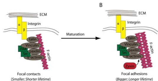
MATRIX ADHESION STRUCTURESform an heterodimers through transmembrane interaction of an α and β subumits. The extracellular domain of Integrin recognizes and binds to ECM components, whereas the intracellular domain interacts with the actin cytoskeleton through adaptor proteins such as Talin, Tensin and α-actinin. Due to these interactions, Integrins provide a linkage between the underlying actin filaments and the ECM. Integrins also interacts with various signalling molecules, including kinases and phosphatases such as Focal Adhesion Kinase (FAK) (reviewed in Worth and Parsons, 2010), (Fig.1.2.).
Figure 1.2 Cartoons representing the major components of cell-matrix adhesion structures and the process of their maturation.
Cell-matrix adhesion structures are composed of Integrins (yellow rectangles) that link actin filaments (pink helix) to the components of the Extracellular matrix (ECM). Actin filaments
interact with the cytoplasmic domain of β-Integrin through the adaptor proteins (grey squares)
that form the cytoplasmic plaque. Many components of the cytoplasmic plaque have signalling properties (green rectangles). (A) Focal contacts are small and have a short life-span. They can mature into Focal adhesions (B) that are larger and more stable. The process of cell-matrix adhesion maturation occurs by the addition of the new proteins into the cytoplasmic plaque (red rounded rectangle) and can be initiated by the application of mechanical forces (adapted from Worth and Parsons, 2010).
protein composition (Fig.1.2.). In Drosophila epithelial tissues, the composition of the cytoplasmic plaques of C-ECM adhesions can change during development (Delon and Brown 2009) thus, for simplification, I will refer to C-ECM adhesions as FAs in this manuscript.
1.4.
T
HEC
ROSS-
TALK BETWEENA
DHERENSJ
UNCTIONS AND CELL-
MATRIXADHESION STRUCTURES
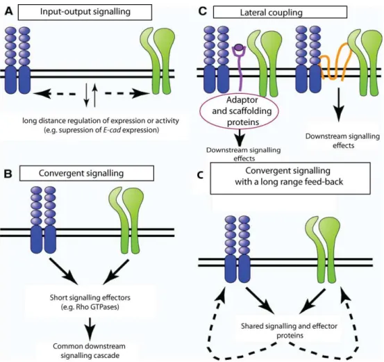
Figure 1.3 The Schematic representation of the types of interactions between Adherens Junctions and Focal adhesions.
First, a “long-range- input-output” signalling in which signalling initiated downstream of
adhesion junctions influences the organization of adhesion junctions elsewhere in the cell (Fig.1.3.A.). For example, adhesion of Integrins to distinct ECM proteins can alter expression of genes encoding for AJs components or regulators. During the formation of the mouse embryonic salivary glands, adhesion of Integrins reduce E-cad expression, thus altering AJs organization and cell morphology (Onodera et al., 2010). Another example is the activation of Src signalling within FAs that leads to either strengthening or loss of E-cad-mediated adhesion, depending on the levels of Src activation (Martinez-Rico et al., 2010; Avizienyte et al., 2002). A moderate level of Src activation within FAs enables acto-myosin contractility that supports E-cad adhesion in epithelial cells (Martinez-Rico et al., 2010). In contrast, signalling resulting from activation of a constitutively active form of Src (vSrc), propagated upon Integrins clustering, leads to E-cad misslocalization and loss of adhesion in colon cancer cells (Avizienyte et al., 2002). A second way by which AJs and FAs interact with each other is by sharing common components, such as the scaffolding proteins Vinculin and small GTPases such as Rac
that allows for “convergent signalling” (Fig.1.3.B). For example, activation of Rac GTPase
occurs in the redundant manner downstream of Integrins or E-cad clustering, which in turn triggers synthesis of cyclin D and higher rate of proliferation in epithelial cells (Fournier et al., 2008). Moreover, scaffolding structures such as the actin cytoskeleton, which allows for the short-range interactions, are also shared between AJs and FAs. For example, FAs clustering induces actin cytoskeleton contractility that can lead to loss of E-cad mediated adhesion (reviewed in Weber et al., 2011).
AJs and FAs can also interact through “lateral coupling”, which involves short-range
Activation of EGF1R signalling leads to loss of Integrin and E-cad mediated adhesion (Fig.1.3.C), (reviewed in Weber et al., 2011).
Finally each of these interactions may converge on common signalling pathways giving rise to complex feed-back loops that modulate both initiating signals (Fig.1.3.D). For instance, Src signalling can be activated downstream of both AJs and FAs in mammalian cells and as a result, can alter adhesion mediated by E-cad and Integrins (reviewed in Weber et al., 2011).
ACTIN FILAMENTS ORGANIZATION AND DYNAMICS
2.
Figure 1. 4 Diagram representing the mechanism of the actin filament treadmilling.
(A) At the physiological concentration of Actin monomers, ATP-G-actin preferentially binds to the barbed end of F-actin (depicted in red), while G-actin-ADP dissociates from the point end of F-actin (depicted in yellow). (B) The actin monomers can flow through the filament by attaching preferentially to the barbed end of the filament and dissociating from the pointed end. The oldest subunits of the filaments are in the highest proximity to point end (adapted from a webpage of Sichuan University http://jpkc.scu.edu.cn/).
N
UCLEATION OF ACTIN FILAMENTS2.1.
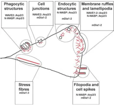
Both WASP and WAVE enable the coupling of F-actin branched networks into the membrane. (reviewed in Campellone and Welch, 2010). Arp2/3, together with NPFs, has been described to contribute to cell junctions assembly, endocytosis, membrane ruffling, lamellipodia dynamics and filopodia formation (reviewed in Campellone and Welch, 2010), (Fig.1.5.).
Figure 1.5 Schematic representation of the localization and function of the Arp2/3 complex and Formins in mammalian cells.
Actin filaments (red lines) are nucleated and organized into networks by the Arp2/3 complex and its nucleation promoting factors (WASP, N-WASP and WAVE1-2) or are generated into unbranched structures by Formins. Functional roles for Arp2/3 and Formins are depicted: during phagocytosis, cell junction assembly, membrane ruffling and lamellipodia dynamics, filopodia and stress fibers formation. Abbreviations used: Arp2/3, actin-related protein 2/3; mDia, murine Diaphanous; N WASP, neuronal-WASP, WASP, Wiskott-aldrich syndrome protein, WAVE, WASP verprolin homologous (adapted from Campellone and Welch, 2010).
feature is the presence of conserved Formin-homology (FH) domains, FH1 and FH2 (reviewed in Mattila and Lappalainen, 2008). The best characterized mammalian Formins are Diaphanous related Formins (DRFs), named based on their similarity to Diaphanous, the first Formin identified in Drosophila. DRSFs are commonly named murine Diaphanous (mDia, mDia1 to mDia3) and they precipitate in formation of various cellular structures (Fig.1.5.). The best studied examples are stress fibres that underlie FAs, membrane ruffles involved in cell migration, and contractile ring formed during cytokinesis (reviewed in Campellone and Welch, 2010). Moreover DRSF contribute to organization of cell junctions and coordinate F-actin assembly during endocytosis (reviewed in Campellone and Welch, 2010), (Fig.1.5).
D
EPOLYMERIZATION OF ACTIN FILAMENTS AND SEQUESTRATION OF ACTIN2.2.
MONOMERS
In addition to F-actin nucleators, the activities of actin depolymerizing factors also regulate the extent and spatial pattern of F-actin organization. The best described examples are Actin depolymerizing factor (ADF)/Cofilin family proteins that severe F-actin and increase the rate of G-F-actin dissociation from a point end that ultimately increase F-actin turn-over (Mazur et al., 2010). G-actin binding proteins such as Adenylate Cyclase Associated Proteins (CAPs) ensure the availability of G-actin by sequestrating them and preventing from incorporation into F-actin. On a contrary, Profilin, that binds to G-actin, increase the rate of F-actin elongation by facilitating the exchange rate of G-actin ADP to G-actin ATP (Witke, 2004).
conserved hetrodimer consisting of Capping Protein α (Cpa) and Capping Protein β
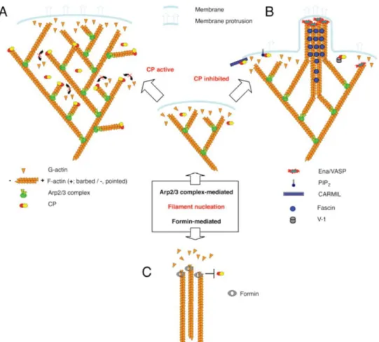
(Cpb). CP binds to the barbed end of F-actin, thereby preventing addition and loss of G-actin. CP inhibition leaves the barbed end of F-actin uncapped allowing for incorporation of new G-actin and binding of actin nucleators allowing for the formation of the long actin filaments (reviewed in Cooper and Sept, 2008).
C
APPINGP
ROTEIN AND ITS ROLE IN ORGANIZATION OF ACTIN FILAMENTS2.4.
interactions (reviewed in Cooper and Sept, 2008). Furthermore, other molecules that bind to the barbed end of F-actin can compete with CP and indirectly inhibit its function (reviewed in Mattila and Lappalainen, 2008).
Figure 1.6 Cartoon representing the actin binding domains and predicted hydrophobic domains in Cpa and Cpb.
Representation of the aminoacid sequence of Cpb and Cpa. Both Cpa and Cpb had two hydrophobic domains (HB, green) that had been predicted to interact with the cellular membrane lipids and the actin binding domains (ABD, pink). The hydrophobic domains predicted in silico; located in (134-151; 215-232 aa residues) Cpb or (113-130;225-242 aa residues) Cpa. The localization of the ABDs established in the structure function analysis in yeast and chicken cells; located in the (264-287 aa residues) for Cpb and (259-286aa residues) for Cpa.
Representation of the aminoacid sequence of Cpb and Cpa. Both Cpa and Cpb had two hydrophobic domains (HB, green) that had been predicted to interact with the cellular membrane lipids and the actin binding domains (ABD, pink). The hydrophobic domains predicted in silico; located in (134-151; 215-232 aa residues) Cpb or (113-130;225-242 aa residues) Cpa. The localization of the ABDs established in the structure function analysis in yeast and chicken cells; located in the (264-287 aa residues) for Cpb and (259-286aa residues ) for Cpa.
competing with Formins at filament barbed ends (Mattila and Lappalainen, 2008). When CP activity is inhibited, it enables binding of Formins and F-actin elongation (Fig.1.7.C). This mechanism has been proposed to contribute to filopodia formation (reviewed in Cooper and Sept, 2008). Since Formins also promote the formation of other actin-based structures, such as stress fibres, CP might also be involved in this process (reviewed Campellone and Welch, 2010), (Fig.1.5) .
Figure 1.7 Schematic representations of the contribution of Capping Protein into the formation of distinct actin filaments- based structures.
inhibited Formins can bind to the barbed ends of F-actin and promote their elongation (Cooper and Sept, 2008).
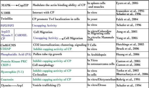
Table 1Capping Protein interactors
ROLE OF ACTIN FILAMENTS IN ORGANIZATION AND REMODELING OF EPITHELIAL
3.
JUNCTIONS
A
CTIN FILAMENTS INA
DHERENSJ
UNCTIONS STABILITY3.1.
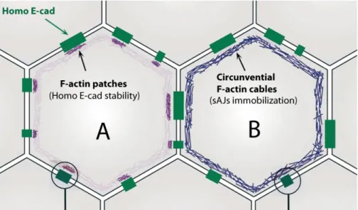
Tomas Lecuit had described two distinct F-actin pools that contribute to AJs stability in the early Drosophila embryo (Cavey et al., 2008).. They show that a pool of stable actin filaments, localizes in small patches adjacent to spots AJs (sJAs), contributes to the maintenance of homophilic DE-cad dimmers (Fig.1.8.A), whereas, a pool of F-actin, which forms the circumferential F-actin cable, is necessary to immobilize sJAs within AJs and restricts their proper localization (Cavey et al., 2008), (Fig.1.8.B).
Figure 1.8 Diagram representing two different F-actin pools that contribute to Adherens Junctions stability.
Schematic representation of the cellular apical sites. Homo E-cadherin (green) represents the E-cadherin homophilic complexes from 2 adjacent cells. Cell A presents the organization of small F-actin spots that contribute to the stability of E-cadherin homophilic interactions. Cell B represents the circunvential F-actin cables that immobilize AJs spots (sAJs) within AJs (Cavey et al., 2008).
R
OLE OF ACTIN FILAMENTS IN MATURATION OF CELL–
MATRIX ADHESION3.2.
STRUCTURES
.
Worth and Parsons, 2010). Application of mechanical forces triggers Myosin II and mDia1 mediated contractility of actin filaments, which contributes to FAs maturation (Riveline et al., 2001; Geiger and Yamada, 2011). Moreover, SFs have been proposed to serve as platform on which components of the cytoplasmic plaque assemble to contribute to FAs maturation (Oakes et al., 2012).
THE WING IMAGINAL DISC AS A MODEL SYSTEM
4.
S
TRUCTURE OF THE WING IMAGINAL DISC4.1.
The wing imaginal disc had emerged as a powerful model to study mechanisms of epithelial cell polarity, tissue integrity and homeostasis (reviewed in Hariharan and Bilder, 2006). Wing imaginal disc forms during embryogenesis from clusters of 20 to 50 cells that proliferate during larval development to reach a final size of 20,000 to 50,000 cells (reviewed in Herranz and Milán, 2008). They organized into a columnar monolayer epithelium, covered by a thin layer of squamous epithelium called the peripordial membrane (reviewed in Hariharan and Bilder, 2006).
During larval development, wing imaginal discs are subdivided into spatially distinct compartments of different genetic identities, established by the expression of selector genes. The identity of each compartment is maintained as cells keeps dividing through processes that prevent cells of distinct compartments to intermingle. (reviewed in Herranz and Milán, 2008). At the Anterior-Posterior (A-P) and Dorsal-Ventral (D-V) boundaries, two signalling pathways through the long-range morphogen
Decapentaplegic (Dpp) and Wingless (Wg) respectively, act as “organizer”, to establish
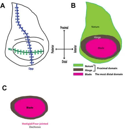
Figure 1.9 Schematic representation of the structure of the 3rd instar wing imaginal disc of
Drosophila melanogaster.
(A) The schematic representation of the wing imaginal disc with the indicated domains of expression of Dpp (blue) and Wg (green). The expression of Dpp partitions the disc into the posterior and the anterior domain while Wg partitions the wing disc into the dorsal and the
ventral domain. (B) Schematic representation of the distinct compartments of the 3rd instar wing
imaginal disc along the dorso-vental border. Notum (light green) and hinge (grey) are referred as proximal wing disc domain and blade (magenta) is referred as the most distal wing disc domain. (C) The blade is characterized by the expression of the Vestigial transcription factor, which
promotes the expression of four-jointed and represses dachsous. The gradient between
Four-jointed and Dachsous promotes Yki-dependent growth of the wing disc blade.
P
ATTERNING OF THE WING IMAGINAL DISC4.2.
Figure 1.10 Cartoon representing the major cellular events triggered by Decapentaplegic or Wingless signalling.
Schematic representation of the interactions of (A) Dpp or (B) Wg with their receptors and the cellular outcomes of receptors clustering. (A) In the absence of Dpp (cell on the left (OFF)) the level of the Brinker (Brk) transcriptional repressor is high. Brk represses most Dpp target genes
including omb and salm. Dpp binding to its receptor promotes phosphorylation and the
cytoplasm, it enters the nucleus and displaces Gro from TCF. As a part of Adherens Junctions, Arm/β-Cat binds to DE-cadherin. The negative and positive regulators of Wg signaling are represented in red and green respectively (adapted from (Lin, 2004; Städeli et al., 2006).
W
ING IMAGINAL DISC AS A MODEL TO STUDY TISSUE GROWTH AND4.3.
HOMEOSTASIS
Figure 1.11 Schematic representation of the model of the interactions of the Hippo pathway components in Drosophila and mammals.
Figure 1.12 A Schematic representation of the major components of the JNK pathway in
Drosophila.
(reviewed Igaki, 2009). Activation of JNK signaling has distinct outcomes depending on the tissue type and developmental context (reviewed in Igaki, 2009). It is crucial for cell migration during dorsal closure and wound healing (Martin and Parkhurst, 2004). JNK has also important pro-apoptotic functions (reviewed in Igaki, 2009; Kanda and Miura, 2004). Activated JNK triggers the expression of reaper, which in turn leads to degradation of the caspase inhibitor DIAP1 (Kuranaga et al., 2002). As a result, activated caspases mediate apoptosis. JNK pathway has been also proposed to induce apoptosis independently of caspases (reviewed in Igaki, 2009). By eliminating developmentally aberrant cells from a tissue, JNK signalling has been proposed to maintain tissue homeostasis, as well as protect organisms against tumor development (Brumby and Richardson, 2003; Igaki et al., 2006; Uhlirova et al., 2005) (Uhlirova and Bohmann, 2006).
TUMORIGENESIS AS AN EXAMPLE OF THE LOSS OF EPITHELIAL TISSUE
5.
HOMEOSTASIS
T
HE HALLMARKS OF EPITHELIAL DERIVED CANCERS5.1.
Figure 1.13 Schemat representing major hallmarks of cancer cells.
Tumour cells need to acquire distinct features to survive, multiply and invade distinct tissues. These features can be referred as hallmarks of cancer and include: self-sufficiency in growth signals; insensitivity to anti-growth signals; ability to evade apoptosis; sustained angiogenesis; limitless replication potential; tissue invasion and metastasis (adapted from Hanahan and Weinberg, 2011).
by distinct means; the most common strategy is the loss of P53, which induces apoptosis upon cellular stresses. Fourth, cancer cells need to enable the replicative immortality (limitless replication potential), (Fig.1.13.). Normally, most healthy cells undergo only a limited number of cell divisions before going into a non- proliferative viable state and finally senescence. However, cancer cells keep dividing without these restrictions. One of the most common means by which cancer cells keep proliferating is by expressing telomerase, which elongates telomere ends. The fifth hallmark of cancer cells is their ability to induce angiogenesis (sustained angiogenesis), (Fig.1.13.). The multiplying cancer cells reach blood vessels in order to get nutrients, oxygen and remove wastes and carbon dioxide. This brings a necessity for a formation of new blood vessels, able to feed highly proliferating cancer cells. Finally, cancer cells get the ability to invade and metastases (tissue invasion and metastasis) into distant tissues to form secondary tumours (Fig.1.13), (reviewed in Hanahan and Weinberg 2011).
Missregulation of most genes or pathways involved in cancer progression result in only one or two of the hallmarks of cancer and in most cases net tissue-overgrowth is not observed, because there is also induction of compensatory apoptosis. Loss or gain of function mutations in genes that give rise to at least one of the hallmark of cancer are named tumour suppressor genes (TSGs) and oncogenes respectively (reviewed in Hanahan and Weinberg 2011). Therefore, most mammalian cancers are likely to originate from cooperative interactions between both TSGs and oncogenes (Brumby and Richardson, 2005).
D
ROSOPHILA AS A MODEL TO STUDY TUMORIGENESIS,
ADVANCES AND5.2.
LIMITATIONS
tumorigenesis. First, growth of imaginal disc is mainly autonomous (Bryant and Simpson, 1984). Second, signalling pathways involved in human cancers are conserved in Drosophila (reviewed in Bilder, 2004). Indeed, our understanding of Hpo, Wnt, Dpp and
JNK signalling, which all are missregulated in cancer has been mainly extended from studies in flies (reviewed in Miles et al., 2011). Finally, because the Drosophila genome is smaller and much less redundant than in humans, it allows for large scale screening for new TSGs and oncogenes (reviewed in Miles et al., 2011). Larval tumours can occur either spontaneously or may be induced in mutagenesis screens. They can develop in the brain, imaginal discs or both tissues (reviewed in Bilder, 2004). Larval epithelial tumours are classified as hyperplastic, when the tissue overgrowth but cells are still arranged into a monolayer. In contrast, neoplastic tumours are characterised by the loss of cell morphology, cells appear rounder and tissue architecture is affected. The induction of neoplastic tumours usually results in the death of the animal after prolonged larvae stages. Moreover when transplanted into the abdomen of an adult fly tumour cells continue to proliferate, cross the basal lamina and invade distant tissues (reviewed in Bilder, 2004; Hariharan and Bilder, 2006). However, as any model systems, Drosophila also has several limitations. The process of angiogenesis cannot be modelled in Drosophila since its lymphatic and hematopoietic systems are completely different than in mammals. In addition, Drosophila does not contain Telomerase.
T
UMOUR SUPPRESSORS GENES AND ONCOGENES IND
ROSOPHILA EPITHELIAL5.3.
TISSUES
rate of cell proliferation. In some cases, mutant cells even proliferate at slower rate than wildtype cells. Due to this property, when cells homozygote mutant for neoplastic TSGs are induced in small patches of cells (clones) in an heterozygote background, they are outcompeted by wildtype cells around and undergo JNK-mediated apoptosis (reviewed in Hariharan and Bilder, 2006). Moreover, in contrast to hyperplastic TSGs, mutant cells for neoplastic TSGs fail to terminate proliferation and do not differentiate. However, when the whole tissue is mutant for a neoplastic TSG, it form hightly proliferative tumours, characterized by the loss of epithelial tissue architecture and an invasive behaviour (reviewed in Bilder, 2004; Hariharan and Bilder, 2006). Neoplastic TSGs consist of two major groups, polarity markers of the scrib, dlg and lgl group and genes that encode for components of the endocytic machinery (including avalanche (avl), rab-protein 5 (rab5)). The best-described oncogenes in flies are the activated forms of Ras (RasV12), Notch (NotchAct) and Src. They cooperate with mutations in TSGs in
oncogenic transformation. For example, overexpression of RasV12 triggers hyperplastic overgrowth but when combined with a mutation in scrib, dlg or lgl promotes massive tissue overgrowth, loss of tissue architecture and invasive behaviour (reviewed in Miles et al., 2011).
T
HE COMPONENTS OFH
IPPO PATHWAY AS EXAMPLES OF TUMOUR5.4.
SUPPRESSOR GENES
Yki overexpressing tissues. Moreover by promoting transcription of dMyc that acts as an inducer of ribosome biogenesis, Yki promotes cell growth. Furthermore, the expression of cyclinE and of the bantam microRNA increases the proliferation rate. bantam has also a role in blocking apoptosis. Yki also stimulates transcription of the EGFR ligands vein, Karen and Spitz (Zhao et al., 2011). The Hpo pathway is conserved from fly to human (Fig.1.11.A, B). Human components of the Hpo pathway, including YAP (ortholog of Yki), Large tumor suppressor 1 and 2 (Lats1/2, orthologs of Wts), mammalian STE-20 kinase 1 and 2 (Mst1/2; orthologs of Hpo) and Msp-one-binder (Mob1, ortholog of Mats) are able to functionally rescue the corresponding mutations in Drosophila. Deregulation of the Hpo pathway components is often associated with cancer in humans. For example NF2, the human ortholog of Merlin is a known TSG implicated in various signalling pathways including Rac1 GTPase and Src that together with the control of Yki nuclear localization serves as a tumour suppressor mechanism in mammals. Mutations of NF2 are found in patients with neurofibromatosis, associated with higher risks of developing tumours in the nervous system. The loss of activatory post-trascriptional modifications of Mst1 and Mst2, together with decrease of YAP phosphorylation, is observed in ~30% of human hepatocellular carcinoma (reviewed in Chan et al., 2011). Finally nuclear YAP accumules in samples from distinct types of cancers, including colonic adenocarcinoma, lung adenocarcinoma, and ovarian cysadenocarcinoma, when compared to the healthy tissues (reviewed in Chan et al., 2011).
R
OLE OFJNK
SIGNALLING IN EPITHELIAL-
DERIVED TUMOURS5.5.
has been linked to proliferative and metastatic features of tumours associated with disruptions of apical-basal polarity in epithelial cells, induced by lgl, dlg and scrib mutations (Igaki et al., 2006; Uhlirova and Bohmann, 2006). Loss of lgl triggers JNK signalling, which in turn, promotes Yki nuclear localization, resulting in neoplastic tissue overgrowth (Sun and Irvine, 2011). In addition, JNK signalling contributes to the invasive behaviour of tumour cells induced by lgl loss by promoting mmp1 transcription (Uhlirova and Bohmann, 2006).
C
ONTRIBUTION OF LOSS OF CELL POLARITY AND ADHESION TO5.6.
TUMORIGENESIS
The loss of epithelial polarity and tissue architecture is the primary diagnostic of
malignant cancers in tissues such as breast, prostate, and colon (reviewed in Bilder, 2004). Many of the know TSGs belong to the group of adhesions and polarity markers, including lgl, dlg and scrib (reviewed in Bilder, 2004). Moreover, αPKC that directly phosphorylates and restricts Lgl localization to the subapical domain, when overactivated in lgl mutant clones promotes tumorigenesis (reviewed in Lee and Vasioukhin, 2008). Moreover, overactivation or promotion of αPKC cortical localization induces larval tumour formation indicating that αPKC acts as an oncogene (Grifoni et al., 2007). Lgl, αPKC and Crb are all upstream upstream regulators of the Hpo pathway and negatively regulate Yki nuclear localization (reviwed Genevet and Tapon, 2011). Moreover, loss of apico-basal polarity also induces activation of JNK pathway that can contribute to tumourgenesis (reviewed in Igaki, 2009). Human orthologs of Scrib, Dlg, Lgl, αPKC have been found missregulated in several human cancer types (reviewed in Martin-Belmonte and Perez-Moreno, 2012)
to induce tumorigenesis, it contributes to tumour formation and cancer progression by distinct means. First, loss of E-cad triggers β-Cat release into the cytoplasm and consequently ectopic activation of Wnt signalling pathway, which has been associated with early events of tumuorigenesis such as in colorectal cancers (reviewed in Jeanes et al., 2008). Second, loss of E-cad leads to increase EGF-R signalling (reviewed Jeanes et al., 2008). Increased EGF-R signalling has been also associated with poor prognosis and decreased patients survival and resistance to chemotherapy and radiation treatment in tumour cells (reviewed in Herbst, 2004). However, it is still not clear if E-cad directly inhibits EGF-R activation upon E-cad clustering or down-regulates EGF-R signalling activity (reviewed in Jeanes et al., 2008). Independent of the nature of their interactions E-cad clustering can inhibit cell responsiveness to EGF stimulation, revealed by a decrease in cell proliferation and Ras signalling (Qian et al., 2004; Perrais et al., 2007).
Therefore, the ability of polarity markers to interact with signalling pathways may be a general mechanism by which loss of polarity can contribute to tumorigenesis (reviewed in (Martin-Belmonte and Perez-Moreno 2012; Genevet and Tapon 2011).
S
RC AS AN EXAMPLE OF ONCOGENE5.7.
Figure 1. 14 Schematic representation of Src activation, functions and contribution to tumour formation and progression.
Although the role of Src in tumorigenesis has been extensively studied, the exact mechanisms by which Src contributes to tumourigenesis and tumour progression is still not clear. For instance, overactivation of Src results in distinct cellular outcomes ranging from cell proliferation, differentiation and apoptosis depending on the cell lines used (reviewed in Brown and Cooper, 1996). Moreover, mutations of Src in cancers are rarely identified while the elevated levels of Src protein and its activities is often observed in distinct types of cancer including those of colon, lung, breast and pancreas (reviewed in Irby and Yeatman, 2000) . However, studies from Cagan´s laboratory have extended our understanding on how Src signalling contributes to distinct stages of tumour progression. In this studies Vidal and co-authors used Drosophila imaginal discs to assayed the effect of increased Src signalling activity, either by knocking down dCsk or by overexpressing Src (Vidal et al., 2006; Vidal et al. 2007). They have demonstrated that mild overexpression of Src64B or hypomorphic mutations of dCsk triggers cell proliferation and inhibition of apoptosis (Vidal et al., 2006; Vidal et al. 2007). In contrast, strong Src42A overexpression or null mutation for dCskQ156Stop triggers apoptosis. Interestingly, when combined with RasV12, only high Src signalling activity cooperate with
RasV12 overexpression giving rise to neoplastic overgrowth associated with loss of the
REFERENCES
6.
Affolter, M., Basler, K., 2007. The Decapentaplegic morphogen gradient: from pattern formation to growth regulation. Nat. Rev. Genet. 8, 663–674.
Avizienyte, E., Wyke, A.W., Jones, R.J., McLean, G.W., Westhoff, M.A., Brunton, V.G., Frame, M.C., 2002. Src-induced de-regulation of E-cadherin in colon cancer cells requires integrin signalling. Nat. Cell Biol. 4, 632–638.
Badouel, C., Gardano, L., Amin, N., Garg, A., Rosenfeld, R., Le Bihan, T., McNeill, H., 2009. The FERM-domain protein Expanded regulates Hippo pathway activity via direct interactions with the transcriptional activator Yorkie. Dev. Cell 16, 411–420. Baeg, G.-H., Selva, E.M., Goodman, R.M., Dasgupta, R., Perrimon, N., 2004. The Wingless
morphogen gradient is established by the cooperative action of Frizzled and Heparan Sulfate Proteoglycan receptors. Developmental Biology 276, 89–100. Bilder, D., 2004. Epithelial Polarity and Proliferation Control: Links from the Drosophila
Neoplastic Tumor Suppressors. Genes Dev. 18, 1909–1925.
Brown, M.T., Cooper, J.A., 1996. Regulation, substrates and functions of src. Biochim. Biophys. Acta 1287, 121–149.
Brumby, A.M., Richardson, H.E., 2005. Using Drosophila melanogaster to map human cancer pathways. Nat. Rev. Cancer 5, 626–639.
Bryant, P.J., Simpson, P., 1984. Intrinsic and extrinsic control of growth in developing organs. Q Rev Biol 59, 387–415.
Campellone, K.G., Welch, M.D., 2010. A nucleator arms race: cellular control of actin assembly. Nat. Rev. Mol. Cell Biol. 11, 237–251.
Chan, S.W., Lim, C.J., Chen, L., Chong, Y.F., Huang, C., Song, H., Hong, W., 2011. The Hippo pathway in biological control and cancer development. J. Cell. Physiol. 226, 928–939.
Cooper, J.A., Sept, D., 2008. New insights into mechanism and regulation of actin capping protein. Int Rev Cell Mol Biol 267, 183–206.
Delon, I., Brown, N.H., 2007. Integrins and the actin cytoskeleton. Curr. Opin. Cell Biol. 19, 43–50.
Fournier, A.K., Campbell, L.E., Castagnino, P., Liu, W.F., Chung, B.M., Weaver, V.M., Chen, C.S., Assoian, R.K., 2008. Rac-dependent cyclin D1 gene expression regulated by cadherin- and integrin-mediated adhesion. J Cell Sci 121, 226–233. Geiger, B., Yamada, K.M., 2011. Molecular architecture and function of matrix
adhesions. Cold Spring Harb Perspect Biol 3.
Georgiou, M., Marinari, E., Burden, J., Baum, B., 2008. Cdc42, Par6, and aPKC Regulate Arp2/3-Mediated Endocytosis to Control Local Adherens Junction Stability. Current Biology 18, 1631–1638.
Gremm, D., Wegner, A., 2000. Gelsolin as a calcium-regulated actin filament-capping protein. Eur. J. Biochem. 267, 4339–4345.
Grifoni, D., Garoia, F., Bellosta, P., Parisi, F., De Biase, D., Collina, G., Strand, D., Cavicchi, S., Pession, A., 2007. aPKCzeta cortical loading is associated with Lgl cytoplasmic release and tumor growth in Drosophila and human epithelia. Oncogene 26, 5960–5965.
Hamaratoglu, F., Willecke, M., Kango-Singh, M., Nolo, R., Hyun, E., Tao, C., Jafar-Nejad, H., Halder, G., 2006. The tumour-suppressor genes NF2/Merlin and Expanded act through Hippo signalling to regulate cell proliferation and apoptosis. Nat. Cell Biol. 8, 27–36.
Hanahan, D., Weinberg, R.A., 2000. The hallmarks of cancer. Cell 100, 57–70.
Hariharan, I.K., Bilder, D., 2006. Regulation of Imaginal Disc Growth by Tumor-Suppressor Genes in Drosophila. Annual Review of Genetics 40, 335–361. Herbst, R.S., 2004. Review of epidermal growth factor receptor biology. Int. J. Radiat.
Oncol. Biol. Phys. 59, 21–26.
Herranz, H., Milán, M., 2008. Signalling molecules, growth regulators and cell cycle control in Drosophila. Cell Cycle 7, 3335–3337.
Hug, C., Miller, T.M., Torres, M.A., Casella, J.F., Cooper, J.A., 1992. Identification and characterization of an actin-binding site of CapZ. J. Cell Biol. 116, 923–931.
Huh, J.R., Guo, M., Hay, B.A., 2004. Compensatory Proliferation Induced by Cell Death in the Drosophila Wing Disc Requires Activity of the Apical Cell Death Caspase Dronc in a Nonapoptotic Role. Current Biology 14, 1262–1266.
Igaki, T., 2009. Correcting developmental errors by apoptosis: lessons from Drosophila JNK signaling. Apoptosis 14, 1021–1028.
Igaki, T., Pagliarini, R.A., Xu, T., 2006. Loss of cell polarity drives tumor growth and invasion through JNK activation in Drosophila. Curr. Biol. 16, 1139–1146.
Irby, R.B., Yeatman, T.J., 2000. Role of Src expression and activation in human cancer. Oncogene 19, 5636–5642.
Jeanes, A., Gottardi, C.J., Yap, A.S., 2008. Cadherins and cancer: how does cadherin dysfunction promote tumor progression? Oncogene 27, 6920–6929.
Kikuchi, A., Yamamoto, H., Sato, A., Matsumoto, S., 2011. New insights into the mechanism of Wnt signaling pathway activation. Int Rev Cell Mol Biol 291, 21–71. Kuranaga, E., Kanuka, H., Igaki, T., Sawamoto, K., Ichijo, H., Okano, H., Miura, M., 2002.
Martin-Belmonte, F., Perez-Moreno, M., 2012. Epithelial cell polarity, stem cells and cancer. Nat. Rev. Cancer 12, 23–38.
Martinez-Rico, C., Pincet, F., Thiery, J.-P., Dufour, S., 2010. Integrins stimulate E-cadherin-mediated intercellular adhesion by regulating Src-kinase activation and actomyosin contractility. J. Cell. Sci. 123, 712–722.
Matter, K., Balda, M.S., 2003. Signalling to and from tight junctions. Nat Rev Mol Cell Biol 4, 225–237.
Mattila, P.K., Lappalainen, P., 2008. Filopodia: molecular architecture and cellular functions. Nat. Rev. Mol. Cell Biol. 9, 446–454.
Mazur, A.J., Gremm, D., Dansranjavin, T., Litwin, M., Jockusch, B.M., Wegner, A., Weeds, A.G., Mannherz, H.G., 2010. Modulation of actin filament dynamics by actin-binding proteins residing in lamellipodia. Eur. J. Cell Biol. 89, 402–413.
Michelot, A., Drubin, D.G., 2011. Building distinct actin filament networks in a common cytoplasm. Curr. Biol. 21, R560–569.
Miles, W.O., Dyson, N.J., Walker, J.A., 2011. Modeling tumor invasion and metastasis in Drosophila. Dis Model Mech 4, 753–761.
Neto-Silva, R.M., Wells, B.S., Johnston, L.A., 2009. Mechanisms of growth and homeostasis in the Drosophila wing. Annu Rev Cell Dev Biol 25, 197–220.
Oakes, P.W., Beckham, Y., Stricker, J., Gardel, M.L., 2012. Tension is required but not sufficient for focal adhesion maturation without a stress fiber template. J. Cell Biol. 196, 363–374.
Oh, H., Irvine, K.D., 2011. Cooperative regulation of growth by Yorkie and Mad through bantam. Dev Cell 20, 109–122.
Onodera, T., Sakai, T., Hsu, J.C., Matsumoto, K., Chiorini, J.A., Yamada, K.M., 2010. Btbd7 regulates epithelial cell dynamics and branching morphogenesis. Science 329, 562–565.
Pérez-Garijo, A., Shlevkov, E., Morata, G., 2009. The role of Dpp and Wg in compensatory proliferation and in the formation of hyperplastic overgrowths caused by apoptotic cells in the Drosophila wing disc. Development 136, 1169–
1177.
Pinho, S.S., Seruca, R., Gärtner, F., Yamaguchi, Y., Gu, J., Taniguchi, N., Reis, C.A., 2011. Modulation of E-cadherin function and dysfunction by N-glycosylation. Cell. Mol. Life Sci. 68, 1011–1020.
Read, R.D., Bach, E.A., Cagan, R.L., 2004. Drosophila C-terminal Src kinase negatively regulates organ growth and cell proliferation through inhibition of the Src, Jun N-terminal kinase, and STAT pathways. Mol. Cell. Biol. 24, 6676–6689.
dos Remedios, C.G., Chhabra, D., Kekic, M., Dedova, I.V., Tsubakihara, M., Berry, D.A., Nosworthy, N.J., 2003. Actin binding proteins: regulation of cytoskeletal microfilaments. Physiol. Rev. 83, 433–473.
Riveline, D., Zamir, E., Balaban, N.Q., Schwarz, U.S., Ishizaki, T., Narumiya, S., Kam, Z., Geiger, B., Bershadsky, A.D., 2001. Focal contacts as mechanosensors: externally applied local mechanical force induces growth of focal contacts by an mDia1-dependent and ROCK-inmDia1-dependent mechanism. J. Cell Biol. 153, 1175–1186. Robinson, B.S., Huang, J., Hong, Y., Moberg, K.H., 2010. Crumbs regulates
Salvador/Warts/Hippo signaling in Drosophila via the FERM-domain protein Expanded. Curr. Biol. 20, 582–590.
Ryoo, R.H., Gorenc, G.T., Steller, S.H., 2004. Apoptotic cells can induce compensatory cell proliferation through the JNK and the Wingless signaling pathways. Dev. Cell 7, 491–501.
Städeli, R., Hoffmans, R., Basler, K., 2006. Transcription under the control of nuclear Arm/beta-catenin. Curr. Biol. 16, R378–385.
Sun, G., Irvine, K.D., 2011. Regulation of Hippo signaling by Jun kinase signaling during compensatory cell proliferation and regeneration, and in neoplastic tumors. Dev. Biol. 350, 139–151.
Taylor, M.A., Lee, Y.-H., Schiemann, W.P., 2011. Role of TGF-β and the tumor microenvironment during mammary tumorigenesis. Gene Expr. 15, 117–132. Tepass, U., Gruszynski-DeFeo, E., Haag, T.A., Omatyar, L., Török, T., Hartenstein, V.,
1996. shotgun encodes Drosophila E-cadherin and is preferentially required during cell rearrangement in the neurectoderm and other morphogenetically active epithelia. Genes & Development 10, 672–685.
Uhlirova, M., Bohmann, D., 2006. JNK- and Fos-regulated Mmp1 expression cooperates with Ras to induce invasive tumors in Drosophila. EMBO J. 25, 5294–5304.
Vidal, M., Larson, D.E., Cagan, R.L., 2006. Csk-deficient boundary cells are eliminated from normal Drosophila epithelia by exclusion, migration, and apoptosis. Dev. Cell 10, 33–44.
Wartlick, O., Mumcu, P., Jülicher, F., Gonzalez-Gaitan, M., 2011. Understanding morphogenetic growth control — lessons from flies. Nat Rev Mol Cell Biol 12, 594–604.
Weber, G.F., Bjerke, M.A., DeSimone, D.W., 2011. Integrins and cadherins join forces to form adhesive networks. J. Cell. Sci. 124, 1183–1193.
Willecke, M., Hamaratoglu, F., Sansores-Garcia, L., Tao, C., Halder, G., 2008. Boundaries of Dachsous Cadherin Activity Modulate the Hippo Signaling Pathway to Induce Cell Proliferation. PNAS.
Witke, W., 2004. The role of profilin complexes in cell motility and other cellular processes. Trends Cell Biol. 14, 461–469.
Zecca, M., Struhl, G., 2010. A feed-forward circuit linking wingless, fat-dachsous signaling, and the warts-hippo pathway to Drosophila wing growth. PLoS Biol. 8, e1000386.
Zhao, B., Tumaneng, K., Guan, K.-L., 2011. The Hippo pathway in organ size control, tissue regeneration and stem cell self-renewal. Nat. Cell Biol. 13, 877–883.
C
HAPTER
2
–
C
APPING
P
ROTEIN RESTRICTS APICAL ACTIN
SUMMARY
1.
INTRODUCTION
2.
Capping Protein (CP) is composed of the two highly conserved α (Cpa) and β (Cpb)
contractile F-actin network tethers homophilic DE-cad within AJs and limits their lateral mobility. These two F-actin pools are distinct in term of dynamics and function and are probably intermingled at AJs (Cavey et al., 2008). Moreover, a F-actin population has also been described to regulate DE-cad endocytosis, enabling AJs turn-over (Georgiou et al., 2008). Thus, CP might specifically by required to regulate one of these F-actin pools in the distal but not in the proximal wing disc domains.
F-actin linked to FAs in the distal but not the proximal wing disc domains or CP affects F-actin linked to FAs in the whole wing disc epithelium but only cells located in the distal domain are sensitive to change at FAs.
In this chapter, I aimed to analyze if CP restricts different F-actin populations along the Proximo-Distal axis of the wing imaginal disc. To do so, I investigated the effects of reducing CP levels using RNA interference (RNAi) on apical and basal F-actin populations, and on the stability of AJs and FAs in the distal wing domain.
MATERIAL AND METHODS
3.
G
ENERATION OFUAS-CPA
WT ANDUAS-CPA
ΔABD TRANSGENIC FLIES3.1.
Both UAS-cpaWT and UAS-cpaΔABD fly strains were obtained through the site-specific φC31
integrase method (Bischof et al., 2007).The pUASattB vector (GenBank EF36240) was used.