EVALUATION OF THE ASSOCIATION BETWEEN
TEMPOROMANDIBULAR DISORDER AND VERTICAL
DIMENSION OF OCCLUSION IN CHILDREN AND
ADOLESCENTS AGED SEVEN TO 12 YEARS
Avaliação da relação entre disfunção temporomandibular
e dimensão vertical de oclusão em crianças de 7 a 12 anos
Rubia Garcia Lopes(1), Camila H. L. de Godoy(2), Lara Jansiski Motta(3),
Daniela Aparecida Biasotto-Gonzalez(4), Kristianne Porta Santos Fernandes(5),
Lilian Giannasi(6), Raquel Agnelli Mesquita Ferrari(7), Sandra Kalil Bussadori(8)
(1) University Nove de Julho - UNINOVE, Sao Paulo, SP,
Brazil.
(2) University Nove de Julho - UNINOVE, Sao Paulo, SP,
Brazil.
(3) Master’s Program in Management of Health Systems and
Pediatric Dental Clinic, University Nove de Julho - UNI-NOVE, Sao Paulo, SP, Brazil.
(4) Master’s Program in Rehabilitation Sciences, University
Nove de Julho - UNINOVE, Sao Paulo, SP, Brazil.
(5) Master’s Program in Rehabilitation Sciences, University
Nove de Julho - UNINOVE, Sao Paulo, SP, Brazil.
(6) Master’s Program in Rehabilitation Sciences, University
Nove de Julho - UNINOVE, Sao Paulo, SP, Brazil.
(7) Master’s Program in Rehabilitation Sciences, University
Nove de Julho - UNINOVE, Sao Paulo, SP, Brazil.
(8) Master’s Program in Rehabilitation Sciences, University
Nove de Julho - UNINOVE, Sao Paulo, SP, Brazil.
Funding: FAPESP process [2009/54628-5]
Conlict of interest: None
INTRODUCTION
Centric occlusion is characterized by maximum intercuspation. When within the pattern of normality, the entire stomatognathic system responds adequately, with muscles, bones and joints functioning correctly1. When the proper balance
occurs between the positioning of the teeth and the forces of elevation, contraction and resting of the muscles, the temporomandibularjoint (ATM) performs optimally2,3.
ABSTRACT
Purpose: to establish the relationship between the presence of temporomandibular disorders and variability of vertical dimension of occlusion aged between 7 and 12 years. Methods: it was analyzed 96 children and adolescents of Rogacionista Institute in Sao Paulo, Brazil. The diagnostic investigation of temporomandibular disorders was through Helkimo index and subsequent clinical examination. To measure the data on the vertical dimension were employed distances labial - corner of his eye and nose base - ment. To compare the mean values of anthropometric measurements between genders and the groups with and without TMD, we used analysis of variance (ANOVA) complemented by least signiicance diference test. Results: the vertical dimension of occlusion showed distinct measures in children and adolescents in all age groups studied, signiicant changes were observed for this measure at ages 10 and 12 years of both genders. Conclusion: we can conclude that in this sample there was a direct relationship between TMD and DVO, positive correlation between the measures lip commissure – corner of eye and nasium - mentum in females and signiicant changes in the vertical dimension of occlusion at the ages of 10 and 12 years for both sexes.
METHODS
The present observational, cross-sectional study was carried out in compliance with the guidelines of Resolution 196/96 of the Brazilian National Board of Health and received approval from the Human Research Ethics Committee of the Universidade Nove de Julho (Brazil) under process number 249781/2009. The parents/guardians of the partici-pants were duly informed regarding the objectives and procedures of the study and agreed to the participation of their child or adolescent by signing a statement of informed consent.
Among the 105 children enrolled at the Rogationist Institute in the city of Sao Paulo (Brazil), 48 fulilled the eligibility criteria and participated in the study. The inclusion criterion was the presence of four erupted permanent irst molars without carious lesions. The exclusion criteria were neurological problems and past or current history of orthodontic or orthopedic treatment of the jaws.
The investigation of TMDwas performed using the Helkimo Index, followed by a clinical exam involving an extraoral and intraoral inspection of the teeth and bite, palpation of the trapezius, sternocleidomastoid, temporal, masseter, digastricand medial pterygoid muscles, palpation of the temporomandibular joints and an analysis of mandibular movement with the aid of a digital caliper for measures of maximum mouth opening and lateral movements.
A clinical examination was performed for the measurement of data referring to VDO. For such, the child remained seated under natural light, with the head aligned with the body and gaze ixed on a predetermined point. The following measures were performed during centric occlusion using a digital caliper (Mytutoyo):20
1 – distance from lip commissure to outer corner of eye (LC – CE);
2 – distance from base of nose (nasium) to chin (mentum) (Na – Me).
The data were tabulated and submitted to statis-tical tests. Descriptive statistics (mean and standard deviation) were used for the characterization of the sample. Analysis of variance (ANOVA) comple-mented by the least signiicant difference (LSD) test was used for the comparison of mean anthropo-metric measures between genders and groups with and without TMD. The level of signiicance was set to 5%.
RESULTS
Among the 48 children and adolescents evaluated, 15 (31%) were male and 33 (69%) were female. Mean age was 9.8 ± 1.56 years. The The vertical dimension of occlusion (VDO)
is the main determinant in the establishment of occlusal and facial balance. A number of studies have demonstrated that any change in VDO can compromise the activity of the hyoid and digastric muscles, predisposing individuals to temporoman-dibular disorder (TMD)4-6. The analysis of VDOis all
the more important in the pediatric population, as young patients are in the growth phase, with all joint structures, bone bases and apical bases undergoing development. However, few papers offer clinical protocols or standard measures of VDOfor children and adolescents in speciic age groups7-9.
Geerts9 measured VDOin children and
adoles-cents using a digital caliper. The author states that this fast, noninvasive, low-cost method offers good reliability and no risk to the patient and is therefore considered the gold standard for this type of measure.
A change in VDO is accompanied by the most varied types of changes in one’s bite. Such changes can cause pathogenic muscle and joint development indicative of TMD4,10,11. TMD is a term applied to
functional abnormalities in the temporomandibular jointand associated structures1,12,13.The signs of this
disorder are joint noises and limited range of motion or deviation during mandibular function, whereas the symptoms include pain in the pre-auricular region, temporomandibular jointand/or muscles of mastication5,6,14-16.
Signs and symptoms of TMDare found in all age groups. However, the prevalence of this disorder is considered low among children and increases with age in adolescence and early adulthood17,18.
Since the temporomandibular jointundergoes remodeling in adolescence, there is a need for the direct, accurate evaluation of dental condi-tions, the temporomandibular joint and associated neuromuscular apparatus in this period of life17. As
TMD can arise at the onset of craniofacial growth, a high percentage of adolescents exhibit signs and symptoms of this disorder18,19.
Epidemiological studies have reported an increase in signs and symptoms of TMDin children beginning at six years of age through to adoles-cence, when the prevalence of this disorder is similar to that found in adults8,9. Moreover, characteristics of
malocclusion are believed to predispose individuals to signs and symptoms of TMDand are also related to a reduction in VDO9.
Na-Me distance was 61.39 ± 4.48 mm. Table 1 displays the descriptive statistics for LC-CEand Na-Me at all ages analyzed.
diagnostic exams revealed what 35 (72.9%) of the participants exhibited TMDand 13 (27.2%) did not have the disorder. Considering all ages, mean LC-CE distance was 59.30 ± 4.52 mm and mean
Table 1 - Correlation between LC-CEand Na-Me for all ages
Variable Mean ± SD Minimum Maximum Median
Cl - Co 59.30±4.52 47.70 70.20 59.10
Na- Me 61.39±4.48 52.30 70.60 61.30
Analysis of correlation between lip commissure to corner of eye (LC-CE) and nasiumto mentum (Na-Me) through Pearson’s correlation test (0.47)
Descriptive statistics of LC-CEand Na-Me considering all ages
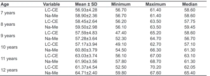
Stratifying the data by age, the minimum LC-CE distance was 52.30 mm at nine years of age and maximum LC-CE was 70.20 mm at 12 years of age. Minimum Na-Me distance was 56.70 mm at seven
years of age and maximum Na-Me was 68.70 mm at 11 years of age (Table 2).Pearson’s correlation was signiicant (0.47) for the correlation between the LC-CEand Na-Me measures.
Table 2 - LC-CE and Na-Me measures according to age
Age Variable Mean ± SD Minimum Maximum Median
7 years LC-CE 56.93±4.28 56.70 61.40 58.60
Na-Me 58.90±2.36 56.70 61.40 58.60
8 years LC-CE 58.45±2.64 56.20 63.50 57.75
Na-Me 59.50±2.98 56.10 63.50 59.45
9 years LC-CE 57.59±4.83 47.40 65.20 58.60
Na-Me 57.28±3.64 52.30 64.70 56.70
10 years LC-CE 57.17±3.94 49.10 62.70 57.10
Na-Me 60.80±3.79 54.50 66.30 61.30
11 years LC-CE 63.03±3.74 56.10 67.00 63.10
Na-Me 61.90±3.56 57.80 68.70 61.30
12 years LC-CE 61.37±4.54 52.50 70.20 62.05
Na-Me 64.71±2.40 59.80 67.60 65.40
Descriptive statistics of measures stratiied by age
LC-CE: lip commissure to corner of eye; Na-Me: nasium to mentum
Stratifying by gender, the correlation between LC-CEand Na-Me was non-signiicant for the male gender (p = 0.60959), but signiicant for the female gender (p=0.01126) (Table 4).
A statistically signiicant difference in mean VDO was found between participants with and without TMD in both genders (Table 5).
Table 3 - Difference between LC-CE and Na-Me for all ages together and separately
Age Variable t p-value
All ages LC-CE _Na-Me -2.60 0.0135
7 years LC-CE _Na-Me -0.53 0.6516
8 years LC-CE _Na-Me -0.69 0.4642
9 years LC-CE _Na-Me 0.19 0.8571
10 years LC-CE _Na-Me -2.47 0.0390*
11 years LC-CE _Na-Me 0.73 0.4949
12 years LC-CE _Na-Me -2.25 0.0509*
Paired t-test at 5% signiicance level
LC-CE: lip commissure to corner of eye; Na-Me: nasium to mentum
Table 4 - Comparison of mean LC-CE and Na-Me according to gender and age
Gender Age LC-CE (mm) Na-Me (mm)
Female
7 59.2±3.2 56.7±2.0
8 63.5±2.5 61.4±3.9
9 57.3±5.1 56.9±3.7
10 57.0±4.9 60.7±2.9
11 62.6±3.9 62.3±3.7
12 62.8±3.9 64.7±2.5
Male
7 55.8±5.4 57.5±3.2
8 57.4±1.0 60.0±2.0
9 60.0±2.9 59.1±3.2
10 57.6±1.4 60.2±2.3
11 65.8±1.0 61.0±6.0
12 58.1±5.0 59.3±3.7
ANOVA complemented by LSD test
LC-CE: lip commissure to corner of eye; Na-Me: nasium to mentum
Table 5 - Mean vertical dimension of occlusion in participants with and without temporomandibular disorder
N Mean Standard
deviation 95% conidence interval
Minimum mm
Maximum Mm
Without TMD 13 61.623 2.7860 59.940 63.307 56.1 65.7
With TMD 35 60.854 4.5072 59.306 62.403 52.0 70.0
Total 48 61.062 4.0984 59.872 62.253 52.0 70.0
ANOVA complemented by LSD test TMD: temporomandibular disorder
DISCUSSION
The results of the present study demonstrate different measures of VDOin all age groups analyzed(7 to 12 years). Signiicantly signiicant changes in VDOwere found in both genders at 10 and 12 years of age, which is likely due to the insta-bility in growth that occurs when the VDO increases
due to the eruption of the permanent premolars and second molars caused by active bone apposition and reposition21.
There is no consensus in the literature on the relation of VDO measures in the deciduous and mixed dentition phases21,22. In adults, however, this
may be caused by occlusal macro-traumas and micro-traumasthat affect chewing function and cause functional asymmetry of the stomatognathic system9,25,26.A number of studies have
demon-strated that there is no standardized method for the detection of TMD. One should also bear in mind that individual variation is an important factor to the development of TMD. In a recent systematic review involving 37 papers, the craniomandibular index was used in one paper, the Research Diagnostic Criteria for Temporomandibular Disorders was used in nine, a clinical patient-history protocol was used in 21 papers and Helkimodisorder index was used in six papers27.
TMD is a multifactor disorder with a psychogenic inluence that affects quality of life27and increases
with age in adolescence and young adulthood28.In
a recent study, Pereira et al.29 evaluated risks for the
development of TMDin adolescents and concluded that the gestational behavior of the mother, maloc-clusion, menarche and harmful oral habits were not associated with TMDand that gender alone was correlated with the incidence of TMDin 12-year-old patients.
Another study concluded that it is not possible to establish an association between TMDand orthodontic treatment, as occlusion may not be the main factor in the development of the disorder. In contrast, Laucis-Pinto30 and Bevilacqua31 state that
occlusion is indeed one of the etiological agents of TMD. In the present study, 12-year-old patients exhibited a statistically signiicant change in the difference between LC-CEand Na-Me measures, demonstrating a positive correlation between TMDandVDOin this age group.
For an effective diagnosis of TMD, it is important to evaluate all variables capable of inluencing the harmony of the stomatognathic system, including the variability in VDO, to prevent this disorder and allow craniofacial growth and development within the patterns of normality. Further studies with a larger number of individuals in each age group are needed to establish the actual mean VDOin children and adolescent during craniofacial development.
CONCLUSION
In the present study, a direct association was found between TMD and VDO, with positive correla-tions between the lip commissure to outer corner of eye and the nasium to mentum in the female sex as well as signiicant changes in the vertical dimension of occlusion at 10 and 12 years of age in both genders.
can function correctly, thereby avoiding future problems with malocclusion23,24.
In the present study, the smallest LC-CE distance was 47.40 mm at nine years of age and the largest was 70.20 mm at 12 years of age, whereas the smallest Na-Me distance was 52.30 mm at seven years of age and the largest was 68.70mm at 11 years of age. As each 3 mm in change in VDOleads to an approximate change of 0.8 mm in the central relation,20 it is important to maintain the ideal VDOin
accordance with age for a correct central relation, thereby avoiding problems with the occlusion and temporomandibular joint, as such problems can cause changes in the muscles and joints indicative of TMD3,11,21,22.
A previous study demonstrated that changes in the bite of more than or less than 2 mm can cause a 50% interincisal increase during intercuspation and a 40% increase in the resting position23, which can
impair facial esthetics19. Deiciencies in VDOcan
also have an negative impact on facial attractiveness and the development of malocclusions22.
A statistically signiicant association between VDOand age was found in both genders and an association was found with TMDin 35 children (72.91% of the sample). Moreover, children with TMD demonstrated greater variations in VDOin comparison to those without TMD. This variation ranged from 52 to 70 mmin children and adoles-cents with TMD and 56.1 to 65.70 mm among those without the disorder.
As the temporomandibular jointundergoes constant remodeling in adolescence, high preva-lence rates of signs and symptoms of TMD are found in this age group22,23. The present data are
in agreement with these indings, as 72.91% of the sample exhibited signs and symptoms of TMD. The results underscore the importance of an early diagnosis to impede the progress of the disorder and limit the harm caused to the stomatognathic system. On the other hand, the fact that the temporo-mandibular jointundergoes constant remodeling in childhood and adolescence may imply that TMDis not related to changes in VDO. A previous longi-tudinal study involving children treated with ortho-dontics/orthopedics of the jaws (which sometimes increases the VDO)followed up for a 30-year period concluded that one cannot afirm whether the treatment had a beneicial or harmful effect on the presence of TMD24.
REFERÊNCIAS
1. Knelbeman S. Method for determining vertical dimension. Patent Storm. 2006;1:14-8.
2. Spers F. Occlusion the new millennium: the controversy continuous. Spears Perspect. 2007;3(2):64-9.
3. Ciavarella D, Parziale V, Mastrovincenzo M, et al. Condylar Position Indicator and T-Scan system II in clinical evaluation of temporomandibular intracapsular disease. J Craniomaxillofac Surg. 2012;40(5):449-55.
4. Al-Ninri KS. Vertical Changes in class II division I maloclussion afterpremolar extraction. The Angle Ortho. 2008;76(1):52-8.
5. Ohnuki Y, Kawai N, Tanaka E, Langenbach GE, Tanne K, Saeki Y. Effects of increased occlusal vertical dimension on daily activity and myosin heavy chain composition in rat jaw muscle. Arch Oral Biol. 2009;54(8):783-9.
6. Krey KF, Dannhauer KH. Morphometric analysis of facial proile in adults. J Orofac Orthop. 2008;69(6):424-36.
7. Misch CE. Guidelines for maxillary incisal edge position-a pilot study: the key is the canine. J Prost. 2008;17(2):130-4.
8. Cutbirth ST. Increasing vertical dimension: considerations and steps in reconstruction of the severely worn dentition. Pract Proced Aesthet Dent. 2008;20(10):619-26.
9. Geerts GA, Stuhlinger ME, Nel DG. A comparison of the accuracy of two methods used by pre-doctoral students to measure vertical dimension. J Prosthet Dent. 2004;91(1):59-66.
10. Torii K, Chiwata I. Occlusal adjustment using the bite plate-induced occlusal position as a reference position for temporomandibular disorders: a pilot study. Head Face Med. 2010;6:5.
11. Manfredini D, Castrolorio T, Perinetti G, Guarda- Nardini L. Dental occlusion, body posture and temporomandibular disorders: where we are now and where we are heading for. J Oral Rehabil. 2012;39(6):463-71.
12. Marini I, Gatto MR, Bonetti GA. Effects of superpulsed low-level laser therapy on temporomandibular joint pain. Clin J Pain. 2010;26(7):611-6.
13. Andrade TNC, Frare JC. Estudo comparativo entre os efeitos de técnicas de terapia manual isoladas e associadas à laserterapia de baixa potência sobre a dor em pacientes com disfunção temporomandibular. Rev Gauch Odontol. 2008;56 (3):287-95.
14. Frare JC, Nicolau RA. Análise clínica do efeito da fotobiomodulação laser (GaAs – 904 nm) sobre a disfunção temporomandibular. Rev Bras Fisioter. 2008;12(1):37-42.
15. Okson JP. Tratamento das desordens temporomandibulares e oclusão. Rio de Janeiro-RJ: Ed. Elsevier; 2008.
RESUMO
Objetivo: veriicar se existe relação entre a presença de disfunção temporomandibular (DTM) relacio -nada à variabilidade da dimensão vertical de oclusão em crianças e adolescentes na faixa etária de 7 a 12 anos. Métodos: foram avaliadas 96 crianças e adolescentes do Instituto Rogacionista em São Paulo. A pesquisa diagnóstica da disfunção temporomandibular foi por meio do Índice de Helkimo e exame clínico posterior.Para a mensuração dos dados referentes à dimensão vertical foram empre-gadas as distâncias comissura labial - canto externo do olho e base do nariz – mento. A comparação dos valores médios das medidas antropométricas entre os gêneros e os grupos com e sem disfunção temporomandibular foi realizada empregando-se a análise de variância (ANOVA) complementada pelo teste least signiicance diference. Resultados: a dimensão vertical de oclusão demonstrou medidas distintas nas crianças e adolescentes em todas as faixas etárias avaliadas, foram obser-vadas alterações signiicantes dessa medida nas idades de 10 e 12 anos de ambos os gêneros.
Conclusão: pode -se concluir que na amostra estudada houve relação direta entre a presença de DTM e Dimensão vertical de oclusão (DVO), correlação positiva entre as medidas comissura labial – canto do olho externo e Násio – Mento no sexo feminino e alterações signiicantes na dimensão vertical de oclusão nas idades de 10 e 12 anos para ambos os sexos.
23. Fonseca DM, Bonafante G, Valle AL, Freitas SFT. Diagnóstico pela anamnese da disfunção craniomandibular. RGO. 1994;42(1):23-8.
24. Strajnić L, Stanisić-Sinobad D, Marković D, Stojanović L. Cephalometric indicators of the vertical dimension of occlusion. Coll Antropol. 2008; 32(2):535-41.
25. Lucena LBS, Kosmisky M, Costa LJ, Góes PSA. Validation of the portuguese version of theRDC/ TMD axis II questionnaire. Braz Oral Res. 2006; 20(4):312-7.
26. Dworkin SF, Huggins KH, Leresche L, von Korff M, Howard J, Truelove E, et al. Epidemiology of signs and symptoms in temporomandibular disordens: clinical signs in case and controls. J Am Dent Assoc. 1990;120(3):273-81.
27. Moyers RE. Etiologia da Maloclusão. In: Moyers, RE. Ortodontia. 4. ed. Rio de Janeiro: Guanabara Koogan. 1991. p. 212-37.
28. Toscano P, Defabianis P Clinical evaluation of temporomandibular disorders in children and adolescents: a review of the literature. Eur J Paediatr Dent. 2009;10(4):188-92.
29. Pereira LJ, Pereira-Cenci T, Del Bel Cury AA, Pereira SM, Pereira AC, Ambosano GM, Gavião MB. Risk indicators of temporomandibular disorder incidences in early adolescence. Pediatr Dent. 2010;32(4):324-8.
30. Laucis-Pinto S, Diegues MB, Ferreira SLM, Simonato CASS. Bruxismo em odontopediatria e sua correlação com hábitos orais. Rev Paul Odontol. 2000; 22(5):10-8.
31. Bevilaqua DG, et al. Atividade Eletromiográica dos músculos masseter e temporal anterior de crianças com mordida cruzada posterior unilateral (MCPu). Rev. Bras. Fisioter. 2005;9(2):257-63. 16. Carrasco TG, Mazzetto MO, Mazzetto
RG, Mestriner W. Low intensity laser therapy in temporomandibular disorder: a phase II double – blind study. J Craniomandibular Pract. 2008;26(4):274-81.
17. Motta LJ, Guedes CC, De Santis TO, Fernandes KP, Mesquita-Ferrari RA, Bussadori SK. Association between parafunctional habits and signs and symptoms of temporomandibular dysfunction among adolescents. Oral Health Prev Dent. 2013;11(1):3-7. 18. Motta LJ, Martins MD, Fernandes KP, Mesquita-Ferrari RA, Biasotto-Gonzalez DA, Bussadori SK. Craniocervical posture and bruxism in children. Physiother Res Int. 2011;16(1):57-61.
19. Egermark-Eriksson I, Carlsson GE, Magnusson T. A 20-year longitudinal study of subjective symptoms of temporomandibular disorders from childhood to adulthood. Acta Odontol Scand. 2001;59(1):40-8.
20. Thilander B, Rubio G, Pena L, de Mayorga C. Prevalence of temporomandibular dysfunction and its association with malocclusion in children and adolescents: an epidemiologic study related to speciied stages of dental development. Angle Orthod. 2002;72(2):146-54.
21. Sant’Ana, E. Avaliação comparativa do padrão de normalidade do peril facial em pacientes Brasileiros leucodermas e em Norte-Americanos. Rev Dent Press Ortodon Ortop Facial. 2009;14(1):80-9. 22. Chaves TC, Oliveira AS, Grossi DB. Principais instrumentos para avaliação da disfunção temporomandibular, parte II: critérios diagnósticos; uma contribuição para a parte clínica e de pesquisa. Fisio e Pesq. 2008;15(1):101-6.
Received on: February 12, 2013 Accepted on: May 22, 2013
Mailing address: Sandra Kalil Bussadori
Avenida Pompéia, 2186 - Sumarezinho São Paulo – SP – Brasil
CEP: 05022-001