135
Jornal Brasileiro de Patologia e Medicina Laboratorial
Diagnosis of the human fetal age based on the development
of the normal kidney
Diagnóstico da idade fetal humana baseado no desenvolvimento normal do rim
Helena Maria Lizardo-Daudt1 Maria Isabel Albano Edelweiss2 Fernanda Teixeira dos Santos3 Rita de Cassia Alves Schumacher4
y
key words
unitermos
abstract
Background and aims: The diagnosis of human fetal age is usually estimated based on the measurement of crown-rump length or crown-heel length and the weight of the fetus. However, this estimate is not totally accurate and sometimes is necessary to combine other data to determine the fetal age. An analysis of the normal embryological development of the kidney may assist in this determination. The histology of this process, although well described, lacks photographic documentation. We intend to fill this gap by providing histologists and pathologists, especially inexperienced ones, with information about the staging of the renal development through microphotography. The objective of the present study was to achieve greater accuracy for the diagnosis of human fetal age through the proposed classification and the photographic documentation presented. Material and methods: Normal embryological development of the human kidney was studied by light microscopy. The fetal period from 6 to 40 weeks of gestation was observed according the stage of maturity of glomeruli and tubules; localization of glomeruli, occurrence of nephrogenic tissue and cortico-medullary
differentiation. At least 5 different exams were observed from each week of development. Two hundred four exams were analyzed in the whole study. The histological characteristics were quantified and the process was documented by microphotography. Results and final considerations: The fetal development of the kidney was divided into 8 stages, which was documented through microphotography. Nephron structural formation occurred until the 34th week of prenatal development. From the 35th week on, tubules and glomeruli continued to mature without the formation of new nephrons. The proposed classification intends to improve the accuracy of the fetal age diagnosis.
Human fetal age
Fetal kidney
Staging of nephrogenesis
resumo
Introdução e objetivos: O diagnóstico da idade fetal humana é usualmente estimado com base em medidas de tamanho e peso do feto. No entanto, esta estimativa não é totalmente acurada, e muitas vezes é necessário combinar outros dados para determinar a idade fetal. A análise do desenvolvimento embriológico normal do rim pode auxiliar nesta determinação. A histologia deste processo, apesar de bem descrita, apresenta uma documentação fotográfica escassa. Pretende-se preencher esta lacuna fornecendo a histologistas e patologistas, especialmente aos inexperientes, informações sobre o desenvolvimento renal no período pré-natal através de microfotografias. O objetivo do presente estudo foi conceder maior acurácia ao diagnóstico da idade fetal humana através da classificação proposta e da documentação apresentada. Material e métodos: Necropsias de fetos humanos de 6 a 40 semanas de gestação foram estudadas através de microscopia óptica. O tecido renal foi analisado segundo a ocorrência de glomérulos e túbulos rudimentares, em diferenciação e maduros; distribuição espacial dos glomérulos no parênquima; presença de tecido nefrogênico; e diferenciação corticomedular. Foram analisados no mínimo cinco exames diferentes para cada semana de desenvolvimento, perfazendo um total de 204 exames. As características foram quantificadas e o processo documentado através de microfotografias. Resultados e considerações finais: O desenvolvimento fetal do rim humano foi dividido em oito estágios com base nas características histológicas e a quantificação realizada. Os estágios foram documentados através de microfotografias. A neoformação de néfrons ocorreu até a 34ª semana. A partir da 35ª semana, os túbulos e glomérulos continuam a amadurecer, não ocorrendo, no entanto, a formação de novos néfrons. A classificação proposta pretende conceder maior acurácia ao diagnóstico da idade fetal humana.Rio de Janeiro, v
. 38, n. 2, p. 135-139, 2002
1. Bióloga; mestre em Ciências Veterinárias; professora assistente do Centro de Ciências da Saúde, Universidade do Vale do Rio dos Sinos. 2. Doutora em Patologia; professora adjunta de Patologia da Universidade Federal do Rio Grande do Sul; médica patologista do Serviço de Patologia do Hospital de Clínicas de Porto Alegre (HCPA). 3. Bióloga; mestranda em Zoologia pela Pontifícia Universidade Católica do Rio Grande do Sul. 4. Médica residente do Serviço de Patologia do HCPA. Trabalho realizado no Serviço de Patologia do HCPA.
Recebido em 19/04/01 Aceito para publicação em 05/12/01
Idade fetal humana
Rim fetal
136
Rio de Janeiro, v
. 38, n. 2, 2002
Jornal Brasileiro de Patologia e Medicina Laboratorial
Introduction
The age of the human embryo after the first month is usually determined based on the crown-rump length (CRL – measurement from the vertex of the skull to the midpoint between the apices of the buttocks). The period from the beginning of the third month until the birth, called fetal period, is estimated using the CRL or the crown-heel length (CHL – measurement from the vertex of the skull to the heel) and the weight of the fetus. However there is consi-derable variation in embryo and fetal length and weight, and many times the estimate is not accurate. By combining other data such as morphological characteristics and other measurements such as head circumference and femur length, a reasonable determination of the age of the fetus can be formulated (1, 13). The analysis of the embryo-logical development of the organs may provide useful data to estimate the embryo and fetal age.
During human fetal development, the formation of the urinary system exhibits singular characteristics. Three slightly overlapping kidney systems are formed, two of them will disappear during the fetal life and the last one will be the permanent kidney. The pronephros, the first kidneys, are formed at the beginning of the fourth week of development and disappear at the end of the same week. They are rudimentary and nonfunctional. The second ones, the mesonephros, appear during the regression of pronephros. By the end of the eighth week they degenerate in the females. In males some caudal tubules and the mesonephric duct persist and participate in the formation of the genital system (9, 13). The metanephros appears after the fifth week as the final stage of kidney development, exhibiting a highly developed excretory function.
The metanephros presents a double embryological origin: the corpuscules and tubules derive from the metanephrogenic mesoderm, and the excretory structures, including the collectin tubules, calyces, pelvis and ureter, come from the ureteral bud. Nevertheless, both systems originate from the mesoderm (9, 13).
The complex events that accompany the development of each nephron start with the condensation of mesen-chymal cells of the metanephrogenic mesoderm in the terminal branches of the collecting conduits. Each cell cluster forms “heads” that end in ampoules (terminal dilatation) of the conduits. The accumulation of cells gives origin to metanephric vesicles around the ampoule mesh. A mantle of cells delimits the lumens of these vesicles as epithelial cells, forming primitive urinary tubules (10).
As the primitive urinary tubules form, each ampoule begins to undulate and to branch off. These branches, called arched collecting conduits, attach themselves to the primitive urinary tubules forming the future urinary tract. For each arched collecting conduit, a new larger ampoule forms that eventually attaches to another primitive urinary tubule (12). The cephalic part of the primitive tubule cur-ves in the shape of an “S”, and becomes the site of the future glomerulus.
The majority of the metanephrogenic cells differentiate into tubular cells and a few of these become mesenchymal cells distributed in the stroma of the metanephros.In humans, the formation of the nephrons continues until the final stages of intrauterine life (9). In the tenth fetal month, the metanephrogenic tissue disappears, suggesting that there is no postnatal nephron formation (12).
The embryological development of the organs of the human body has been studied throughout the modern era. With the advent of the light microscope, and later the electron microscope, many advances have been made. The histology of this development, although well described, lacks photographic documentation. We intend to fill this gap by providing histologists and pathologists with this information about the staging of this process through microphotography which may represent an additional tool for assisting in the estimation of fetal age.
Material and methods
All of the autopsy reports and anatomo-pathological exams filled by the Pathological Anatomy Sector of the University Hospital of Porto Alegre from January, 1982 to December, 1992 were reviewed. Reports of normal kidneys from patients aged 6 to 40 weeks were selected for study (at least 5 different cases from each week of development. Two hundred four exams were analyzed in the whole study). Fetal developmental age was estimated based on size, including crown-rump, crown-heel and weight. Organ histology was evaluated by examining two slides of each case stained with HE.
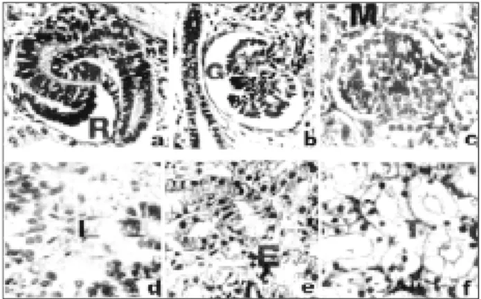
Grades ranging from 1 (absence) to 4 (maximum quantity) were assigned to each characteristic described as follows:
• rudimentary glomeruli (RG): cephalic part of the primitive urinary tubule in the process of curvature; • glomeruli in differentiation (EG): compacted capillary
137
Rio de Janeiro, v
. 38, n. 2, 2002
Jornal Brasileiro de Patologia e Medicina Laboratorial
• mature glomeruli (MG): normal histological aspect; • subcapsular glomeruli (SG): glomeruli forming a
subcapsular layer;
• cortical glomeruli (CG): glomeruli forming a cortical layer; • dispersed glomeruli (DG): glomeruli dispersed in the
parenchyma, not forming a distinct layer;
• rudimentary tubules (RT): absence of morphological differentiation between proximal and distal tubules; • tubules in differentiation (ET): brush border in
forma-tion, initiation of differentiation between proximal and distal tubules;
• mature tubules (MT): normal histological aspects; • nephrogenic tissue (NT): undifferentiated tissue; • cortico-medullary differentiation (D): sharp
differen-tiation between the cortical and medullary layer.
Figure 1 provides a general view of the different types of glomeruli and tubules described above.
The fetal period from 6th to 40th weeks was divided into different stages according the similarities observed between the analyzed exams. A normal histological pattern was established for each stage based on the microscopic observations. Each stage was documented by micropho-tography.
Results
The histological analysis of the metanephros based on the observation of the maturity of glomeruli and tubules, localization of glomeruli , presence of nephrogenic tissue and cortico-medullary differentiation combined with the quantification of these characteristics allowed the descri-ption of eight different stages of development.
Table shows the quantified characteristics taken from the histological analysis.
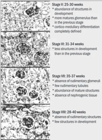
Figures 2 and 3 show a general view of the findings from the microscopic analysis of the various stages.
The rudimentary glomeruli, numerous in stage I, become scarce from stage IV on, disappearing after 35 weeks. The glomeruli in differentiation, present but few in number since stage I, are numerous in the 25 to 30 weeks period (stage V), after which they diminish in number. The mature glomeruli, absent in stage I are rare up to 24 weeks, are abundant only after 35 weeks, coinciding with the disappearance of rudimentary glomeruli.
Only in stage I do the glomeruli arrange so as to form a subcapsular layer. After stage II the glomeruli are disper-se in the parenchyma and a cortico-medullary differen-tiation starts, becoming very distinct after stage V. Rudi-mentary structures and nephrogenic tissue are absent in stage VIII, indicating proximity to the time of birth.
In general, the maturation of tubules and glomeruli are similar in time. However, the histological findings indicate that tubules start and finish their maturation qui-te earlier than glomeruli.
Discussion
The diagnosis of human fetal age is usually estimated based on the measurement of crown-rump length or crown-heel length and the weight of the fetus. However, this estimate is not totally accurate and sometimes is necessary to combine other data such as biparietal diameter, head and abdominal circumference and femur length to determine the fetal age (7, 13). The histological analysis may provide an additional tool, especially in fetuses from abortion or necropsy. On the other hand, the histological analysis may present variations, depending on the observer. In fact, many people have allowed the standardization of approaches to biopsy histology and reduced inter-observer and inter-departmental variations (6, 8, 14) It is very important, specially when inexperienced pathologists are the observers. The quantified charac-teristics combined with the photographic documentation presented in this paper will provide an additional tool for assisting in the estimation of fetal age more accurately.
Fetal developmental age was estimated in this report based on size, including crown-rump, crown-heel and weight. Considering this is the usual method to determine Figure 1 – Human fetal kidney. Detail of the different stages of maturation of
138
Rio de Janeiro, v
. 38, n. 2, 2002
Jornal Brasileiro de Patologia e Medicina Laboratorial
STG Weeks RG EG MG RT ET MT SG CG DG NT D
I 6-10 4 2 1 4 2 1 4 1 1 4 1
n = 25
II 11-15 3 3 2 3 3 2 1 3 2 3 2
n = 25
III 16-19 3 3 2 2 3 2 1 3 2 2 3
n = 20
IV 20-24 2 3 2 2 3 3 1 4 1 2 3
n = 25
V 25-30 2 4 3 2 4 3 1 4 1 2 4
n = 34
VI 31-34 2 3 3 2 3 3 1 4 1 2 4
n = 25
VII 35-37 1 3 4 2 3 4 1 4 1 1 4
n = 25
VIII 38-40 1 2 4 1 2 4 1 4 1 1 4
n = 25
STG – stage; weeks – weeks of gestation; RG – rudimentary glomeruli; EG – glomeruli in differentiation; MT – mature glomeruli; RT – rudimentary tubules; ET – tubules in differentiation; MT – mature tubules; SG – subcapsular glomeruli; CG – cortical glomeruli; NT – nephrogenic tissue; D – cortico-medullary differentiation, n – number of exams observed.
Quantitative values for each characteristic analyzed grouped by stage
Table
Figure 2 – Human fetal kidney. General view of the stages I, II, III and IV (6-24 weeks). R – rudimentary glomeruli; G – glomeruli in differentiation; L – rudimentary tubules; E – tubules in differentiation; N – nephrogenic tissue; D – cortico-medullary differentiation (HE 600x)
Figure 3 – Human fetal kidney. General view of the Stages V, VI, VII, VIII (26-40 weeks). R – rudimentary glomeruli; G – glomeruli in differentiation; M – mature glomeruli; L – rudimentary tubules; E-tubules in differentiation; T – mature tubules; D – cortico-medullary differentiation (HE 600x)
Stage I: 6-10 weeks
* numerous rudimentary structures * glomerulus in a subcapsular layer * abundance of nephrogenic tissue
Stage II: 11-15 weeks
* many structures in development * many rudimentary structures * start of differentiation between
cortical and medullar layers * few mature structures
Stage III: 16-19 weeks
* few rudimentary tubules * cortico-medullary differentiation
more evident than in the previous stage
* nephrogenic tissue becames sparse
Stage IV: 20-24 weeks
* few rudimentary glomeruli and tubules
* absence of dispersed glomeruli * more mature tubules than in the
previous stage
Stage V: 25-30 weeks
* abundance of structures in development
* more matures glomerulus than in the previous stage
* cortico-medullary differentiation completely defined
Stage VI: 31-34 weeks
* less structures in development than in the previous stage
Stage VIII: 28-40 weeks
* absence of rudimentary structures * few structures in development
Stage VII: 35-37 weeks
* absence of rudimentary glomeruli * few rudimentary tubules * abundance of mature structures * absence of nephrogenic tissue b
c
d a
b
c a
139
Rio de Janeiro, v
. 38, n. 2, 2002
Jornal Brasileiro de Patologia e Medicina Laboratorial
the fetal age in fetuses from abortion and necropsy, we assumed that the different authors employed the same method but other method had been cited.
The formation of nephrons is continuous until the fi-nal stages of intrauterine life in humans (9). According to Nash & Edelman (11), nephrogenesis is completed during the 35th week of gestation. Carlotti et al. (2) state that
nephrogenesis is completed until 32nd-34th week of
gestation. Our findings demonstrate that rudimentary tubules can be found up to the 37th week, but rudimentary
glomeruli are absent since the 35th week. Thus, the
neoformation of nephrons seems to be blocked from stage VI on, at least one month before the birth, a significant period of time. According to Netter (12), in the 10th fetal month, nephrogenic fetal tissue disappears, suggesting that there is no postnatal nephron development. Our observations show that nephrogenic tissue is absent from the 35th week, reinforcing our position that after this time there is not neoformation of nephrons.
Studies by various authors have stated that nephron
formation begins approximately on the 15th day of
gestation in rats (3) and during the 8th week in humans
(2, 4). Our observations demonstrate that from the 6th
week on, rudimentary glomeruli already exist, although the amount of nephrogenic tissue is highest.
James (5) states that the formation of new glomeruli and tubules occurs at the periphery of the growing kidney. Our findings agree with this observation. In stage I rudimentary structures form a subcapsular layer. In stage II one sees that the majority of rudimentary structure is indeed at the periphery of the organ and that the developing structures, at first dispersed through the kidney, form characteristic layers starting from stage IV. Probably, the dispersion is associated with the rapid growth of the organ during this phase.
The development of tubules and glomeruli occurs simultaneously (15). Our data indicate that the maturation of tubules begins a little before the maturation of glomeruli. Nevertheless, the presence of rudimentary glomeruli suggests an involution of the tubules formed after 34 weeks.
The staging of the formation process of the human kidney here presented shows neoformation of nephron structures up to the 34th week of prenatal development. After the 34th week, tubules and glomeruli continue maturing, but without the formation of new nephrons. This study, based on quantification of determined characteristics combine with photographic documentation permits better accuracy in the diagnosis of fetal age and should act as an additional reference in the study of renal malformations.
Refererences
1. Benson, C.B. & Doubilet, P.M. Sonographic prediction of gestational age ’91: accuracy of second-trimester and third trimester fetal measurements. AJR Am. J. Roentgenology, 157: 1275, 1991. 2. Carlotti, A.P.C.P. et al. O desenvolvimento renal: aspectos
anatômicos e funcionais. Medicina-R Preto, 26: 375-86, 1993. 3. Cheigon, M. et al. Localization of basement membrane glycoproteins in rat kidney during fetal development. Biol. Cell, 60: 49-56, 1987. 4. Clapp, W.L. & Tisher, C.C. Gross anatomy and development of the kidney. In: Tisher, C.C. & Brenner, B.M. (eds.) Renal pathology with clinical and functional correlations. Philadelphia: Lippincott, 1989. 5. James, J.A. Nefrourologia infantil. Barcelona: Salvat, 1974. 6. Kazi, J.I. et al. Diagnosis of early acute renal allograft rejection by
evaluation of multiple histological features using Bayesian belief network. J. Clin. Pathol., 51: 108-13, 1998.
7. Magro, G. et al. Immunohistochemical distribution of type VI collagen in developing human kidney. Histochemical Journal, 28: 385-90, 1996.
8. Marcussen, N. et al. Reproductibility of the Banff classification of renal allograft pathology: inter- and intra-observer variation.
Transplantation, 60: 1083-9, 1995.
9. Moore, K.L. Embriologia básica. Rio de Janeiro: Interamericana, 1984. 10. Mounier, F. & Gubier, M.C. Developement embriofetal du rein.
In: Habib, R. et al. (ed.) Nephrologie pediatrique. Paris, 1983.
11. Nash, M.A. & Edermann, C.M. The developing kidney. Nephron, 11: 71-90, 1973.
12. Netter, F.H. Riñones, ureteres y vejiga urinaria. Barcelona: Salvat, 1986. 13. Sadler, T.W. Langman’s medical embryology. Baltimore: Lippincott
Williams & Wilkins, 2000.
14. Solez, K. et al. International standardization of criteria for the histologic diagnosis of renal allogratf rejection: the Banff working classification of kidney transplant pathology. Kidney Intern., 44: 411-22, 1993.
15. Zamboni, L. Anatomy of the kidney, ureter, bladder and urethra: morphology and embriology. In: Massry, S.G. & Glassock, R.J.
Textbook of nephrology. Baltimore: Williams & Wilkins, 1989.
Endereço para correspondência
Helena Maria Lizardo-Daudt Centro de Ciências da Saúde Av. Unisinos 950