Appearance at the Proximal Carpal Tunnel
Ping Yeap Loh1*, Satoshi Muraki2
1Department of Human Science, Graduate School of Design, Kyushu University, Minami-ku, Fukuoka, Japan,2Department of Human Science, Faculty of Design, Kyushu University, Minami-ku, Fukuoka, Japan *[email protected]
Abstract
This study investigated the effects of wrist angle, sex, and handedness on the changes in the median nerve cross-sectional area (MNCSA) and median nerve diameters, namely lon-gitudinal diameter (D1) and vertical diameter (D2). Ultrasound examination was conducted to examine the median nerve at the proximal carpal tunnel in both dominant and nondomi-nant hands of men (n = 27) and women (n = 26). A total of seven wrist angles were exam-ined: neutral; 15°, 30°, and 45° extension; and 15°, 30°, and 45° flexion. Our results indicated sexual dimorphism and bilateral asymmetry of MNCSA, D1 and D2 measure-ments. MNCSA was significantly reduced when the wrist angle changed from neutral to flex-ion or extensflex-ion positflex-ions. At flexflex-ion positflex-ions, D1 was significantly smaller than that at neutral. In contrast, at extension positions, D2 was significantly smaller than that at neutral. In conclusion, this study showed that MNCSA decreased as the wrist angle changed from neutral to flexion or extension positions in both dominant and nondominant hands of both sexes, whereas deformation of the median nerve differed between wrist flexion
and extension.
Introduction
Over the past few decades, computers have become an essential tool in the workplace. The in-crease in computer use in daily life has caused an inin-crease in discomfort at the neck, shoulder, elbow, wrist, and hand due to repetitive and awkward joint positions [1]. Approximately 20% of computer users experience musculoskeletal disorders of the upper extremities, such as carpal tunnel syndrome (CTS) [2,3]. CTS—one of the most commonly reported work-related musculoskeletal disorders of the upper extremities—is a peripheral nerve compression syn-drome affecting the median nerve at the wrist carpal tunnel region [4]. The etiology of CTS is multifactorial, with mechanical compression stress on the median nerve considered one of the relevant factors. Bonfiglioli et al. [5] suggested that biomechanical stress and long hours of in-tensive manual work without adequate rest might cause impairment of the median nerve. The median nerve contributes to cutaneous sensation of the hand and fingers, as well as innervation to multiple hand muscles that control fine motor manipulation, including pinch and grip.
a11111
OPEN ACCESS
Citation:Loh PY, Muraki S (2015) Effect of Wrist Angle on Median Nerve Appearance at the Proximal Carpal Tunnel. PLoS ONE 10(2): e0117930. doi:10.1371/journal.pone.0117930
Academic Editor:Luigi Cattaneo, Università di Trento, ITALY
Received:July 16, 2014
Accepted:January 2, 2015
Published:February 6, 2015
Copyright:© 2015 Loh, Muraki. This is an open access article distributed under the terms of the
Creative Commons Attribution License, which permits unrestricted use, distribution, and reproduction in any medium, provided the original author and source are credited.
Data Availability Statement:All relevant data are within the paper and its Supporting Information files.
Funding:The authors received no specific funding for this work.
Competing Interests:The authors have declared
Thus, individuals with CTS demonstrate weakened grip and pinch strength and decreased thumb and finger dexterity, which can affect daily life and work-related activities [6].
The carpal tunnel is formed by carpal bones as the floor and the transverse carpal ligament as the roof; the edge of the retinaculum at the pisiform defines the proximal carpal tunnel [7,8]. The carpal tunnel is a confined space comprising the median nerve and a total of nine tendons, including the flexor pollicis longus, four flexor digitorum superficialis, and four flexor digi-torum profundus. The median nerve is located beneath and near the transverse carpal liga-ment, and is vulnerable to compression stress from intratunnel pressure and the surrounding structures. The carpal tunnel pressure at wrist normal posture was less than 15 mm Hg and the carpal tunnel pressure is elevated in flexion and extension wrist posture among non-CTS par-ticipants [8]. Previous studies have shown that individual or combined finger and thumb dy-namic movements impose compression stress on the median nerve due to gliding motion of the tendons, leading to deformation of the median nerve [9]. On the other hand, radiocarpal and midcarpal joints contribute to such wrist movements as flexion, extension, radial devia-tion, and ulnar deviation. Carpal tunnel volume is influenced by dynamic kinematic move-ments of the carpal bones during wrist flexion and extension motion [10].
Well-designed equipment and workspace are necessary for computer users to perform daily work in a good posture so as to reduce the risk of work-related musculoskeletal disorders. Poor ergonomics of the wrist during daily life and computer work causes varying degrees of com-pression over the carpal tunnel. Computer users show a various range of wrist flexion and ex-tension angles during typing work; the most commonly observed angle is 20° wrist exex-tension with 20° ulnar deviation [11]. Therefore, it is important to understand the relationship between wrist angle and compression stress on the median nerve at the carpal tunnel.
A previous feasibility study of 12 male participants showed significant reduction of the me-dian nerve cross-sectional area (MNCSA) at wrist flexion and extension positions compared with that at neutral [12]. In view of the small sample size and inclusion of only male partici-pants, the current study is an extension of the research by Loh et al.[12], which increases the sample size, includes both male and female participants, and examines the median nerve in both dominant and nondominant hands. Duncan et al. [13] described an ellipse formula to cal-culate MNCSA by using longitudinal and vertical diameters of the median nerve; however, it is unknown whether wrist angle affects the median nerve diameters.
The objective of this study was to investigate the effects of wrist angle, sex, and handedness on changes in MNCSA and median nerve diameters, namely longitudinal diameter (D1) and vertical diameter (D2).
Materials and Methods
Participants
Ultrasound examination
Ultrasound examinations were performed using the LOGIQ e ultrasound system with transducer 12L-RS with imaging frequency bandwidth is 5–13 MHz (GE Healthcare, USA). A 7.0-mm-thick sonar pad (Nippon BXI Inc., Tokyo, Japan) was used as a coupling medium to standardize coupling thickness among all participants. B-mode with 12 MHz was used during the examination, and depth resolution of the ultrasound beam was adjusted accordingly to obtain clear images of the me-dian nerve at the proximal carpal tunnel. The ultrasound images were obtained by same examiner.
Participants were examined in a seated position with the forearm in supination and rested on an arm support on the table. Participants were instructed to relax the forearm, wrist, and fingers during the examination. Examiner performed ultrasound examination on the passive wrist angle. During wrist angle positioning, examiner hold the palm across the heads of meta-carpals and applied gentle force to maintain the wrist angle.
The pisiform bonymark was used as a landmark to identify the proximal carpal tunnel. An L-shaped plastic frame was placed along the radius bone with the perpendicular point located at the pisiform; the frame served as a location marker for the ultrasound probe during the ex-amination. Ultrasound examination was performed for both dominant and nondominant wrists, and three images were taken for each wrist position. The ultrasound probe was removed and repositioned the probe for each image taking. The examination sequence of wrist position was as follows: neutral (0°); 15°, 30°, and 45° extension; and 15°, 30°, and 45° flexion. A 180° wrist goniometer was used to determine wrist angle. The triquetrum was used as an axis point for the goniometer, while the static arm of the goniometer was placed parallel to the ulnar bone and the moveable arm was placed parallel to the fifth metacarpal bone.
The median nerve was identified in the transverse plane across the proximal carpal tunnel. During ultrasound examination, the examiner identify the median nerve at the superficially level by a hypoechogenic rim which contained of the hypoechogenic nerve fascicles while the boundary of extraneural was recognized by hyperechogenic and thickened [18]. MNCSA was measured by a tracing method along the hypoechogenic boundary of the median nerve, and D1 and D2 were measured as described by Duncan et al. [13]. The D1 and D2 were identified by the longest perpendicular diameter of the median nerve.ImageJ[19] was used to calculate MNCSA, D1, and D2 (Fig. 1). Mean value of three images was calculated to represent the MNCSA, D1 and D2 at each wrist angles.
Deformation percentage
Deformation percentages were calculated to determine differences in MNCSA, D1, and D2 at different wrist angles (15°, 30°, and 45° of both flexion and extension) compared with that at
Table 1. Demographic data of participants (n = 53).
Male (n = 27) Female (n = 26)
Age (years) 24.9±2.8 24.5±3.2
Height (cm) 171.4±5.8 159.8±5.2
Weight (kg) 68.4±13.1 51.6±7.8
BMI (kg/m2) 23.3±4.2 20.3±3.1
Wrist Circumference (mm) Right 160.6±7.5 144.9±8.1
Left 158.9±7.0 142.5±8.9
Handedness Right hand dominant 23 23
Left hand dominant 4 3
neutral. The following equation was used:
deformation percentage ¼ wrist neutralwrist neutraldifferent wrist angles 100%
Statistical analysis
Statistical analysis was performed using SPSS version 21.0 software (IBM Corporation, Chi-cago, IL). All results were expressed as mean ± SD. Inter- and intrarater reliability were calcu-lated via reliability analysis in SPSS using 20 randomly selected ultrasound images. The Shapiro-Wilk normality test was conducted to examine the sample characteristics of MNCSA for both male and female participants in both dominant and nondominant hand group.
The pairedttest was used to analyze differences in MNCSA, D1, and D2 at neutral position between dominant and nondominant hands in each male and female group. Subsequently, an in-dependent samplesttest was used to analyze differences in MNCSA, D1, and D2 at neutral posi-tion between male and female participants in each dominant and nondominant hand group.
Three-way repeated analysis of variance (2 x 7 x 2 factorial) was conducted with wrist side (dominant and nondominant), wrist flexion-extension positions (neutral; 15°, 30°, and 45° flex-ion; and 15°, 30°, and 45° extension), and sex as factors to examine difference in MNCSA at seven wrist positions. The assumption of sphericity was violated, as indicated by Mauchly’s test; therefore, Greenhouse-Geisser correction was used in the analysis of variance. Post-hoc pairwise Bonferroni-corrected comparison was used to examine mean differences in the factors.
Results
Inter- and intrarater reliability
Inter- and intrarater reliability for MNCSA, D1, and D2 measurements were good to excellent, according to Fleiss et al. [20] (Table 2).
Fig 1. Ultrasound image of the median nerve at the proximal carpal tunnel.A) median nerve cross-sectional area by tracing method; B) longitudinal diameter; C) vertical diameter.
Sample characteristics
The Shapiro-Wilk’s test (p>0.05) [21,22] and visual inspection of histograms, normal Q-Q
plots, and box plots showed that the MNCSAs were approximately normally distributed and slightly skewed and kurtotic (Table 3) for both male and female participants in both dominant and nondominant hands [23–25].
Comparison of median nerve between dominant and nondominant
hands at neutral
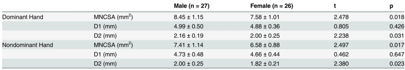
MNCSA, D1 and D2 of the dominant hand were significantly larger than those of the nondom-inant hand in both male and female participants (Table 4).
Comparison of median nerve between male and female participants at
neutral
MNCSA and D2 of male participants were significantly larger than those of female partici-pants, but there was no significant difference in D1 between sexes in each group of dominant and nondominant hands (Table 5).
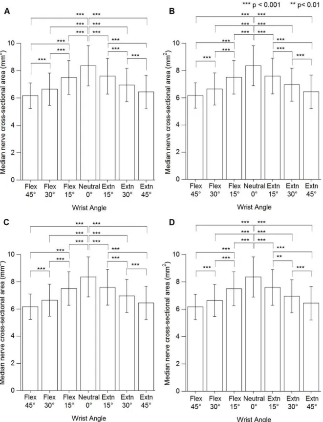
Change in MNCSA at different wrist angles
MNCSA at different wrist angles are presented inS1 Table. No significant interaction was found between wrist angle × handedness × sex (F [4.1, 208.8] = 1.355, p = 0.250). However, there was a significant interaction effect of wrist angle × handedness (F [4.1, 208.8] = 10.135, p<0.001) and wrist angle × sex (F [3.4, 174.0] = 4.772, p<0.01). For both male and female
participants, wrist angle had a significant effect on MNCSA, which became smaller when the wrist changed from neutral to flexion or extension positions in both dominant and nondomi-nant hands. A significant difference in MNCSA was found when comparing neutral position (0°) to 15°, 30°, and 45° flexion and to 15°, 30°, and 45° extension (Fig. 2).
Changes in D1 and D2 at different wrist angles
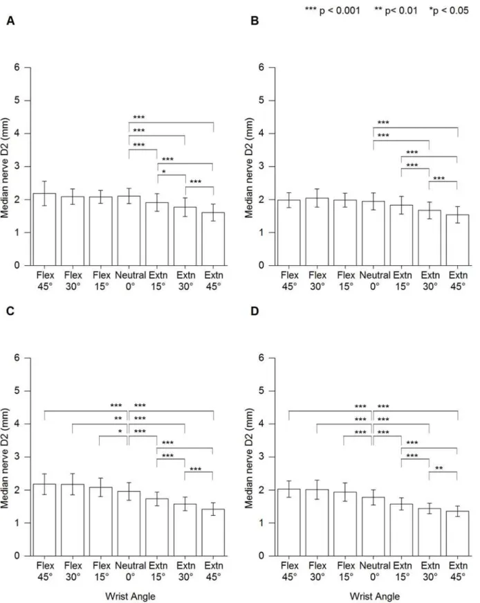
Mean D1 and D2 at different wrist angles are presented inS2andS3Tables, respectively. The re-sults showed no significant interaction between wrist angle × handedness × sex for both D1 and D2 (D1: F [4.0, 202.8] = 0.704, p = 0.590; D2: F [4.6, 233.6] = 1.313, p = 0.262). However, a significant interaction was found for the effect of wrist angle × handedness (D1: F [4.0, 202.8] = 2.429, p<0.05; D2: F [4.6, 233.6] = 2.389, p<0.05) and wrist angle × sex (D1: F [2.5, 146.0] =
7.817, p<0.001; D2: F [2.532, 129.1] = 9.621, p<0.001). Wrist flexion had a significant
influ-ence on D1, which became smaller compared with that at neutral for all participants. However,
Table 2. Inter- and intrarater reliability.
Inter-rater reliability Intra-rater reliability
MNCSA (mm2) 0.838 0.904
D1 (mm) 0.671 0.855
D2 (mm) 0.706 0.890
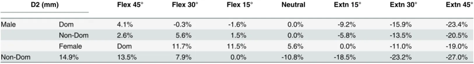
wrist 15° and 30° extension showed a significant increase in D1 at nondominant hand of female participants (Fig. 3). Comparatively, wrist flexion showed a significant increase in D2 compared with that neutral among female participants. However, wrist extension caused a significant de-crease in D2 for all participants (Fig. 4).
Deformation percentages of MNCSA, D1 and D2
Deformation percentage of the MNCSA at 15°, 30°, and 45° flexion were approximately-8%, -15%, and-21%, respectively, while that at 15°, 30°, and 45° extension were-7%, -14%, and-20%, respectively (Table 6). Furthermore, an increase of 15° in wrist flexion or extension caused signif-icant reduction and higher deformation percentage of the MNCSA (Fig. 2&Table 6).
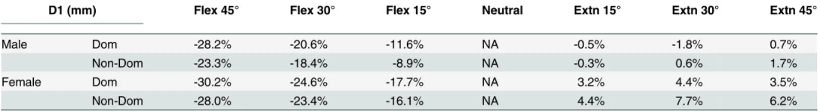
At 15°, 30°, and 45° extension, both increase percentages of D1 and decrease percentages of D2 among female participants were higher than male participants (Tables7&8). In contrast to wrist extension, at 15°, 30°, and 45° flexion, the decrease percentages of D1 and the increase per-centages of D2 among female participants were higher than male participants (Tables7&8).
Discussion
Ultrasound imaging of the median nerve at carpal tunnel level
Various imaging techniques such as ultrasound, magnetic resonance imaging (MRI) and com-puterized tomography have been used in understand the carpal tunnel anatomy and the char-acteristic of median nerve among healthy person and CTS patients meanwhile [26–28]. MRI demonstrate high ability to observe the pathological changes of median nerve characteristic and the bowing of transverse carpal ligament among CTS patients [29–31]. Duymus et al. [26] found no significant differences of the MNCSA between both ultrasound and MRI
Table 3. Normality test for median nerve cross-sectional area.
Wrist Skewness (M±SE) Kurtosis (M±SE) Shapiro-Wilk Test (p value)
Male Dominant 0.08±0.45 0.53±0.87 0.637
Nondominant -0.15±0.45 -0.07±0.87 0.816
Female Dominant 0.01±0.46 -1.04±0.89 0.463
Nondominant 0.06±0.46 -0.89±0.89 0.500
M = mean; SE = standard error doi:10.1371/journal.pone.0117930.t003
Table 4. Comparison between dominant and nondominant hands.
Dominant hand Nondominant hand t p
Male MNCSA (mm2) 8.45±1.15 7.41±1.14 5.045 0.000
D1 (mm) 4.99±0.50 4.73±0.48 2.070 0.052
D2 (mm) 2.16±0.19 2.00±0.25 2.472 0.023
Female MNCSA (mm2) 7.58±1.01 6.58±0.88 6.035 0.000
D1 (mm) 4.88±0.36 4.66±0.44 2.080 0.053
D2 (mm) 2.00±0.25 1.82±0.21 4.245 0.001
measurement. The mean value of MNCSA at wrist neutral position in this study was close to the results reported in the MRI, which were 8.8 mm² at distal carpal tunnel and 10.0 mm² at proximal carpal tunnel [27]. Ultrasound imaging is one of the inexpensive and easy method to understand the behavior of median nerve under most circumstances such as real time evalua-tion during dynamic wrist and finger joints changes.
Sex differences and bilateral asymmetry of the median nerve
It is well known that sex differences affect physical anthropological measurements, such as wrist circumference, arm perimeter, height, waist circumference, and body mass [32–35]. However, there has not been much research on sexual dimorphism of the upper limb peripher-al nervous system. For one, the cross-sectionperipher-al area of the ulnar nerve at the cubitperipher-al tunnel in male adults is larger than that in female adults [36]. In this study, through ultrasound examina-tion of the median nerve, our results showed that MNCSA and D2 of male participants were significantly larger than those of female participants (Table 5). Although D1 of men was larger than that of women, the difference was not significant. On the other hand, Moriyama [37] sug-gested no significant difference in peripheral nerves between men and women by microscopic examination of number of myelinated axons, average transverse area, and circularly ratios of myelinated axons. Based on cross-sectional area measurement, our results suggest sexual di-morphism of the median nerve at the proximal carpal tunnel.
Human upper limb anthropological measurements, such as arm length, elbow breadth, and hand bone sizes, show bilateral asymmetric features regardless of sex differences [38,39]. Our results showed significant bilateral asymmetry in MNCSA, D1 and D2 measurements in both male and female participants. Overall, our results of MNCSA, D1, and D2 measurements sug-gest bilateral asymmetric features of the median nerve.
Relationship between wrist angle and MNCSA
Tendon excursion during active finger motion can cause deformation of the median nerve within the carpal tunnel [40]. However, the effect of passive wrist posture on the median nerve when the fingers are relaxed has not been clarified. During wrist motion, the contribution of radiocarpal and midcarpal joints in wrist flexion are 40% and 60%, respectively, while that in wrist extension are 66.5% and 33.5%, respectively [41]. The interaction between carpal bones and ligaments during flexion and extension causes the carpal bones to change position [42]; as a result, the relative locations of tendons and the median nerve at the carpal tunnel region may
Table 5. Comparison between male and female participants.
Male (n = 27) Female (n = 26) t p
Dominant Hand MNCSA (mm2) 8.45±1.15 7.58±1.01 2.478 0.018
D1 (mm) 4.99±0.50 4.88±0.36 0.805 0.426
D2 (mm) 2.16±0.19 2.00±0.25 2.238 0.031
Nondominant Hand MNCSA (mm2) 7.41±1.14 6.58±0.88 2.497 0.017
D1 (mm) 4.73±0.48 4.66±0.44 0.462 0.647
D2 (mm) 2.00±0.25 1.82±0.21 2.380 0.023
Fig 2. Median nerve cross-sectional area at different wrist angles.(A) Male dominant hand. (B) Male nondominant hand. (C) Female dominant hand. (D) Female nondominant hand. Extn = extension, Flex = flexion.
Fig 3. Median nerve longitudinal diameter (D1) at different wrist angles.(A) Male dominant hand. (B) Male nondominant hand. (C) Female dominant hand. (D) Female nondominant hand. Extn = extension, Flex = flexion.
Fig 4. Median nerve vertical diameter (D2) at different wrist angles.(A) Male dominant hand. (B) Male nondominant hand. (C) Female dominant hand. (D) Female nondominant hand. Extn = extension, Flex = flexion.
change. Therefore, we examined the effects of passive wrist flexion and extension angles on MNCSA.
FromS1 Table, neutral position showed the largest MNCSA compared with that at all flex-ion and extensflex-ion positflex-ions in both dominant and nondominant hands of both sexes. MNCSA decreased significantly as the wrist moved from neutral to extension or flexion positions. Mogk and Keir [28] reported that carpal tunnel volume was largest at neutral position compared with that at 30° flexion or extension. Carpal tunnel volume decreased by approximately 7.0% and 6.4% when the wrist moved from neutral to 30° extension and 30° flexion, respectively [10]. At 45° flexion or extension, deformation ratio was highest and MNCSA became smallest com-pared with that at other wrist angles. This could be due to carpal tunnel volume further de-creasing as the wrist angle changed to 45° flexion or extension. Based on these results, it is important to keep the wrist near neutral position during daily work to prevent compression stress at the carpal tunnel.
Relationships between wrist angle and D1 and D2
Peripheral nerves, such as the median nerve, show different unique characteristics in response to biomechanical stress, such as tensile stress and compression stress [43]. Biomechanical stress can be caused by joint angle, muscle contraction, and/or external compression force over the region [43]. The epineurium—the outermost layer of a peripheral nerve where there is loose connective tissue contained between the fascicles—allows the nerve to glide among the sur-rounding tissues, such as tendons. Consequently, the median nerve can be elongated and short-ened during wrist extension and flexion, respectively. During extension, the median nerve is under strain stress, resulting in elongation into the palm area, while the surrounding tendons may impose shear force on the nerve [43]. The loose connective tissues are important for vol-ume adaptation of the nerve during transverse compression stress [44]. Such volume adapta-tion also causes the shape of the MNCSA to change in response to biomechanical stress.
Table 6. Deformation percentage of median nerve cross-sectional area at different wrist positions compared with at neutral.
MNCSA (mm2) Flex 45° Flex 30° Flex 15° Neutral Extn 15° Extn 30° Extn 45°
Male Dom -25.6% -20.2% -11.8% NA -8.8% -16.5% -22.6%
Non-Dom -20.9% -12.8% -7.6% NA -6.5% -14.0% -19.1%
Female Dom -21.6% -15.4% -7.6% NA -8.1% -14.9% -23.4%
Non-Dom -16.9% -12.0% -5.1% NA -7.0% -11.1% -17.8%
Dom = dominant hand; Non-Dom = nondominant hand; Flex =flexion; Extn = extension; NA = Not applicable doi:10.1371/journal.pone.0117930.t006
Table 7. Deformation percentage of median nerve longitudinal diameter at different wrist positions compared with at neutral.
D1 (mm) Flex 45° Flex 30° Flex 15° Neutral Extn 15° Extn 30° Extn 45°
Male Dom -28.2% -20.6% -11.6% NA -0.5% -1.8% 0.7%
Non-Dom -23.3% -18.4% -8.9% NA -0.3% 0.6% 1.7%
Female Dom -30.2% -24.6% -17.7% NA 3.2% 4.4% 3.5%
Non-Dom -28.0% -23.4% -16.1% NA 4.4% 7.7% 6.2%
At wrist extension positions, the carpal tunnel pressure increased due to the incursion of finger flexor muscles into the proximal carpal tunnel which carpal tunnel volume decreased due to the flattening effect [8]. Furthermore, the transverse contraction stress and longitudinal tensile stress cause median nerve excursion and elongation [45]. Therefore, the biomechanical stress during passive wrist extension may cause the median nerve deformed in which the diam-eter increases longitudinally and decreases vertically, as found in this study.
During wrist flexion and finger flexion positions, the median nerve is exposes to the compression from the nine tendons within the carpal tunnel due to decrease carpal tunnel volume and the incur-sion of lumbrical muscles into the carpal tunnel as a result of finger flexion [8,10,28]. Carpal tunnel depth at wrist flexion the carpal tunnel depth is larger and the carpal tunnel width is smaller com-pare to wrist neutral [28]. The result of narrowed width of carpal tunnel may cause the decrease of longitudinal diameter of the median nerve due to the compression in longitudinal direction.
In all groups, wrist flexion positions showed a significant influence on D1, while wrist exten-sion positions showed a significant influence on D2. Markedly, changes in D2 among female participants were significant at both wrist extension and flexion positions (Figs.2&3) could be due to the female has smaller proximal carpal tunnel compare to male [8]. Therefore, change in D1 during wrist flexion and change in D2 during wrist extension can use as an indicator for median nerve compression during changes in wrist angle since D1 is more sensitive to wrist flexion and D2 is more sensitive to wrist extension.
CTS has a multifactorial etiology and mechanical compression stress on the median nerve is one of the relevant factors. Several studies reported that the pathophysiology of CTS involves a combination of mechanical and changes in synovial tissues within carpal tunnel [8,46,47]. The carpal tunnel pressure can be affected by both external and/or internal compression. Ex-ternal compression associated with such as low force for a long period, repetitive joint move-ments, direct contact pressure over volar wrist and vibration exposure [8,46,47]. On the other hand, the morphological and biochemical changes in synovial tissues such as transverse carpal ligament thickened, inflammation of tendon synovial sheath, edema and fibrosis [8,46].
Accumulated strain and stress over the wrist can lead to musculoskeletal disorders. Although we have not yet concluded the most suitable wrist angles to minimize stress on the median nerve during work, these findings could be applied in future research to investigate changes in the me-dian nerve during specific work tasks. Furthermore, future research could be conducted on elder-ly individuals in the workplace. Better understanding of these changes will help to identify risk factors for musculoskeletal disorders and to apply preventive measures among computer users.
Conclusion
The effect of wrist angle on deformation of the median nerve at the proximal carpal tunnel was investigated in this study. Our results showed that 15°, 30°, and 45° of wrist flexion or extension
Table 8. Deformation percentage of median nerve vertical diameter at different wrist positions compared with at neutral.
D2 (mm) Flex 45° Flex 30° Flex 15° Neutral Extn 15° Extn 30° Extn 45°
Male Dom 4.1% -0.3% -1.6% 0.0% -9.2% -15.9% -23.4%
Non-Dom 2.6% 5.6% 1.5% 0.0% -5.8% -13.5% -20.5%
Female Dom 11.7% 11.5% 5.6% 0.0% -11.0% -19.0%
Non-Dom 14.9% 13.5% 7.9% 0.0% -10.8% -18.5% -23.2% -27.0%
causes various changes in MNCSA and median nerve diameter measurements compared with those at neutral. It was shown that at wrist flexion positions, D1 became shorter and D2 be-came longer, whereas at extension positions, D1 bebe-came longer and D2 bebe-came shorter.
Supporting Information
S1 Table. Median nerve cross-sectional area (MNCSA) (mm2) at different wrist positions.
(DOCX)
S2 Table. Median nerve longitudinal diameter (D1) (mm) at different wrist positions.
(DOCX)
S3 Table. Median nerve vertical diameter (D2) (mm) at different wrist positions.
(DOCX)
Acknowledgments
The authors thank all those who volunteered to participate in this study.
Author Contributions
Conceived and designed the experiments: PYL SM. Performed the experiments: PYL. Analyzed the data: PYL. Contributed reagents/materials/analysis tools: PYL SM. Wrote the paper: PYL SM.
References
1. Toosi KK, Impink BG, Baker NA, Boninger ML (2011) Effects of computer keyboarding on ultrasono-graphic measures of the median nerve. Am J Ind Med 54: 826–833. doi:10.1002/ajim.20983PMID:
21739468
2. Baker NA, Cham R, Cidboy EH, Cook J, Redfern MS (2007) Kinematics of the fingers and hands during computer keyboard use. Clin Biomech (Bristol, Avon) 22: 34–43. PMID:17052825
3. Sauter SL, Schleifer LM, Knutson SJ (1991) Work posture, workstation design, and musculoskeletal discomfort in a VDT data entry task. Hum Factors 33: 151–167. PMID:1860702
4. Pratt NE (2011) Chapter 1—atlas on regional anatomy of the neck, axilla, and upper extremity. In: Skir-ven TM, Osterman AL, Fedorczyk J, Amadio PC, editors. Rehabilitation of the hand and upper extremi-ty. Philadelphia: Elsevier/Mosby.
5. Bonfiglioli R, Mattioli S, Fiorentini C, Graziosi F, Curti S, et al. (2007) Relationship between repetitive work and the prevalence of carpal tunnel syndrome in part-time and full-time female supermarket ca-shiers: A quasi-experimental study. Int Arch Occup Environ Health 80: 248–253. PMID:16865405
6. Viera AJ (2003) Management of carpal tunnel syndrome. Am Fam Physician 68: 265–272. PMID: 12892346
7. Presazzi A, Bortolotto C, Zacchino M, Madonia L, Draghi F (2011) Carpal tunnel: Normal anatomy, ana-tomical variants and ultrasound technique. J Ultrasound 14: 40–46. doi:10.1016/j.jus.2011.01.006 PMID:23396809
8. Keir PJ, Rempel DM (2005) Pathomechanics of peripheral nerve loading: Evidence in carpal tunnel syndrome. Journal of Hand Therapy 18: 259–269. PMID:15891983
9. van Doesburg MH, Yoshii Y, Villarraga HR, Henderson J, Cha SS, et al. (2010) Median nerve deforma-tion and displacement in the carpal tunnel during index finger and thumb modeforma-tion. J Orthop Res 28: 1387–1390. doi:10.1002/jor.21131PMID:20225286
10. Mogk JPM, Keir PJ (2009) The effect of landmarks and bone motion on posture-related changes in car-pal tunnel volume. Clin Biomech 24: 708–715. doi:10.1016/j.clinbiomech.2009.05.012PMID:
19656596
11. Donoghue MF, O’Reilly DS, Walsh MT (2013) Wrist postures in the general population of computer users during a computer task. Appl Ergon 44: 42–47. doi:10.1016/j.apergo.2012.04.009PMID:
12. Loh PY, Nakashima H, Muraki S (2014) Effect of different wrist positions on median nerve cross-sectional area at proximal carpal tunnel. In: Bridging Research and Good Practices towards Patients Welfare: Proceedings of the 4th International Conference on Healthcare Ergonomics and Patient Safe-ty (HEPS).: CRC Press. pp. 149–154.
13. Duncan I, Sullivan P, Lomas F (1999) Sonography in the diagnosis of carpal tunnel syndrome. AJR Am J Roentgenol 173: 681–684. PMID:10470903
14. Bakhsh H, Ibrahim I, Khan W, Smitham P, Goddard N (2012) Assessment of validity, reliability, respon-siveness and bias of three commonly used patient-reported outcome measures in carpal tunnel syn-drome. Ortop Traumatol Rehabil 14: 335–340. doi:10.5604/15093492.1005085PMID:23043056
15. LaJoie AS, McCabe SJ, Thomas B, Edgell SE (2005) Determining the sensitivity and specificity of com-mon diagnostic tests for carpal tunnel syndrome using latent class analysis. Plast Reconstr Surg 116: 502–507. PMID:16079681
16. Sambandam SN, Priyanka P, Gul A, Ilango B (2008) Critical analysis of outcome measures used in the assessment of carpal tunnel syndrome. Int Orthop 32: 497–504. PMID:17370071
17. Oldfield RC (1971) The assessment and analysis of handedness: The edinburgh inventory. Neuropsy-chologia 9: 97–113. PMID:5146491
18. Kele H (2012) Ultrasonography of the peripheral nervous system. Perspectives in Medicine 1: 417–421.
19. Schneider CA, Rasband WS, Eliceiri KW (2012) NIH image to ImageJ: 25 years of image analysis. Nat Methods 9: 671–675. PMID:22930834
20. Fleiss JL, Levin B, Paik MC (2003; 2004) The measurement of interrater agreement. In: Anonymous Statistical Methods for Rates and Proportions.: John Wiley & Sons, Inc. pp. 598–626.
21. Razali N, Wah YB (2011) Power comparisons of shapiro-wilk, kolmogorov-smirnov, lilliefors and anserson-darling tests. Journal of Statistical Modeling and Analytics 2: 21–33.
22. Shapiro SS, Wilk MB (1965) An analysis of variance test for normality (complete samples). Biometrika 52: 591–611.
23. Cramer D (1998) Fundamental statistics for social research. Step-by-step calculations and computer techniques using SPSS for Windows. London and New York: Routledge.
24. Cramer D, Howitt D, editors. (2004) The SAGE dictionary of statistics. SAGE publications, ltd.
25. Doane DP, Seward LE (2011) Measuring skewness: A forgotten statistic? Journal of Statistics Education 19.
26. DuymuşM, Ulaşli AM, Yilmaz Ö, Asal N, Kacar M, et al. (2013) Measurement of median nerve cross sectional area with ultrasound and MRI in idiopathic carpal tunnel syndrome patients. J Neurol Sci-Turk 30(1): 059–071.
27. Yao L, Gai N (2009) Median nerve cross-sectional area and MRI diffusion characteristics: Normative values at the carpal tunnel. Skeletal Radiol 38: 355–361. doi:10.1007/s00256-008-0626-1PMID: 19132371
28. Mogk JPM, Keir PJ (2007) Evaluation of the carpal tunnel based on 3-D reconstruction from MRI. J Bio-mech 40: 2222–2229. PMID:17166503
29. Jarvik JG, Yuen E, Kliot M (2004) Diagnosis of carpal tunnel syndrome: Electrodiagnostic and MR im-aging evaluation. Neuroimim-aging Clin N Am 14: 93–102, viii. PMID:15177259
30. Pasternack II, Malmivaara A, Tervahartiala P, Forsberg H, Vehmas T (2003) Magnetic resonance imag-ing findimag-ings in respect to carpal tunnel syndrome. Scand J Work Environ Health 29: 189–196. PMID: 12828388
31. Jarvik JG, Yuen E, Haynor DR, Bradley CM, Fulton-Kehoe D, et al. (2002) MR nerve imaging in a prospec-tive cohort of patients with suspected carpal tunnel syndrome. Neurology 58: 1597–1602. PMID:12058085
32. Fragala MS, Clark MH, Walsh SJ, Kleppinger A, Judge JO, et al. (2012) Gender differences in anthro-pometric predictors of physical performance in older adults. Gender Medicine 9: 445–456. doi: 10.1016/j.genm.2012.10.004PMID:23123187
33. Lopes MM, Lawson W, Scott T, Keir PJ (2011) Tendon and nerve excursion in the carpal tunnel in healthy and CTD wrists. Clin Biomech (Bristol, Avon) 26: 930–936. doi:10.1016/j.clinbiomech.2011. 03.014PMID:21550703
34. Perissinotto E, Pisent C, Sergi G, Grigoletto F, ILSA Working Group (Italian Longitudinal Study on Age-ing). (2002) Anthropometric measurements in the elderly: Age and gender differences. Br J Nutr 87: 177–186. PMID:11895170
36. Jacob D, Creteur V, Courthaliac C, Bargoin R, Sassus B, et al. (2004) Sonoanatomy of the ulnar nerve in the cubital tunnel: A multicentre study by the GEL. Eur Radiol 14: 1770–1773. PMID:15258824 37. Moriyama H (2013) The relationship between sexually dimorphic peripheral nerves and diseases,
sexu-al dimorphism, prof. hiroshi moriyama (ed.). In: Moriyama Hiroshi Prof, editor. Sexusexu-al Dimorphism.: InTech. 10.5772/56080. Available: http://www.intechopen.com/books/sexual-dimorphism/the-relationship-between-sexually-dimorphic-peripheral-nerves-and-diseasesviatheInternet.
38. Klauser AS, Halpern EJ, De Zordo T, Feuchtner GM, Arora R, et al. (2009) Carpal tunnel syndrome as-sessment with US: Value of additional cross-sectional area measurements of the median nerve in pa-tients versus healthy volunteers. Radiology 250: 171–177. doi:10.1148/radiol.2501080397PMID: 19037017
39. Roy TA, Ruff CB, Plato CC (1994) Hand dominance and bilateral asymmetry in the structure of the sec-ond metacarpal. Am J Phys Anthropol 94: 203–211. PMID:8085612
40. Lopez PM, Fernandez-Ballesteros R, Zamarron MD, Lopez SR (2011) Anthropometric, body composi-tion and health determinants of active ageing: A gender approach. J Biosoc Sci 43: 597–610. doi:
10.1017/S0021932011000228PMID:21729364
41. Sarrafian SK, Melamed JL, Goshgarian GM (1977) Study of wrist motion in flexion and extension. Clin Orthop Relat Res (126: ): 153–159. PMID:598105
42. Savelberg HH, Kooloos JG, De Lange A, Huiskes R, Kauer JM (1991) Human carpal ligament recruit-ment and three-dimensional carpal motion. J Orthop Res 9: 693–704. PMID:1870033
43. Topp KS, Boyd BS (2012) Peripheral nerve: From the microscopic functional unit of the axon to the bio-mechanically loaded macroscopic structure. Journal of Hand Therapy 25: 142–152. doi:10.1016/j.jht. 2011.09.002PMID:22133662
44. Millesi H, Zoch G, Reihsner R (1995) Mechanical properties of peripheral nerves. Clin Orthop Relat Res (314: ): 76–83. PMID:7634654
45. Topp KS, Boyd BS (2006) Structure and biomechanics of peripheral nerves: Nerve responses to physi-cal stresses and implications for physiphysi-cal therapist practice. Phys Ther 86: 92–109. PMID:16386065
46. Uchiyama S, Itsubo T, Nakamura K, Kato H, Yasutomi T, et al. (2010) Current concepts of carpal tunnel syndrome: Pathophysiology, treatment, and evaluation. J Orthop Sci 15: 1–13. doi:
10.1007/s00776-009-1416-xPMID:20151245