Image-guided percutaneous procedures in deep pelvic
sites: review of the main approaches*
Procedimentos percutâneos pélvicos guiados por imagem: revisão das principais vias de acesso
Rodrigo Gobbo Garcia1
, Carlos Leite de Macedo Filho2
, Alexandre Maurano2
, Miguel José Francisco Neto3
, Mauro Miguel Daniel3
, Laercio Alberto Rosemberg3
, Marcelo Buarque de Gusmão Funari4
Image-guided percutaneous procedures have increasingly been established as safe and effective interventional tools in the diagnosis and management of masses and collections in several body segments. However, lesions in deep pelvic sites still pose a challenge for radiologists because of overlying anatomic structures. The success of a percutaneous biopsy depends on a safe access route planning based on a deep understanding of cross sectional anatomy of the pelvis. Anterior and lateral transabdominal, anterolateral extraperitoneal, transvaginal, transrectal and transgluteal approaches are described. The present study was aimed at reviewing the normal pelvic cross-sectional anatomy, demonstrating the different access routes for ultrasonography and computed tomography guided pelvic biopsies and drainages as well as discussing the main advantages and complications associated with these approaches.
Keywords: Biopsies; Percutaneous drainage; Interventional procedures; Pelvic anatomy; Pelvic abscesses.
Os procedimentos percutâneos orientados por imagem têm ganhado espaço crescente na radiologia inter-vencionista, constituindo ferramenta eficaz para a abordagem diagnóstica e terapêutica de massas e cole-ções nos diversos segmentos corporais. No entanto, localizacole-ções pélvicas profundas ainda representam grande desafio para o radiologista, por causa da interposição de estruturas anatômicas. Para que o procedimento seja bem sucedido é fundamental o planejamento da via de acesso baseado no conhecimento detalhado da anatomia radiológica da pelve. As principais vias de acesso para a abordagem destas lesões são: transabdo-minais (anterior e lateral), extraperitoneal ântero-lateral, transvaginal, transretal e transglútea. O objetivo deste trabalho é fazer uma revisão da anatomia seccional pélvica normal, demonstrando as diversas vias de acesso para biópsias e drenagens guiadas pela ultra-sonografia e pela tomografia computadorizada, bem como discutir as principais vantagens e complicações potenciais de cada uma delas.
Unitermos: Biópsias; Drenagem percutânea; Procedimentos intervencionistas; Anatomia pélvica; Abscessos pélvicos.
Abstract
Resumo
* Study developed in the Department of Radiology and Image-Guided Procedures at Hospital Israelita Albert Einstein, São Paulo, SP, Brazil.
1. MD, Radiologist, Physician Assistant at Unit of Nonvascu-lar Interventional Radiology, Hospital Israelita Albert Einstein, and Instituto de Radiologia do Hospital das Clínicas da Faculdade de Medicina da Universidade de São Paulo (InRad/HC-FMUSP), São Paulo, SP, Brazil.
2. MDs, Radiologists, Physician Assistants at Unit of Nonvas-cular Interventional Radiology, Hospital Israelita Albert Einstein, São Paulo, SP, Brazil.
3. PhDs, MDs, Radiologists, Physician Assistants at Unit of Nonvascular Interventional Radiology, Hospital Israelita Albert Einstein, and Instituto de Radiologia, Hospital das Clínicas da Faculdade de Medicina da Universidade de São Paulo (InRad/ HC-FMUSP), São Paulo, SP, Brazil.
4. PhD, MD, Radiologist, Head for the Department of Radiol-ogy at Hospital Israelita Albert Einstein, Physician Assistant at Instituto de Radiologia, Hospital das Clínicas da Faculdade de Medicina da Universidade de São Paulo (InRad/HC-FMUSP), São Paulo, SP, Brazil.
Mailing address: Dr. Rodrigo Gobbo Garcia. Rua Passo da Pátria, 1294, ap. 353, Vila Leopoldina. São Paulo, SP, Brazil, 05085-000. E-mail: [email protected]
Received February 26, 2008. Accepted after revision June 10, 2008.
bone structure. Bowel loops, urinary blad-der, neurovascular structures, uterus and adnexa in female patients represent ob-stacles which should be carefully circum-vented as possible. The successful proce-dure depends essentially on a safe access planning requiring a thorough knowledge of the pelvic anatomy(1).
The main approaches adopted for ac-cessing deep pelvic lesions are the follow-ing: anterior and lateral transabdominal, anterolateral extraperitoneal, transvaginal, transrectal and transgluteal.
The present study was aimed at review-ing the normal pelvic cross-sectional anatomy, demonstrating the different ac-cess routes for ultrasonography (US)- and computed tomography (CT)-guided pelvic
Garcia RG, Macedo Filho CL, Maurano A, Francisco Neto MJ, Daniel MM, Rosemberg LA, Funari MBG. Image-guided per-cutaneous procedures in deep pelvic sites: review of the main approaches. Radiol Bras. 2008;41(5):343–348.
INTRODUCTION
Image-guided percutaneous biopsy and drainage have been established as safe and effective procedures within the practice of interventional radiology, accounting for the majority of nonvascular procedures in de-partments of imaging diagnosis. These methods represent safe and effective tools for a minimally invasive management of different clinical and surgical conditions, if appropriately performed(1–8).
biopsies and drainages, as well as discuss-ing the main advantages and possible com-plications associated with these ap-proaches.
THE NORMAL PELVIC ANATOMY
A safe approach planning for biopsy of deep pelvic lesions requires knowledge of the complex anatomy of the region. Famil-iarity with the location and typical appear-ance of the different anatomic structures at CT is particularly relevant (Figures 1 and 2). The choice of an access route and method to be adopted for the procedure will depend on the site of the lesion and the method availability. Whenever possible, US should be the method of choice, con-sidering its lower expensiveness, besides
allowing the management of the procedure with real-time images and without the ne-cessity of ionizing radiation. In cases where the lesion is unapproachable by US, CT and, most recently, CT fluoroscopy may be utilized as alternative methods.
ANTERIOR OR LATERAL TRANSABDOMINAL APPROACH
In this type of approach, the needle is in-serted through the muscles of the lower abdominal wall and peritoneum. Inferior epigastric vessels adjacent to the rectus abdominis muscle, as well as deep circum-flex iliac vessels ascending along the an-terior abdominal wall near the iliac crest should be avoided. Additionally, it is im-portant to consider that bowel loops occupy
a major portion of the upper pelvis and constitute quite mobile structures that may frequently change their position during the course of the procedure. Bowel transgres-sion should be avoided. Thin gauge needles (20 G or 22 G) can be safely utilized in cases where the bowel loops cannot be bypassed. Target-lesions to be reached by this approach are those located superior, anterior or laterally to the urinary bladder. The greatest advantage of this approach is that the patient can frequently remain su-pine that is a more comfortable positioning for long-lasting procedures. This technique is illustrated with the drainage of an abscess secondary to a complicated diverticulitis, where a US-guided procedure was per-formed with subsequent tomographic fol-low-up. (Figures 3 and 4).
Figure 1. View through a female midpelvis. Uterus (ut) and adnexa (an), external iliac vessels (vie), internal iliac vessels (vvii), colic loop (ac), sig-moid loop (sg), ureter (ur) and iliopsoas muscle (ilps).
Figure 2. View through the greater sciatic foramen (fim) showing the internal obturator (oi) and piriform (prf) muscles, sciatic nerve (nc), iliac vessels (vvii), inferior epigastric vessels (vvei), inferior gluteal vessels (vvgi), internal pudental vessels (vvpi), obturator nerve (no), rectum (re) and urinary bladder (bex).
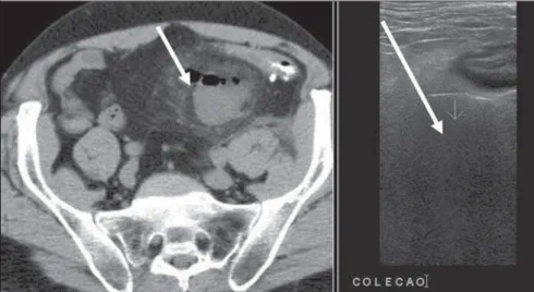
Figure 3. Extraluminal abscess with air-fluid level adjacent to the sigmoid colon at left. At right, an equivalent US image is shown. US-guided drainage was performed.
ANTEROLATERAL
EXTRAPERITONEAL APPROACH
This technique involves the iliopsoas muscle transfixion to avoid injuries to deep circumflex iliac vessels, and should be preferentially utilized for target-lesions located medially to the iliopsoas muscle, such as lymphadenomegaly in deep iliac and obturator chains.
Another advantage to be considered is that this approach is performed with the patient in dorsal decubitus, which is quite comfort-able for patients who are uncomfort-able to lie prone because of obesity, colostomy bags or in-cisions/wounds on the anterior abdominal wall.
A potential risk of this approach in-volves injuries to the femoral nerve which courses in the fat plane between the iliac and psoas muscles. However, no similar complication was observed by the authors in their experience involving 15 cases. The disadvantage of this approach is that this procedure is usually more painful as com-pared with the transabdominal approach, requiring deeper sedation or analgesia.
Biopsy of deep pelvic lesions is per-formed by transfixion of the iliopsoas muscle by means of coaxial needles, allow-ing repeated biopsy samplallow-ing through a
same access route, and avoiding contact with vascular structures (Figures 5 and 6).
TRANSGLUTEAL APPROACH
The patient is usually placed in ventral or lateral decubitus. This approach also is
called transsciatic approach because the needle is inserted through the great sciatic foramen (Figure 2). If possible, the needle should transfix the sacrospinous ligament localized below the level of the piriform muscle to avoid injury to gluteal vessels and the sacral plexus which lie anterior to
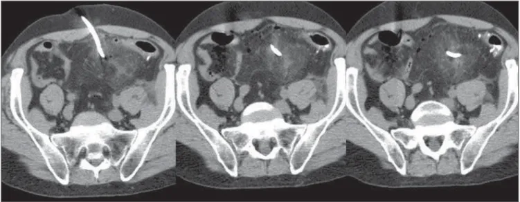
Figure 4. Tomographic follow-up after drainage demonstrating a complete drainage of the collection with a drain placed inside the abscess cavity.
the muscle. Target-lesions to be approached by this method are those located posterior to the urinary bladder and adnexal masses (Figures 7 and 8).
This approach presents the advantage of avoiding transfixion of the peritoneum, besides minimizing risks for injury to bowel loops, bladder and iliac vessels. Additionally, in the transgluteal approach, the needle is more stable for transfixing a static muscular mass in the absence of
res-piratory movements of the abdominal wall. However, the prone position is uncomfort-able for the patients, making the ventilation and anesthetic management more difficult.
TRANSVAGINAL AND
TRANSRECTAL APPROACHES
These are simple and safe techniques, and frequently represent the sole access route for the management of some pelvic
diseases. Aspiration of cystic masses and biopsy of solid masses can be easily per-formed by transvaginal or transrectal ap-proach, with an endocavitary transducer and a biopsy guide (Figures 9, 10, 11 and 12).
The major advantage of these tech-niques is that both are easy to perform through natural pelvic orifices, constitut-ing an extremely useful access in the ap-proach of cul-de-sac lesions, as well as
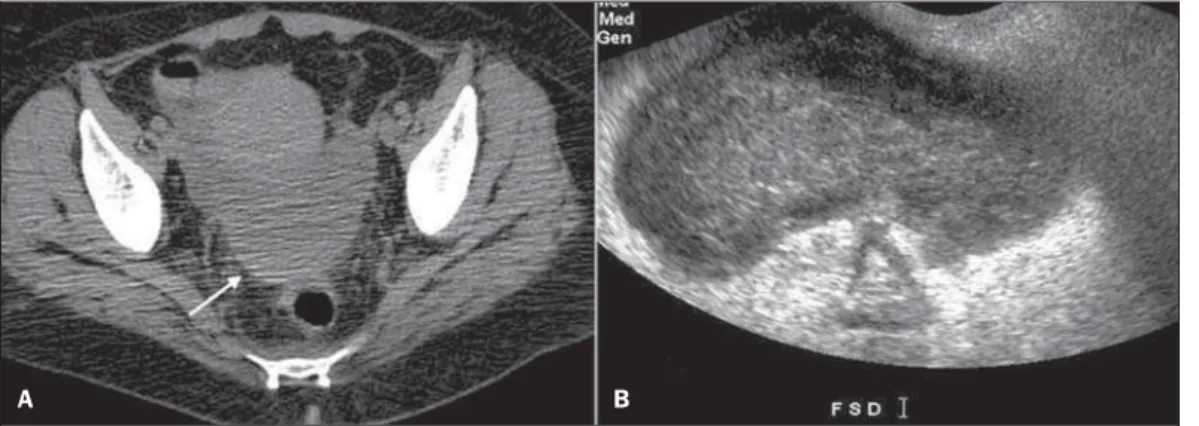
le-Figure 9. Pelvic cul-de-sac abscess. CT (A) and transvagi-nal US (B). Axial images of the pelvis demonstrate pelvic cul-de-sac abscess with a thick aspect (arrow).
A B
Figure 8. 3D CT image reconstruction demonstrating the final positioning of a drainage tube.
Figure 10. Collection drainage by the Seldinger technique. A,B: Positioning of the needle and guide-wire for later inser-tion of the drainage tube.
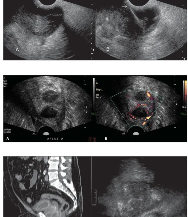
Figure 11. Bulging prostatic abscess. A,B: Transrectal color
Doppler US. A B
Figure 12.A: Computed to-mography, sagittal reconstruc-tion. B: Transrectal puncture and drainage. The arrow indi-cates a 18 G needle inserted
into the collection. A B
sions in the adnexal and prostatic regions. Considering that this is a semi-sterile pro-cedure, even involving risk for contamina-tion (transrectal approach), addicontamina-tional care should be taken in relation to the patient preparation. Antisepsis of the vaginal and rectal cavities with povidine iodine is rec-ommended, besides fleet-enema in cases of transrectal procedures. Prophylactic in-travenous antibiotic-therapy (cephoxitin 1g or clindamycin 1g + gentamycin 80 mg
+ ampicillin 1g) is mandatory. Addition-ally a 300 mg-dose of clindamycin each six hours for five days may be recom-mended(5).
Collections and cysts must be com-pletely drained in order to avoid hyperin-fection by the rectovaginal flora(5).
The transvaginal approach also is use-ful in cases of tubo-ovarian abscess, post-operative abscess and complications result-ing from intestinal conditions (Crohn’s
dis-ease, apendicitis or diverticulitis) for allow-ing the insertion of drains.
COMPLICATIONS
Familiarity with the pelvic anatomy and preprocedural laboratory evaluation to ex-clude patients at high risk of bleeding mini-mize the chance of clinically significant bleeding. Piriform muscle transfixion with the transgluteal approach may result in ir-ritation of the sciatic nerve or sacral plexus. This reaction is usually self-limiting and resolve within 24 hours following the pro-cedure.
CONCLUSIONS
The knowledge of cross-sectional pel-vic anatomy facilitates the choice of an ap-propriate approach in US- or CT-guided interventional procedures, increasing the
effectiveness of the method. Familiarity with interventional percutaneous tech-niques and materials, in association with risk factors evaluation reduce the incidence of complications.
REFERENCES
1. Gupta S, Nguyen HL, Morello FA Jr, et al. Various approaches for CT-guided percutaneous biopsy of deep pelvic lesions: anatomic and technical considerations. Radiographics. 2004;24:175–89. 2. McDowell R, Mueller P. Pelvic fluid collections: anatomy for interventional procedures. Semin Intervent Radiol. 1995;12:177–90.
3. Higgins J, Letourneau J. Percutaneous biopsy of abdominal masses. In: Casteñeda-Zuniga W, editor. Interventional radiology. Philadelphia: Williams & Wilkins; 1997. p. 1691–718. 4. Schweiger GD, Yip VY, Brown BP. CT
fluoro-scopic guidance for percutaneous needle
place-ment into abdominopelvic lesions with difficult access routes. Abdom Imaging. 2000;25:633–7.
5. O’Neill MJ, Rafferty EA, Lee SI, et al. Transvagi-nal interventioTransvagi-nal procedures: aspiration, biopsy, and catheter drainage. Radiographics. 2001;21: 657–72.
6. Harisinghani MG, Gervais DA, Hahn PF, et al. CT-guided transgluteal drainage of deep pelvic abscesses: indications, technique, procedure-related complications, and clinical outcome. Radiographics. 2002;22:1353–67.
7. Harisinghani MG, Gervais DA, Maher MM, et al. Transgluteal approach for percutaneous drainage of deep pelvic abscesses: 154 cases. Radiology. 2003;228:701–5.