349
Leiomyomatosis peritonealis disseminata: a case report
Radiol Bras. 2008 Set/Out;41(5):349–351 Case Report • Relato de Caso
Leiomyomatosis peritonealis disseminata: a case report*
Leiomiomatose peritoneal disseminada: relato de casoMarco Antonio Marini Gedda1, Giuliano Arrivabeni Piantavinha1, Túlio Rebello Coutinho1, Silvana Machado Mendonça1, Gustavo de Vasconcelos Bello1
The present study reports the case of a 46-year-old female patient complaining of mild premenstrual pain and increase in the volume of the periumbilical region. The condition progressed with a gradual increase of the pain intensity. Ultrasonography, computed tomography and magnetic resonance imaging were performed. Biopsy of tissue collected during exploratory laparotomy confirmed the diagnosis of leiomyomatosis peritonealis disseminata.
Keywords: Leiomyomatosis peritonealis disseminata; Oral contraceptive method; Computed tomography; Magnetic resonance imaging.
Neste trabalho relata-se o caso de uma paciente de 46 anos de idade com queixa de aumento de volume periumbilical e dor leve antes da menstruação. O quadro evoluiu com aumento gradativo da dor. Foram reali-zados ultra-som, tomografia computadorizada e ressonância magnética. Após laparotomia exploradora, foi retirado material para biópsia que confirmou o diagnóstico de leiomiomatose peritoneal disseminada. Unitermos: Leiomiomatose peritoneal disseminada; Anticoncepcional oral; Tomografia computadorizada; Imagem por ressonância magnética.
Abstract
Resumo
* Study developed at Clínica Radiológica Emílio Amorim, Rio de Janeiro, RJ, Brazil.
1. MDs, Residents in Radiology and Imaging Diagnosis at Clí-nica Radiológica Emílio Amorim, Rio de Janeiro, RJ, Brazil.
Mailing address: Dr. Marco Antonio Marini Gedda. Rua São Manuel, 20, ap. 801, Botafogo. Rio de Janeiro, RJ, Brazil, 22290-010. E-mail: [email protected]
Received March 27, 2007. Accepted after revision June 14, 2007.
when she went to another health unit, un-dergoing a new US scan that, again, as re-ported, demonstrated uterine fibroids. Oral contraceptives were prescribed, but no improvement was observed. Then, the pa-tient was referred to our institution to be submitted to computed tomography (CT) and magnetic resonance imaging (MRI).
Eight years ago, the patient had been submitted to myomectomy, when four uter-ine fibroids were removed. She reported hypertension being treated with hydro-chlorotiazide and atenolol for three years and had already utilized oral contraceptives between her 18 and 37 years of age. Gyne-cologic history: GII, P0, AII (both by curet-tage), menarche at 16 years of age and regu-lar menstrual cycles at 29-day intervals.
At clinical examination, the patient pre-sented abdominal distention with the abdo-men depressable upon superficial or deep palpation, with reflex and diffuse pain most intensely observed in the right iliac fossa. A median inferior surgical scar and a bulg-ing, poorly defined, palpable mass occupy-ing a great part of the lower abdomen were observed. Dullness to percussion was ob-served in the periumbilical region as well as in the lower abdomen. Painful cul-de-sac under two-handed palpation.
Gedda MAM, Piantavinha GA, Coutinho TR, Mendonça SM, Bello GV. Leiomyomatosis peritonealis disseminata: a case report. Radiol Bras. 2008;41(5):349–351.
disseminata in a patient who presented with periumbilical swelling and diffuse abdomi-nal pain.
CASE REPORT
A female, 46-year-old patient born in São Gonçalo dos Campos, BA, Brazil, where she lived up to her 18 years of age when she moved to Rio de Janeiro. The patient was single, housewife who com-plained of the presence of a mass in her umbilicus during the premenstrual period. The patient reported that three years ago she had observed a mild periumbilical swelling appearing invariably in the pre-menstrual period, generally 7 to 15 days before the first day of menstruation. Con-comitantly, the patient reported an initially diffuse mild abdominal pain, progressively becoming localized in the right iliac fossa. The pain disappeared at the first day of menstruation and the periumbilical swell-ing decreased in the followswell-ing days, al-though without disappearing completely. Two years ago, she had undergone ultra-sonography (US) in a public health service and, as reported, uterine myomatosis had been found. Her clinical picture had re-mained stable up to three months ago,
0100-3984 © Colégio Brasileiro de Radiologia e Diagnóstico por Imagem INTRODUCTION
Few cases of leiomyomatosis perito-nealis disseminata have been reported in the literature, but there is a possibility that the incidence of this disease may be higher, considering that many patients are asymp-tomatic and the finding of the disease ends up being incidental(1).
Usually, this is a benign condition af-fecting women in childbearing age, and is generally associated with pregnancy or uti-lization of oral contraceptive methods. It is suspected that this disease originates from a metaplasia of submesothelial multipotent mesenchymal cells(2). Some authors sug-gest that exposure to estrogen is the primary mechanism in the development of this dis-ease and, frequently, this condition is asso-ciated with the presence of uterine fi-broids(3,4).
350
Gedda MAM et al.
Radiol Bras. 2008 Set/Out;41(5):349–351 Figure 3. Magnetic resonance imaging. The lesions are isointense in relation to the muscle on non-contrast-enhanced T1-weighted images (A), and homo-geneous enhancement after contrast injection (B). Areas of hyposignal are observed in the center of the lesions. On the T2-weighted image (C) the lesions present heterogeneous signal, with predominance of areas with hypersignal.
A B C
Figure 2. Computed tomography demonstrates several, small nodules with soft tissue densities (A), with homogeneous venous contrast uptake (B), from the pelvis to the upper abdomen.
A B
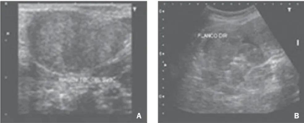
Nodular formations, some of them confluent, were observed at US (Figure 1) with intermediate echogenicity and hypoechoic areas. The lesions were inter-mingled with loops, protruding through the umbilical region, filling the pelvis and at-tached to the uterus. Increased uterine
vol-ume was observed with the presence of myomatous nodules.
At CT, innumerable nodules with soft tissues density and varying in size were ob-served within the abdominal cavity from the pelvis to the upper abdomen. The nod-ules demonstrated homogeneous contrast
uptake. In the pelvis, confluent nodules formed a mass with possible central necro-sis. No sign of ureteral and rectal invasion was observed despite the involvement of these structures (Figure 2).
The nodular characteristic of the lesion as well as its localization in the mesen-terium and omentum were most easily ob-served at MRI. On non-contrast enhanced T1-weighted sequences, the lesions were isointense to the muscle, with lower inten-sity in the center. Contrast-enhancement was observed and the hyposignal remained unchanged in the center of the mass. On T2-weighted images, heterogeneous signal intensity was observed with predominance of hypersignal (Figure 3).
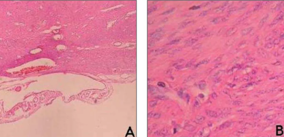
The definite diagnosis was achieved with a biopsy of abdominal nodule (Figure 4).
DISCUSSION
Leiomyomatosis peritonealis disse-minata is a rare disease, with a little more than 100 cases reported. Most of times, the patients are asymptomatic, and the disease is incidentally found, so there is a possibil-ity that the disease incidence is higher than the number of cases described in the litera-ture(1).
The disease etiology still remains un-known. It is suspected that this disease originates from a metaplasia of submeso-thelial multipotent mesenchymal cells(2). Apparently, this is not a familial or inher-ited disease, despite the case of several members of a same family affected by leiomyomatosis peritonealis disseminata(5).
Figure 1. Ultrasonography demonstrates nodular formations varying in sizes, some of them confluent, presenting heterogeneous and predominantly hypoechoic echogenicity, distributed within the peritoneal cavity.
351
Leiomyomatosis peritonealis disseminata: a case report
Radiol Bras. 2008 Set/Out;41(5):349–351
Although 10% of cases may present ma-lignant degeneration, this is usually a be-nign condition, characterized by the pres-ence of multiple smooth-muscle nodules in the peritoneal surface, affecting women in childbearing age. It is generally associated with pregnancy or utilization of oral con-traceptive methods(1,2,4).
According to some authors, exposure to estrogen is the primary mechanism in-volved in the development of leiomyo-matosis peritonealis disseminata(3,4). Two cases have been reported associating the disease with the utilization of tamoxifen for the management of breast cancer(6,7). There are cases of leiomyomatosis peritonealis regression after delivery or suspension of oral contraceptive methods(4,8). Post-treat-ment recurrence is rarely reported.
This condition is frequently associated with uterine fibroids. On the other hand, as-sociation with endometriosis is not fre-quent.
Sometimes the imaging diagnosis may be difficult, considering that radiological findings may suggest the presence of a ma-lignant condition(3,9). At US, the present case showed small, clustered nodules in the lower abdomen and pelvis, simulating a lobulated mass and presenting a variable echogenicity, with apparently cystic areas inside. At CT (Figure 1) and MRI (Figures 2 and 3) a confluent nodular pattern was observed, with soft tissues density, seem-ing a sseem-ingle mass. No sign of ureteral in-vasion was found. Some of the nodules presented areas of cystic degeneration.
CONCLUSION
The patient whose case is reported in the present study was within the age range expected for this disease and presented a history of chronic exposure to estrogen that is characteristically reported in most of re-corded cases. Additionally, imaging
find-Figure 4. Biopsy plate (A). Myocyte bundle presenting ovoid nuclei and bipolar, long and thin cytoplasmatic prolongations (B).
ings were compatible with those described the literature, demonstrating confluent nod-ules seeming a single mass with homoge-neous contrast uptake both at CT and MRI.
Acknowledgements
The authors thank Dr. Marília Maga-lhães, Dr. Rosangela Guerreiro, Dr. Euniro de Macedo Melo, Dr. Leila Freitas, Dr. Carlos Peixoto and Dr. Arno Von Ristow, for their cooperation along the develop-ment of the present study.
REFERENCES
1. Borsellino G, Zante P, Ciraldo MC. Diffuse peri-toneal leiomyomatosis. A clinical case report. Minerva Ginecol. 1997;49:53–7.
2. Abulafia O, Angel C, Sherer DM, et al. Computed tomography of leiomyomatosis peritonealis disseminata with malignant transformation. Am J Obstet Gynecol. 1993;169:52–4.
3. Sobiczewski P, Bidzi½ski M, Radziszewski J, et al. Disseminated peritoneal leiomyomatosis – case report and literature review. Ginekol Pol. 2004;75:215–20.
4. Bekkers RL, Willemsen WN, Schijf CP, et al. Leiomyomatosis peritonealis disseminata: does malignant transformation occur? A literature re-view. Gynecol Oncol. 1999;75:158–63. 5. Halama N, Grauling-Halama SA, Daboul I.
Fa-milial clustering of leiomyomatosis peritonealis disseminata: an unknown genetic syndrome? BMC Gastroenterol. 2005;5:33.
6. Danikas D, Goudas VT, Rao CV, et al. Luteiniz-ing hormone receptor expression in leiomyoma-tosis peritonealis disseminata. Obstet Gynecol. 2000;95(6 Pt 2):1009–11.
7. Lubczyk V, du Bois A, Fisseler-Eckhoff A. Leio-myomatosis peritonealis disseminata and border-line lesion. Pathologe. 2005;26:291–5.
8. Bristow RE, Montz FJ. Leiomyomatosis perito-nealis disseminata and ovarian Brenner tumor associated with tamoxifen use. Int J Gynecol Cancer. 2001;11:312–5.