Article
J. Braz. Chem. Soc., Vol. 22, No. 2, 223-229, 2011. Printed in Brazil - ©2011 Sociedade Brasileira de Química
0103 - 5053 $6.00+0.00
A
*e-mail: [email protected]
Three New Amides from
Streptomyces
sp. H7372
Sarot Cheenpracha,a Robert P. Borris,a Tammy T. Tran,a Jap Meng Jee,b Heng Fong Seow,b Hwen-Yee Cheah,b Coy Choke Hoc and Leng Chee Chang*,a
aDepartment of Pharmaceutical Sciences, College of Pharmacy, University of Hawaii Hilo, 34
Rainbow Drive, 96720 Hilo, HI, USA
bImmunology Unit, Department of Pathology, Faculty of Medicine and Health Sciences,
Universiti Putra Malaysia, 43400 Serdang, Selangor, Malaysia
cBiotechnology Program, School of Science and Technology, University Malaysia Sabah, 88999
Kota Kinabalu, Sabah, Malaysia
As amidas fenatato de metila (1), actifenamida (2) e 1-b-D-glucopiranosídeo-actinofenol (3), juntamente com outros 13 compostos de estruturas conhecidas, foram isolados do extrato de uma cultura de Streptomyces sp H7372. As estruturas destes compostos foram determinadas por análises de RMN uni e bidimensionais e por espectrometria de massas. Cicloheximida (6) e ciclo (∆Ala-L-Val) (8) apresentaram forte atividade inibitória na interação entre Ras-Raf-1 pela técnica de duplo híbrido em leveduras, com halos de inibição de 10 e 25 mm em SD His–, respectivamente, enquanto foram inativos em SD His+, na concentração de 25 μg/disco.
Three new amides, methyl phenatate A (1), actiphenamide (2) and actiphenol 1-b-D -glucopyranoside (3), along with thirteen known compounds, were isolated from the organic extract of a fermentation culture of Streptomyces sp. H7372. The structures were elucidated by spectroscopic methods including 1D- and 2D-NMR techniques, and MS analyses. Cycloheximide (6) and cyclo(DAla-L-Val) (8) gave a clear zone of inhibition of Ras-Raf-1 interaction in the yeast two-hybrid assay which showed high potency with 10 and 25 mm clear ZOIs on SD His− and inactive on SD His+ at 2.5 µg per disk, respectively.
Keywords: Streptomyces sp., yeast two-hybrid screen, Ras-Raf-1 interaction, amides, Cyclo(∆Ala-L-Val)
Introduction
Protein kinases constitute an important group of enzymes that are essential in many physiological processes. Protein phosphorylation is one of the major regulatory mechanisms involved in signal transduction pathways including apoptosis, cell proliferation, cell differentiation, and metabolism.1 Abnormalities in protein phosphorylation
are often associated with human diseases. Thus, the inhibitors of both protein kinases and phosphatases appear to be very promising drug targets in the chemotherapeutic treatment of cancer and have received wide attention.1 The
Ras family GTPases play a central role in the growth factor signaling controlling cell proliferation, differentiation, and survival.2 The interaction of activated Ras with Raf
initiates signaling cascades that contribute to a signiicant percentage of human tumors, suggesting that agents that speciically disrupt this interaction might have desirable chemotherapeutic properties. Recently, the approval of NEXAVAR® (sorafenib) by the FDA for advanced renal
cell carcinoma provides justiication for the development of Ras-Raf-1 kinase inhibitors. Radicicol, is a macrocyclic antibiotic isolated from the fungus Monosporium bonorden that inhibits the Ras-Raf-1 protein interaction at concentrations of 0.1 ca. 1 µg mL-1.3 To identify such
inhibitors, we employed the yeast two-hybrid system in our screening.3
of protein kinase C and also showed antimicrobial activity against fungi and yeast.4 As part of an ongoing search for
novel protein phosphorylation inhibitors, Streptomyces sp. H7372, a novel strain isolated from a mangrove soil of Sabah, Borneo, Malaysia, was studied. This strain was identiied based on the 16S rRNA gene sequence analysis and is a member of the phylogenetically diverse genus Streptomyces. The n-BuOH-soluble extract of fermentation cultures of Streptomyces sp. H7372 was found to disrupt Ras-Raf-1 interaction in the yeast two hybrid assay.5 We
report herein the isolation, structure elucidation, and biological activities of 1-14.
Experimental
General experimental procedures
The optical rotation [α]D values were determined with an Autopol® IV Automatic polarimeter. UV spectra
were measured on a Shimadzu PharmaSpec-1700 UV-Visible spectrophotometer. IR spectra were recorded on a Shimadzu-8400S FT-IR spectrometer. Mass spectra and high-resolution MS spectra were taken with a BioTOF II ESI mass spectrometer. 1D and 2D NMR spectra were recorded in chloroform-d and methanol-d4 on an INOVA Unity (500 MHz) Varian spectrometer equipped with an xyz-shielded gradient triple resonance probe. Reversed-phase HPLC was carried out on a Beckman Coulter Gold-168 system equipped with a photodiode array detector using an Alltech semipreparative Econosil C18 column (10 μm, 10 × 250 mm) run with a low rate of 2.0 mL min-1. Column chromatography (CC) was carried
out on Merck silica gel 60 (70-230 mesh). Precoated plates of silica gel 60 F254 were used for analytical purposes.
Microbial material and sample collection
The organism wasisolated from mud sample (pH 7.30) under the root of Bruguiera sp., located at sea shore near mouth of river into mangrove swamp, Kampung Termunong, Tuaran, Sabah, Malaysia in 2003.5 The strain
was isolated by suspending 0.1 g of soil into 10 mL of sterile distilled water and later diluting this up to 10-3. One
hundred-µL of the suspension is spread on humic-acid (HV)-vitamins B plus cycloheximide,6 with pH 7.2. Isolated
strains were transferred from HV medium onto similar pH oatmeal agar plates and incubated at 28 °C for 14 days until sporulation. The strain H7372 was characterized by microscopy and determination of cell wall DAP (meso or LL). It was further characterized by 16S rRNA gene analysis. A pure culture of strain H7372 was deposited
in National Measurement of Australia and was given an accession number V07/019103. Frozen spore stock of Streptomyces sp. H7372 was stored in 20% glycerol at
−78°C. The vial was thawed and spores spread on an ISP4 plate for complete sporulation.
Seed medium
The composition of the seed medium (20 g L-1 of
D-mannitol, 20 g L-1 of peptone, and 10 g L-1 of dextrose)
was prepared with distilled water, and the pH was adjusted to 7.0 prior to sterilization. The medium was dispensed at 50 mL per 250 mL in Belco bafled shaker lasks. A single colony from the agar plate was used as inoculum into each lask of mannitol-peptone media, cultured at 30 °C at 250 rpm for two days.
Production medium
An aliquot (1%) from the seed medium was inoculated into the production medium. The composition of the production medium was similar to that of the seed medium. The production cultures were incubated at 30°C at 250 rpm for seven days and were then harvested. The supernatants were iltered and partitioned with n-BuOH three times. Both the n-BuOH-soluble and aqueous layers were tested against the yeast two-hybrid assay.
Extraction and isolation
The culture broth of Streptomyces sp. H7372 (120 L) was centrifuged, and the supernatant was extracted with n-BuOH. An organic extract was suspended in H2O (1:1) then partitioned successively with CHCl3, and EtOAc (3 × 250 mL each). The CHCl3 and EtOAc-soluble fractions of the n-BuOH extract of fermented Streptomyces sp. H7372, exhibited signiicant activity in the yeast two-hybrid assay at a concentration of 80 µg per disk with 21 and 27 mm zones of inhibition, respectively, and were subjected to bioassay-guided fractionation. The CHCl3 extract (22.5 g) was fractionated by CC eluting with hexanes and increasing polarity with acetone and MeOH, successively, to give four fractions (C1-C4). Fraction C2 (2.35 g) was further puriied by CC eluting with MeOH-CHCl3 (3:97) to afford compound 7 (2.9 mg). Fraction C3 (580.5 mg) was recrystallized with MeOH-CHCl3 (1:1) to give compound 8 (32.2 mg) and the mother liquor (528.0 mg) was further separated by CC eluting with MeOH-CHCl3 (1:99) and followed by RP-18 HPLC with MeOH-H2O (3:1) to give compounds 10 (12.0 mg, tR 17.6 min) and
Cheenpracha et al. 225 Vol. 22, No. 2, 2011
chromatographed sequentially on silica gel eluting with hexanes and increasing polarity with EtOAc and MeOH, respectively, to give nine fractions (B1-B9). Fraction B3 (1.2 g) was chromatographed over sephadex LH-20 with MeOH:H2O (7:3) to provide three subfractions (B3a-B3c). Recrystallization of subfraction B3a (MeOH:H2O, 7:3) with MeOH:CHCl3 (3:97) gave compound 4 (140.3 mg), while subfraction B3b (10.4 mg) was puriied with preparative TLC with MeOH:CHCl3 (1:49) to give compound 1
(3.4 mg). Compound 9 (20.2 mg) was recrystallized from fraction B5 (MeOH-H2O, 7:3) with MeOH:CHCl3 (3:97). Fraction B6 (1.58 g) was isolated by CC eluting with hexanes:EtOAc (3:7) to give compound 5 (3.5 mg) and 11
(110.3 mg) 14 (3.2 mg). Fraction B7 (2.7 g) was puriied by CC eluting with CHCl3 and increasing polarity with MeOH to afford six subfractions (B7a-B7f). Subfractions B7b, B7c and B7f were isolated by RP-18 HPLC with MeCN:H2O (1:3) to give compounds 12 (4.4 mg, tR 17.5 min), 13
(1.5 mg, tR 18.1 min), and 2 (2.6 mg, tR 23.9 min). Fraction B8 (1.07 g) was puriied by CC eluting with MeOH:CHCl3 (1:9) to provide compound 3 (7.0 mg).
Methyl phenatate A (1)
Colorless viscous oil, [α]D22−18.8 (c 0.01, MeOH); UV
(MeOH) λmax /nm: 207 (log ε 3.16), 262 (2.75), 348 (2.45); IR (neat) νmax/ cm-1: 3450, 1645; 1H and 13C NMR (CDCl
3,
500 MHz) data: see Table 1; HRESIMS m/z 308.1501 [M + H]+ (calc. for C
16H22NO5, 308.1498).
Actiphenamide (2)
Colorless viscous oil, [α]D22−29.7 (c 0.01, MeOH); UV
(MeOH) λmax /nm: 206 (log ε 3.38), 218 (3.28), 282 (2.74); IR (neat) νmax/ cm-1: 3405, 1660; 1H and 13C NMR (CD
3OD,
500 MHz) data: see Table 1; HRESIMS m/z 276.1232 [M+H-H2O]+ (calc. for C
15H20NO5, 276.1230).
Actiphenol 1-b-D-glucopyranoside (3)
Colorless viscous oil, [α]D22 +19.1 (c 0.01, MeOH);
UV (MeOH) λmax /nm: 207 (log ε 4.33), 242 (3.67), 282 (3.25); IR (neat) νmax/ cm-1: 3400, 1690, 1253, 1055; 1H and
13C NMR (CD
3OD, 500 MHz) data: see Table 1; HRESIMS
m/z 461.1629 [M+H+Na]+ (calc. for C
21H28NO9Na,
461.1662).
Yeast two-hybrid assay
The inhibition of yeast two-hybrid assay in Saccharomyces cerevisiae was performed as previously described.2 The LZ strain, H10014 of the genotype MATa
trp1 leu2 his3 LYS: lexA-HIS3 URA3: lexA-lacZ
[pLexA-RASV12 and pVP16-RAF] was grown on the SD medium
agar plate lacking histidine for the generation of stock plate. These cells were grown in the SD medium lacking histidine at 30 °C, 220 rpm, for 2 days. Four hundred microliters of this culture were inoculated into 100 mL of medium containing 1% agar supplemented with (His+) or without
(His−) 130 µmol L-1 histidine. Compounds of known
concentration dissolved in MeOH were dispensed onto disks in 20 µL aliquots. The air-dried disks were applied directly onto the plates. The plates were incubated at 30 °C for 48 h, and the zones of inhibition were measured and compared between those on histidine-minus and histidine plates. The yeast strain carrying both pLexA-RASV12 and
pVP16-RAF (strain LZ) will be to grown on the SD-(His−)
plate and allowed to express b-galactosidase. Ilimaquinone and geldanamycin were employed as positive controls. Active compounds were then tested at lower concentrations (20-2.5 µg per disk). The assays were performed in triplicate.
b-Galactosidase assay
b-Galactosidase activity was assayed according to the literature with modiications.7 One mL culture strains was
taken at A600 between 0.50-1.00 and mixed with 20 μL test compounds. The mixture was kept at 30 °C for 2 h and was harvested by centrifugation in a microfuge tube. Yeast pellets were resuspended in 0.7 mL of Z buffer (60 mmol L-1 Na
2HPO4•7H2O, 40 mmol L -1 NaH
2PO4•H2O,
10 mmol L-1 KCl, 1 mmol L-1 MgSO
4•7H2O, 2.7 μL mL -1 b-mercaptoethanol), and followed by adding 20 μL of freshly prepared 0.1% SDS and 50 μL of chloroform into a microfuge tube with vortexing for 15 seconds. At time zero, the assay is initiated by adding 160 μL of 4 mg mL-1
2-nitrophenyl-b-D-galactopyranoside to each microfuge tube. After 20-60 min at 30 °C, the reactions were terminated by the addition of 0.4 mL of 1mol L-1 Na
2CO3
and the liberated p-nitrophenol was measured at A420 nm. The b-galactosidase activity expressed in Miller Units (MU) which is calculated with formula below:
Miller Unit = (A420 × 1000)/(A600 × t × V)
t = reaction time (minutes); V = reaction volume in mL, equals to 1 if there is no dilution of cells.
Cells treated with extracts with less than ½ MU of the control cells are scored as positive.7
Results and Discussion
with n-BuOH. The extract was suspended in water, and then partitioned successively with CHCl3 and EtOAc to obtain the CHCl3 and EtOAc fractions. These fractions exhibited zones of inhibition on yeast two-hybrid screening which were subjected to repeated column chromatography and followed by semi-preparative HPLC separation to afford three new amides (1-3) along with thirteen known compounds; actiphenol (4),8 AH-135Y (5),9 cycloheximide
(6),10 α-epicycloheximide (7),11 cyclo(DAla-L-Val) (8),12
cyclo(L-Pro-L-Val) (9),12 cyclo(L-Pro-L-Phe) (10),13
cyclo(L-Pro-L-Trp) (11),14 cyclo(L-Trp-L-Phe) (12),15
cyclo(L-Trp-L-Tyr) (13),16 and cyclo(L-Phe-L-Leu) (14).17
The structures of the isolated compounds (Figure 1) were determined by the analysis of 1D- and 2D-NMR, mass spectral information and comparison with reported physical and spectroscopic data.
Compound 1 was isolated as a colorless viscous oil with the molecular formula C16H21NO5, determined from HRESIMS (m/z 308.1501 [M+H]+, calc. 308.1498),
indicating seven degrees of unsaturation. The IR spectrum indicated the presence of OH (3450 cm-1) and carbonyl
(1645 cm-1) functionalities. The UV spectrum showed
maxima absorptions at λmax 207, 262, and 348 nm, which indicated the presence of a conjugated carbonyl-containing chromophore in the structure. The 13C NMR and DEPT
spectra (Table 1) displayed 16 resolved signals, which were classiied into three methyl (dC 51.7, 20.5, and 15.4),
three methylene (dC 41.6, 39.2, and 37.6), three methine
(dC 138.7, 127.7, and 29.0), four quaternary (dC 158.9,
127.2, 127.4, and 118.3), one ketone-carbonyl (dC 205.2),
and two ester-carbonyl (dC 173.6, and 172.9) carbons. Three
carbonyls from the 13C NMR spectra accounted for three
degrees of unsaturation, and the remaining four degrees of unsaturation required the presence of an aromatic ring in 1. The 1H NMR spectrum of 1 (Table 1) displayed two singlet
signals at d 2.28 (3H-15) and 2.22 (3H-14), attributable to aromatic methyl groups, while meta-coupling aromatic protons at d 7.46 (1H, br s, H-5) and 7.18 (1H, br s, H-3)
8 Me OH 1 NH O OR1 R2 Me O O
3; R1 = β-glucose, R2 = Me
4; R1 = H, R2 = Me
5; R1 = H, R2 = CH2OH Me O NH2 O O OMe Me Me OH OH O NH2 O O 2 NH O H OH O O
6;7R
7; 7S
7 7 1 2 3 4 5 9 13 11 14 15 10 12 7 8 9 14 N NH O O
9; R1 = R2 = Me 10; R1 = H; R2 = Ph 11; R1 = H, R2 = 3-indole
12; R = H 13; R = OH
H HN NH O O HN R H H HN NH O O H H HN NH O O H R2 R1 H
Cheenpracha et al. 227 Vol. 22, No. 2, 2011
suggested the presence of 1,2,3,5-tetrasubstituted aromatic ring. The 1H NMR and COSY spectra indicated that the
methine at d 2.95 (1H, sept, J 6.6 Hz, H-9), and three sets of methylene protons α to a carbonyl [d 3.18 (1H, d, J 6.6 Hz, H-8), 2.57 (1H, d, J 6.6 Hz, H-10), and 2.40 (1H, d, J 6.6 Hz, H-12)], were connected as a –CH2-CH-(CH2)2 unit. Additionally, two singlets at d 5.84, and 5.46 were assigned to NH2. The full 1H and 13C NMR spectroscopic
assignments for 1 based on 2D-NMR experiments were closely related to those of phenatic acid A,8 except for
the additional singlet signal of OMe group at d 3.69. The HMBC spectrum of 1 showed correlations from 3H-14 to C-1, C-2, and C-3; 3H-15 to C-3, C-4, and C-5; and H-5 to C-1, C-3, C-7 and C-15, conirming the assignments of substituted aromatic ring. The correlations of the OMe signal to C-11, of 2H-8 to C-6, C-7, C-10 and C-12, of 2H-10 to C-8,C-9, C-11 and C-12, and of 2H-12 to C-8,
C-9, C-10 and C-13 conirmed the structural assignments and the OMe group at C-11. Therefore, compound 1 was determined to be methyl phenatate A.
Compound 2 was isolated as a colorless viscous oil and had a HRESIMS molecular ion peak at m/z 276.1232 [M+H-H2O]+, which was compatible with the molecular
formula C15H19NO5 (calc. 276.1230). The 1H and 13C NMR
spectra (Table 1) of 2 were comparable to those of 1, except for the differences in the aliphatic proton region. The presence of two oxymethine protons at d 5.22 (1H, d, J 3.2 Hz) and 4.58 (1H, dd, J 4.8, 3.2 Hz), and an aliphatic methine proton at d 2.94 (1H, m) were assigned to H-7, H-8 and H-9, respectively. In addition, one AX system methylene protons was found at d 2.70 (1H, dd, J 18.0, 9.8 Hz, H-10a), and 2.26 (1H, dd, J 18.0, 5.6 Hz, H-10b) and another AB system of methylene protons was found at d 2.11 (1H, dd, J 15.0, 9.0 Hz, H-12a), 2.06 (1H, dd,
Table 1.1H (500 MHz) and 13C (125 MHz) NMR Assignments of compounds 1-3 in CD
3OD (d, mult, J in Hz)
Position 1a 2 3
dC dH dC dH dC dH
1 158.9 151.7 151.3
2 127.2 126.6 137.7
3 138.7 7.18 br s 131.8 6.84 br s 136.2 7.20 d (1.7)
4 127.4 130.6 133.4
5 127.7 7.46 br s 129.9 6.99 br s 127.2 7.01 d (1.7)
6 118.3 128.2 136.2
7 205.2 72.1 5.22 d (3.2) 207.6
8 41.6 3.18 d (6.6) 88.2 4.58 dd (4.8, 3.2) 48.4 3.35 m
2.95 dd (17.8, 5.3 )
9 29.0 2.95 sept (6.6) 32.6 2.94 m 27.5 2.70 m
10 37.6 2.57 d (6.6) 35.9 2.70 dd (18.0, 9.8)
2.26 dd (18.0, 5.6)
38.0 2.74 m
2.46 m
11 172.9 179.5 175.0
12 39.2 2.40 d (6.6) 40.2 2.11 dd (15.0, 9.0)
2.06 dd (15.0, 5.5)
38.3 2.70 m
2.37 m
13 173.6 175.9 175.0
14 15.4 2.22 s 16.3 2.15 s 16.4 2.35 s
15 20.5 2.28 s 20.7 2.20 s 20.6 2.28 s
1′ 105.8 4.35 d (7.8)
2′ 75.4 3.46 dd (9.3, 7.8)
3′ 77.8 3.36 t (9.3)
4′ 71.8 3.35 t (9.3)
5′ 78.3 3.07 ddd (9.3, 6.7, 2.3)
6′ 63.0 3.71 dd (12.2, 2.3)
3.60 dd (12.2, 6.7)
OMe 51.7 3.69 s
NH2 5.84 s, 5.46 s
J 15.0, 5.5 Hz, H-12b). The connectivity of H-7/H-8, H-8/H-9, H-9/H10 and H-9/H-12 in a COSY spectrum conirmed the assignments. In the HMBC experiments, the oxymethine proton H-8 correlated with the C-6, C-9, C-10, C-11, and C-12, and conirmed the attachment of a lactone ring at C-8. The small coupling constant of H-8 (J 4.8 Hz) and cross-peaks between H-8/H-9 and H-7/2H-10 from ROESY experiments indicated that H-8 and H-9 were in a cis-orientation. Thus, compound 2 was determined to be actiphenamide.
Compound 3, isolated as a colorless viscous oil, had the molecular ion [M+H+Na]+ whose mass as determined
by HRESIMS was m/z 461.1629, calcd for C21H27NO9. The
1H NMR data (Table 1) displayed a characteristics of a
glutarimide ring9 at d 2.74 (1H, m, H-10a), 2.70 (2H, m, H-9
and H-12a), 2.46 (1H, m, H-10b) and 2.37 (1H, m, H-12b). A methine proton at d 2.70 (H-9) also showed cross-peaks with the methylene protons at d 3.35 (1H, m, H-8a) and 2.95 (1H, dd, J 17.8, 5.3 Hz, H-8b) from the COSY experiments. The remaining resonances observed in the 1H
and 13C NMR spectra of 3 were attributable to a glycosyl
unit. The 1H and 13C NMR spectroscopic data (Table 1)
displayed resonances characteristic for a glycosyl moiety containing ive oxymethine groups at dH/dC 4.35/105.8
(H-1′/C-1′), 3.46/75.4 (H-2′/C-2′), 3.36/77.8 (H-3′/C-3′), 3.35/71.8 (H-4′/C-4′), and 3.07/78.3 (H-5′/C-5′) and one set of oxymethylene protons at dH/dC 3.71, 3.60/63.0
(2H-6′/C-6′). This unit was attached to 1-hydroxy-2,4-dimethylbenzene ring at C-1, as determined by an HMBC correlation from H-1′ to C-1. The vicinal coupling constants, J1′,2′ 7.8 Hz, J2′,3′ 9.3 Hz and J4′,5′9.3 Hz, suggested that H-1′, H-2′, H-3′, H-4′ and H-5′ were axial in orientation in the pyranose ring. NOESY cross signals observed from H-1′ to H-3′ and from H-1′ to H-5′conirmed that a sugar unit of 3 was b-D-glucopyranose. Thus, compound 3 was identiied as actiphenol 1-b-D-glucopyranoside.
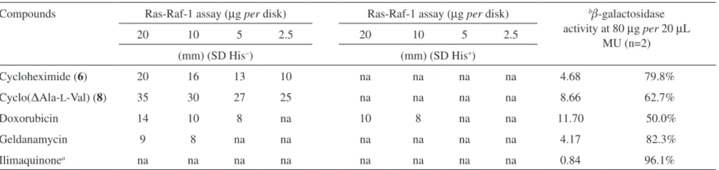
All isolates were evaluated for their inhibitory activities of Ras-Raf-1 interaction in the yeast two-hybrid assay (Table 2) according to a literature method.3 In brief, we
looked for a compound giving a clear zone of inhibition (ZOI) that inhibited growth of LZ cells in the SD His– plates
and not in the SD His+ plates, together with inhibition of b-galactosidase activity. This would indicate the compound is an inhibitor of Ras-Raf-1 interaction. Compounds 6
and 8 showed high potency with 10 and 25 mm clear ZOIs on SD His− and inactive on SD His+ at 2.5 µg per
disk, respectively, whereas all other compounds 1-5, 7,
9-14 were inactive. In addition, both compounds gave a clear ZOI that inhibited growth of LZ cells in the SD His–
plates and not in the SD His+ plates, and together with
inhibition of b-galactosidase activity, indicated 6 and 8 were inhibitors of Ras-Raf-1 interaction. The potency of these compounds was comparable to that of doxorubicin and geldanamycin but higher than those of the positive controls. Cycloheximide (6) showed the strongest activity against
b-galactosidase with 79.8% inhibition of b-galactosidase activity whereas cyclo(DAla-L-Val) (8) exhibited a weaker effect (62.7%). Geldanamycin, a heat shock protein 90 (HSP90) inhibitor and ilimaquinone were employed as positive controls and gave 82.3%, and 96.1% inhibition of b-galactosidase activity, respectively. Ilimaquinone, previously isolated from the marine sponge Hippospongia metachromia, inhibited tyrosine kinase activity by 87% at a concentration of 1 µg µL-1.18 Cycloheximide (6) was
isolated from the cultures of Streptomyces griseus and has been demonstrated to inhibit protein synthesis in mammalian systems.3,19 AH-135Y (5) has been reported
from Streptomyces albovinaceus is reported to have antiherpetic, cytotoxic and antifungal activities.9
Many terrestrial yeast, lichens, and fungi are known to produce diketopiperazines by condensation of two amino acids. Natural diketopiperazines show a remarkable preference for proline as a building block.20 They consist
mostly of two L-amino acids, but DL-, LD-, and DD-isomers can be generated by non-enzymatic epimerization, possibly accelerated by steric effects.21 In the literature there are
two report of DD-enantiomers of diketopiperazines as natural compounds.22,23 The DD-enantiomers exhibited
Table 2. Yeast two-hybrid screening of compounds 6 and 8
Compounds Ras-Raf-1 assay (µg per disk) Ras-Raf-1 assay (µg per disk) bb-galactosidase
activity at 80 µg per 20 µL MU (n=2)
20 10 5 2.5 20 10 5 2.5
(mm) (SD His−) (mm) (SD His+)
Cycloheximide (6) 20 16 13 10 na na na na 4.68 79.8%
Cyclo(DAla-L-Val) (8) 35 30 27 25 na na na na 8.66 62.7%
Doxorubicin 14 10 8 na 10 8 na na 11.70 50.0%
Geldanamycin 9 8 na na na na na na 4.17 82.3%
Ilimaquinonea na na na na na na na na 0.84 96.1%
Cheenpracha et al. 229
Vol. 22, No. 2, 2011 Cheenpracha et al. 229
Vol. 22, No. 2, 2011
potent inhibitory activity against the pathogen Vibrio anguillarum,23 whereas LL-enantiomers as well as cyclo-(L
-Leu-L-Pro), cyclo-(L-Phe-L-Pro), and cyclo-(L-Trp-L-Pro) isolated from a deep sea bacterium exhibited antilarval activity.24 Interestingly, the dehydro-ketopiperazines,
cyclo(∆Ala-L-Val) (8) was recently been reported to be produced by Pseudomonas aeruginosa and act as a new signal ligand in bacterial quorum sensing12. Furthermore,
the dehydro-ketopiperazines are known to be mammalian cell cycle inhibitor25 as such this compound (8) deserved
to be further investigated for its mode of action and application.
Supplementary Information
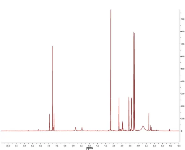
The 1H and 13C NMR of 1-3 are available free of charge
at http://jbcs.sbq.org.br as PDF ile.
Acknowledgments
We would like to thank W. Niemczura and W. Yoshida (University of Hawaii Manoa Chemistry Department NMR Facility) for assistance with Varian Unity Inova 500 MHz NMR measurements, and P. William (University of Hawaii Manoa Chemistry Department) for technical assistance with mass spectra. We are grateful to D. Horgen (Hawaii Paciic University) for NMR and mass spectra. We thank M. Yoshida (Tokyo University), J. Cooper (University of Washington at Seattle), S. W. Ki and S. Lal for help in the yeast two-hybrid system. Financial support from the Start-up fund from College of Pharmacy, UH Hilo (to L. C. C). This work was also supported by a grant in UMS (to H.C.C).
References
1. Cohen, P.; Nat. Rev. Drug Discovery 2002, 1, 309. 2. Wipf, P.; Chem. Rev.1995, 95, 2115.
3. Ki, S. W.; Kasahara, K.; Kwon, H. J.; Eishima, J.; Takesako, K.; Cooper, J. A.; Yoshida, M.; Horinouchi, S.; J. Antibiot.1998,
51, 936.
4. Omura, S.; Iwai, Y.; Hirano, A.; Nakagawa, A.; Awaya, J.; Tsuchiya, H.; Takahashi, Y.; Masuma, R.; J. Antibiot. 1977,
30, 275.
5. Cheah, H. Y.; MScDissertation, University Malaysia Sabah, Malaysia, 2003.
6. Nonomura, H.; Hayakawa, M. In Biology of Actinomycetes’88; Okami, Y.; Beppu, T.; Ogawara, H., eds.; Japan Scientiic Societies Press: Tokyo, 1988, pp. 288-293.
7. Zhang, X.; Bremer, H.; J. Biol. Chem.1995, 270, 11181. 8. Fukuda, T.; Matsumoto, A.; Takahashi, Y.; Tomoda, H.; Omura,
S.; J. Antibiot.2005, 58, 252.
9. Uyeda, M.; Aoki, M.; Nakajima, K.; Shiromoto, C.; Tatsuguchi, N.; Yokomizo, K.; Kido, Y.; Kino, Y.; J. Antibiot.1992, 45, 1370. 10. Jeffs, P.; McWilliams, D.; J. Am. Chem. Soc.1981, 103, 6185. 11. Johnson, F.; Carlson, A. A.; Tetrahedron Lett.1965, 6, 885. 12. Holden, M. T. G.; Chhabra, S. R.; de Nys, R.; Stead, P.;
Bainton, N. J.; Hill, P. J.; Maneield, M.; Kumar, N.; Labatte, M.; England, D.; Rice, S.; Givskov, M.; Salmond, G. P. C.; Stewart, G. S. A. B.; Bycroft, B. W.; Kjelleberg, S.; Williams, P.; Mol. Microbiol.1999, 33, 1254.
13. Jayatilake, G. S.; Thornton, M. P.; Leonard, A. C.; Grimwade, J. E.; Baker, B. J.; J. Nat. Prod.1996, 59, 293.
14. Wang, F.; Fang, Y.; Zhu, T.; Zhang, M.; Lin, A.; Gu, Q.; Zhu, W.; Tetrahedron2008, 64, 7986.
15. Kimura, Y.; Tani, K.; Kojima, A.; Sotoma, G.; Okada, K.; Shimada, A.; Phytochemistry 1996, 41, 665.
16. Grundmann, A.; Li, S. -M.; Microbiology2005, 151, 2199. 17. Kanzaki, H.; Imura, D.; Nitoda, T.; Kawazu, K.; J. Mol. Catal.
B: Enzym.1999, 6, 265.
18. Wessels, M.; König, G. M.; Wright, A. D.; J. Nat. Prod.1999,
62, 927.
19. Ennis, H. L.; Lubin, M.; Science1964, 146, 1474.
20. Bull, S. D.; Davies, S. G.; Parkin, R. M.; Sánchez-Sancho, F.;
J. Chem. Soc., Perkin Trans. 11998, 2313.
21. Smith, G. G.; Evans, R. C.; Baum, R.; J. Am. Chem. Soc.1986,
108, 7327.
22. Adamczeski, M.; Reed, A. R.; Crew, P.; J. Nat. Prod.1995, 58, 201.
23. Fdhila, F.; Vazquez, V.; Sanchez, J. L.; Riguera, R.; J. Nat. Prod. 2003, 66, 1299.
24. Li, X.; Dobretsov, S. ; Xu, Y.; Xiao, X.; Hung, O. S.; Qian, P.-Y.; Biofouling2006, 22, 187.
25. O’Neill, J. C. ; Blackwell, H. E.; Comb. Chem. High Throughput Screening 2007, 10, 857.
Received: January 7, 2010
Supplementary Information
J. Braz. Chem. Soc., Vol. 22, No. 2, 1-4, 2011. Printed in Brazil - ©2011 Sociedade Brasileira de Química
0103 - 5053 $6.00+0.00
S
I
*e-mail: [email protected]
Three New Amides from
Streptomyces
sp. H7372
Sarot Cheenpracha,a Robert P. Borris,a Tammy T. Tran,a Jap Meng Jee,b Heng Fong Seow,b Hwen-Yee Cheah,b Coy Choke Hoc and Leng Chee Chang*,a
aDepartment of Pharmaceutical Sciences, College of Pharmacy, University of Hawaii Hilo, 34
Rainbow Drive, 96720 Hilo, HI, USA
bImmunology Unit, Department of Pathology, Faculty of Medicine and Health Sciences,
Universiti Putra Malaysia, 43400 Serdang, Selangor, Malaysia
cBiotechnology Program, School of Science and Technology, University Malaysia Sabah, 88999
Kota Kinabalu, Sabah, Malaysia
Three New Amides from Streptomyces sp. H7372 J. Braz. Chem. Soc.
S2
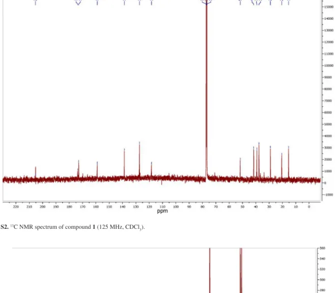
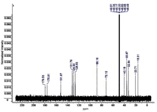
Figure S2. 13C NMR spectrum of compound 1 (125 MHz, CDCl 3).
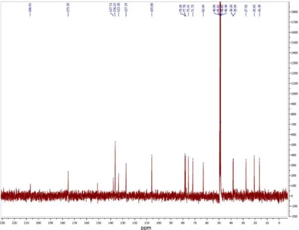
Figure S5. 1H NMR spectrum of compound 3 (125 MHz, CD 3OD).
Three New Amides from Streptomyces sp. H7372 J. Braz. Chem. Soc.
S4