Article
J. Braz. Chem. Soc., Vol. 22, No. 2, 230-238, 2011. Printed in Brazil - ©2011 Sociedade Brasileira de Química 0103 - 5053 $6.00+0.00
A
*e-mail: [email protected]
Bioconcentration of Cd and Pb by the River Crab
Trichodactylus luviatilis
(Crustacea: Decapoda)
Mariana Franchi,a Amauri A. Menegário,*,a Ana L. Brossi-Garcia,a,b Graziela C. Chagas,a Monizze V. Silva,a Antonio C. S. Piãoc and José S. Govonec
aCentro de Estudos Ambientais, Universidade Estadual Paulista, Av. 24-A, 1515,
13506-900 Rio Claro-SP, Brazil
bInstituto de Biociências, Departamento de Zoologia, Universidade Estadual Paulista, Av. 24-A, 1515,
13506-900 Rio Claro-SP, Brazil
cInstitutode Geociências e Ciências Exatas, Departamento de Estatística e Matemática Aplicada e
Computacional, Universidade Estadual Paulista, Av. 24-A, 1515, 13506-900 Rio Claro-SP, Brazil
Foi avaliada a bioconcentração de cádmio e chumbo pelo caranguejo luvial Trichodactylus
luviatilis. Trinta animais foram expostos a 200 µg L-1 de cádmio e chumbo por 7, 14 e 21 dias. Após
dissecação e digestão nitro-perclórica, os metais foram determinados nas brânquias, hepatopâncreas e músculo por espectrometria de emissão óptica com plasma acoplado indutivamente. O chumbo foi detectado apenas nas brânquias, sem diferenças signiicativas entre diferentes períodos de exposição. O cádmio foi encontrado em todos os tecidos após exposição. Diferenças signiicativas entre as concentrações de cádmio em animais expostos por diferentes períodos sugerem um processo de acumulação, com concentração estabilizada após 14 dias. Usando um procedimento de extração em
fase sólida com Saccharomyces cerevisae e com o objetivo de avaliar a transferência e estocagem
de cádmio nos tecidos, foi realizado o fracionamento de cádmio na forma livre (ou lábil) e
cádmio-proteína (possivelmente metalotioneína) (Cd-P) em animais expostos a 200 µg L-1 de cádmio por
21 dias. A determinação de cádmio livre e Cd-P nos tecidos mostrou que nas brânquias o metal foi encontrado principalmente na forma livre, enquanto que no hepatopâncreas o metal foi encontrado principalmente ligado à proteína. Pode-se inferir que, absorvido através das brânquias, o cádmio foi transferido e estocado no hepatopâncreas dos animais.
The bioconcentration of cadmium and lead by the freshwater crab Trichodactylusluviatilis was
evaluated. Thirty animals were exposed to 200 µg L-1 of cadmium and lead for 7, 14 and 21 days.
Both metals were determined in gills, hepatopancreas and muscle after dissection and digestion by inductively coupled plasma optical emission spectrometry. Lead was detected only in gills, but without signiicant difference among different exposure periods. Cadmium was found in all tissues after exposure. Signiicant differences among cadmium concentrations in animals exposed for different periods suggest an accumulation process, with concentration stabilized after 14 days. Fractionation of free (or labile) cadmium and cadmium protein (possibly metallothionein) (Cd-P) in gills and hepatopancreas were carried out to assess the cadmium transference and storage in the
tissues using a solid phase extraction procedure with Saccharomycescerevisae. Fractionation of free
(or labile) cadmium and cadmium protein (possibly metallothionein) (Cd-P) in animals exposed to
200 µg L-1 for 21 days in gills and hepatopancreas were carried to assess the cadmium transference
and storage in the tissues using a solid phase extraction procedure with Saccharomycescerevisae.
In gills, cadmium was found mainly in the free form, while in hepatopancreas the metal was found mainly bound to the protein (Cd-P). It may be inferred that, absorbed through gills, cadmium was transferred and stored in the hepatopancreas.
Introduction
Freshwater environments nearby industries and cities frequently have high levels of contamination by metals. Mining, house waste disposal, efluents from industry and agriculture are the major responsible for these contaminations.1-4
Cadmium and Pb show no functional activity in organism metabolism and their toxic effects on biota are dependent on several factors, such as the chemical speciation in the aquatic environment.3,5
Cadmium is considered one of the most potentially toxic metals in the environment. The anthropogenic sources of environmental contamination of this metal are batteries, synthetic pigments, residues of galvanoplastic factories and fertilizers.5,6 In aquatic environments, Cd has higher motility
and it is found as Cd2+ (hydrated ion II) or as an ionic complex
with other organic or inorganic substances.6
Lead is relatively abundant in the earth crust and it has a tendency to accumulate in sediments. As it has low solubility, the metal can be accessible to the food chain for a long time.6
Lead is found in aquatic environment as Pb(II) in the free form (hydrated ion II) or containing organic or inorganic ligands. Normally, the free form is comparatively more toxic. The main natural sources of lead are volcanic emissions and weathering of rocks, but they are considered insigniicant when compared to anthropogenic ones, like mining and metallurgy activities, chemical industries, electric battery factories, paints and pigments.6 Organic Pb forms are released
in the environment through direct sources (production, transportation and storage of gasoline with Pb and consequent trafic emissions) and chemical/biological methylation of inorganic lead in anaerobic sediments.6 Metals from natural
sources and contaminated efluents can be taken up and subsequently accumulated in aquatic animal tissues according to their physiologic characteristics (species, metabolism, feeding habits, size, sex).3-4,7-12 Besides, characteristics from
the aquatic environment (solubility, concentration and/or different metals interaction, pH, salinity, dissolved oxygen) can also interfere in the accumulation process.3,4,6,13 Bioindicator
organisms can provide real evidence of bioavailability and effects of contaminants in the environment.14 In aquatic
environments, bioindicators can take up and accumulate substances in concentrations several times higher than those found in water.3,11 Some studies show Cd accumulation in
aquatic animals in concentrations from 100 to 1000 times higher than those in the water. The related bioconcentration factors range from 113 to 18,000 for invertebrates and from 3 to 2,213 for ish.6
Many studies have shown that Cd and Pb can accumulate in freshwater decapods.3,4,10,11 Nevertheless, the bioconcentration
of these metals depends on the biological characteristics
of each species and it can vary according to physiological patterns of accumulation, metabolism, size, sex, and individual variability factors of the organisms, like growing, build up or loss of gametes and energy reserves.3,13-15
Invertebrates have many processes of cellular detoxiication which can decrease potentially toxic metal concentrations in circulation, like intracellular mechanisms involving high-afinity binding with low molecular weight proteins, known as metallothioneins.12,13 Metallothioneins are found in almost
any vertebrate including many species of ish and aquatic invertebrates, mainly mollusks and crustaceans.16 In animals,
they are more abundant in parenchymatic tissues (liver, kidney, pancreas and intestine), but their occurrence and biosynthesis have also been shown in other tissues and cellular types.17
The binding of potentially toxic metals with metallothioneins represents a sequestration function which leaves the metals unable to interact with other proteins, like enzymes, conferring protection against metal toxicity at the cellular level.18 Tissues which are directly involved in
metal uptake, storage and excretion have a high capacity of synthesizing metallothioneins.13,16 In aquatic animals, these
proteins have been identiied in hepatopancreas and gills of mollusks and crustaceans.16,18-22
The objective of this study was to investigate Cd and Pb bioconcentration processes using the freshwater crab T. luviatilis as bioindicator species. The effect of sex, time of exposure to metals and their distribution in different tissues of the animal was evaluated. Additionally, the metal storage process in T. luviatilis was evaluated by determination of the concentration of cadmium inorganic forms (possibly Cd(II) or “free-Cd”) and organic forms (possibly Cd-metallotioneins, Cd-P) in gills and hepatopancreas.
Experimental
Field work
Thirty-one Richodactylus luviatilis specimens (Table 1) were caught in Ribeirão Claro river (S 22º 41’35,3” W 47º32’26,1”) - Corumbataí watershed - São Paulo, Brazil, using traps containing cat food, placed on the river bank, near vegetation.
Bioassays
Bioassay 1: Total Cd and Pb in water and tissues
All glassware and plastics used during exposure, sample preparation and determination of Cd and Pb concentrations were previously decontaminated with a 10% v/v nitric acid (HNO3) solution for 8 h, followed by rinsing with distilled and deionised water. All solutions were made with puriied water (18 MΩ cm).
Stock solutions of 1000 mg L-1 Cd and Pb (High-Purity
Standard, Charleston, SC, USA) were used in both bioassays. The digestion process was carried out using HNO3 and HClO4 proanalisi (p.a.) grade (Merck, Darmstadt, Germany). All solutions were made with puriied water (18 MΩ cm).
Ribeirão Claro river water was used for the exposures, once previous analysis has shown Cd and Pb concentrations (dissolved fraction) below the limits of detection (4 and 30 ng mL-1 for Cd and Pb, respectively). All crabs were
individually placed in plastic bottles containing 900 mL of water from the river, with controlled temperature (25 oC). The
exposure to both metals was made by adding 180 µL of each stock solution to the water, resulting in exposure solutions of 200 µg L-1 of Cd and Pb.
Exposure periods were 7 (for 4 males and 6 females), 14 (5 males and 5 females) and 21 (1 male and 7 females) days. For control, 1 male and 1 female were allowed to stand at the same conditions without metals in water for a 7-day-period, since background concentrations for the species is known from previous studies. In these studies Cd and Pb concentrations for T. luviatilis caught in river water without laboratory exposures (16 crabs) were below 0.5 and 8 mg kg-1, respectively. In
addition, river water concentrations of dissolved Cd and Pb were below 4 and 30 ng mL-1, respectively. Analysis of control
individuals and river water used for the exposures conirmed these previous results: animals exposed to low concentrations of Cd and Pb (≤ 4 and 30 ng mL-1) shows tissues concentrations
below 0.5 and 8 mg kg-1.To avoid accumulation of residues
during the exposure period, the water was changed every 72 h. Each crab was fed with small pieces of ish in freshly changed water. In order to prevent metal uptake from food, Cd2+ and
Pb2+ stock solutions were added only after certifying that there
were no ish remains in the water.
Cadmium and Pb concentrations in exposure solutions were monitored by periodic sampling from 0 to 144 h of total exposure. The initial exposure solution (0 to 72 h of total exposure) was called solution 1 and the exposure solution after irst water exchange (72 to 144 h of exposure) was called solution 2. Additionally, the pH of the exposure solutions was measured each 12 h, for 3 days.
After exposure, animals were euthanized by chilling at −10 oC and classiied by sex, mass, carapace length and
width. Using stainless material, gill, hepatopancreas and muscle tissues were removed from each individual, weighed and stored in 2 mL Eppendorf tubes at −10 oC until analysis.
Wet tissues samples (0.08-0.24 g of gills, 0.01-0.19 g of hepatopancreas and 0.03-0.18 g of muscle) were transferred to digestion tubes. The tissues were predigested by adding 2.5 mL of concentrated HNO3 (Merck) in each tube and allowed the material to stand overnight.
The digestion process was completed in a digestion block by heating the tubes at 100 oC (for 1 h) and 160 oC (for
about 2 h), until a solution without suspended fragments was obtained. After cooling, 0.2 mL of concentrated perchloric acid (p.a. grade, Merck) was added in each tube, followed by heating at 160 oC for 15 min, 190 oC for 30 min and 210 oC for
about 2.5 h. The addition of perchloric acid was particularly necessary to ensure effective digestion of hepatopancreas samples. In addition, it gives a residual acid concentration which is similar to all samples. After cooling, the obtained extracts were quantitatively transferred to volumetric lasks and diluted to 20 mL with ultrapure water, produced in a Milli-Q system (Millipore, Billerica, MA, USA).
A GBC model Integra XL ICP OES spectrometer (Melbourne, Australia) with a V-Groove nebulizer (VeeSpray, Glass Expansion, Melbourne, Australia) installed in a cyclonic spray chamber (Glass Expansion) was used for determination of cadmium and lead concentrations in water samples and biological digests/extracts. The spectrometer was operated under the following conditions: forward power = 1200 W; plasma gas flow rate = 10 L min-1; auxiliary gas flow
rate = 0.5 L min-1; nebulizer gas low rate = 0.6 L min-1;
sample introduction low rate = 3.2 mL min-1; observation
height (radial viewing) = 10 mm; sample introduction low rate = 2.8 mL min-1. Cadmium and Pb measurements were
performed at 226.502 and 220.353 nm, respectively. External calibration curves were used for quantification of both metals. Replicates (n = 2) were made to all samples. Standard reference materials (SRM) of Fish Homogenate (SRM MA-A-2/TM, International Atomic Energy Agency, Vienna, Austria) and Copepod Homogenate (SRM MA-A-1/TM, International Atomic Energy Agency) were used to check the accuracy of the analytical procedure.
Bioassay 2: Metallothioneins (Cd-binding biomolecules) and metal speciation
For this work stage other 12 T. luviatilis specimens were caught and exposed to 200 µg L-1 Cd solutions for 21
aimed to evaluate the presence of Cd-binding biomolecules (possibly metallothioneins) in gills and hepatopancreas.
Sample preparation
Based on previously described methods,23-26 the following
procedure was used to extract the cytosol from the selected tissues of exposed T. luviatilis. About 0.1 g of hepatopancreas and gills from each specimen was homogenized using 10 mL 0.05 mol L-1 TRIS (tris(hydroxymethyl)aminomethane) (pH 7.0
and 4 oC) in 30 mL centrifuge tubes. Then, the mixtures were
centrifuged on a 15000 g and 4 oC for 30 min using a JOUAN
centrifuge MR 23i with controlled temperature (St. Herblain, France). The supernatants were transferred to 50 mL tubes and the inal volume of each sample was made up to 50 mL by adding 40 mL of the same 0.05 mol L-1 TRIS solution. The obtained
extracts were used for determining total cadmium concentrations (direct determination on ICP OES) and Cd(II) and Cd-binding biomolecules (Cd-P) concentrations (after fractionation).
The procedure used for Cd fractionation on cytosols was the same as proposed by Menegário et al.,23 where the baker
yeast Saccharomyces cerevisae is used as sorbent material to separate Cd(II) and Cd-P (possibly Cd metallothioneins or Cd bound to other proteins) from cytosols of biological extracts. After contact with the yeast and centrifugation, the inorganic fraction of cadmium (Cd(II) or “free” Cd) is expected to be found in solid phase (retained by the yeast) while the Cd-P is expected to be found in the liquid phase.
Solid phase extraction
Aliquots of 10 mL of the obtained extracts were transferred to 15 mL centrifuge tubes containing 0.0625 g of Saccharomyces cerevisiae. The tubes were manually agitated until complete mixture of yeast and solution (about 30 s) and maintained in water bath at 25 oC for 10 min. Subsequently,
the tubes were centrifuged at 1900 g for 7 min using a JOUAN B4i centrifuge (St. Herblain, France). After phase separation, the liquid phase was transferred to other lask and the solid phase was mixed with 10 mL 2% v/v HNO3.
Liquid and solid phase were agitated and directly introduced into the ICP OES, using a V-Groove nebulizer, to determine Cd-P and Cd(II) concentrations, respectively. The eficiency of V-groove nebulizer to introduce slurry of yeast has been previously demonstrated.23 The yeast slurry basically contains
individual cells of the microorganism with diameter size changing from of 5 to 10 µm, while the tolerance to particulates for the nebulizer is typically up to 300 µm. Acidiication of solid phase results in a slurry characterized by a matrix effect lower than 20% (as compared with nebulization of HNO3 solution), while the matrix effect on liquid phase is insigniicant.
Statistical analysis
Taking into account that the non-parametric test of Kruskal-Wallis allows multiple comparisons among independent samples, it was used to compare differences between concentrations of Cd and Pb due to sexes and exposure periods. When signiicant differences between the variables tested were conirmed (p < 0.05), Dunn’s statistical test was used for multiple comparisons, pair to pair, of the independent samples.
To determine if Cd concentrations found in different tissues were signiicantly different (p < 0.05), Friedman test was used, once it allows comparisons between dependent variables (different tissues from the same animals). The same test was used to compare Cd(II) and Cd-P concentrations in gills and in hepatopancreas (different forms of cadmium in the same tissues) of 21 days exposed animals. Statistical tests were performed using the program BioEstat 5.0, with 95% of conidence.
Results and Discussion
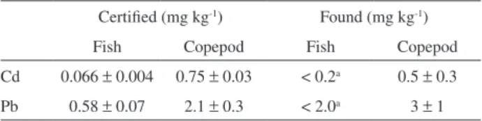
Digestion of reference material was carried out using the procedure described in bioassay 1 item, using 0.5 g of sample. Cadmium and Pb concentrations obtained for standard reference materials by using the digestion procedure described in bioassay 1 are shown in Table 2. For ish sample, concentrations of Cd and Pb were lower than detection limits. For copepod sample, the obtained results were according to certiied values (95% level), which indicate that the procedure was suitable for analysis of biological materials.
Cadmium and Pb concentrations in exposure solutions
There was no signiicant pH alteration in the exposure solution during 72 h of incubation. For this period, values between 7.4 and 8.0 were obtained.
Potentially bioavailable (total dissolved) Cd and Pb were determined in exposure solutions. With this purpose, a iltration in 0.45 µm acetate cellulose membrane was made, followed by acidiication with HNO3.
Table 2. Average concentrations (dry weight) ± conidence interval (n = 4, t = 3.182, 95% level) in standard reference materials
Certiied (mg kg-1) Found (mg kg-1)
Fish Copepod Fish Copepod
Concentrations of analytes in the initial exposure solution (0 to 72 h of total exposure), solution 1, and in the exposure solution after irst water exchange (72 to 144 h of exposure), solution 2, are shown in Figures 1 and 2, respectively.
It is observed a trend towards decreasing concentrations of Cd and Pb in exposure solutions during the incubation period, mainly in the irst 20-25 h. These results indicate metal uptake by the animals with different rates during the incubation period (72 h). Nevertheless, it should be taken into account that this difference can be consequence of possible interactions of Cd and Pb with the carapaces of animals, with particulate material and/or with other compounds present in the river water used for the exposures.
Lead and Cd concentrations in tissues
Three animals died between 19-21 days. After exposure, Pb was detected in gills of 26 individuals. The average metal
concentrations found in gills for different periods of exposure are shown in Figure 3. Lead was also detected in hepatopancreas of only 3 animals (average concentration of 8.42 ± 6.85 mg kg-1)
and muscle of 4 animals (average concentration of 11.33 ± 1.29 mg kg-1). The mean concentration of the metal
in gills was 15.61 mg kg-1. From this value and considering
moisture of 75%,27 a mean concentration of 62.45 mg kg-1 in
dry weight can be estimated for gills. The results obtained for Pb suggest that the transference of the element from gills to other tissues did not occur during the exposure period. There is little information in the literature about interactions of Pb within crabs. For comparison, Turoczy et al.,28 related
signiicant differences in lead concentrations in tissues of some species of marine crabs (Paralithodes camtschatica, Portunus pelagicus, Tachypleus tridentatus and Callinectes spp.).
There were not signiicant differences in Pb concentrations between males and females (Kruskal-Wallis, p = 0.7934). For natural expositions to the metal, similar results were found for the marine crabs Callinectes spp.,29 and Tachypleus tridentatus.28
On the other hand, differences in Pb concentrations between sexes have been reported for Paralithodes camtschatica.30
There were no significant differences among Pb concentrations in gills of animals from different exposure periods (Kruskal-Wallis, p = 0.5067). It shows that, at studied conditions, the maximum concentration of Pb in this tissue occurs on the irst 7 days of exposure. After this period, there is no more lead accumulation by the animals.
Cadmium was detected in gills and hepatopancreas of all exposed animals. In muscle, metal was detected in 25 animals from the 28 exposed ones. Average Cd concentrations (for males and females) obtained in the three periods of exposure for gills, hepatopancreas and muscle are shown in Figures 4, 5 and 6, respectively.
Figure 1. Cadmium and Pb concentrations (mean ± SD) in water samples taken from initial exposure solution (solution 1).
Figure 2. Cadmium and lead concentrations (mean ± SD) in water samples taken from irst changed exposure solution (solution 2).
No signiicant difference was found in Cd concentrations between animals from different sexes (Kruskal-Wallis, p = 0.4442), as previously observed by Schuwerack et al.11
Also, for most crab species exposed to cadmium in natural aquatic systems (as Pseudocarcinus gigas, Callinectes sp., Tachypleus tridentatus) had been reported no differences between Cd concentrations related to sex.3,28 Only Chen
et al.,31 reported differences of cadmium concentrations
between sexes (cadmium in hepatopancreas of Thalamita crenata). The reasons for this difference are still not clear.3
There were significant differences between Cd concentrations of exposed animals from different periods (Kruskal-Wallis, p < 0.05), suggesting an accumulation process. The results obtained for Dunn’s test (Table 3) suggest that in the experimental conditions used, Cd is
accumulated by the animals until 14 days. After this period the concentration was stabilized.
There were significant differences (Friedman, p < 0.05) between Cd concentrations in gills (average of 21.09 mg kg-1) and muscle (average of 2.17 mg kg-1)
and between hepatopancreas (average of 10.52 mg kg-1)
and muscle. Considering moisture of 75%,27 we can
estimate mean Cd concentrations in dry mass of 84.4, 8.7 and 42.1 mg kg-1for gill, muscle and hepatopancreas,
respectively.
For most studied species, differences in bioconcentration of metals among tissues and organs are observed and they show to vary according to metals and crustacean species.3,10,11,14,15,32
For the freshwater crab Potamonautes warreni,11
exposed to Cd in laboratory and for natural expositions for the marine species Carcinus maenas,9 there were
also reported significant differences among metal concentrations in different tissues, following the order: gills > hepatopancreas > muscle.
Similar results to those obtained in this work were reported for Nephrops norvegicus,9 where Cd concentration
was higher as well in gill as in hepatopancreas of males and females, even in metal conditions of 1 µg mL-1 in
Figure 4. Cadmium concentrations (mean ± SD) in gills of T. luviatilis
males and females according to the period of exposure.
Figure 5. Cadmium concentrations (mean ± SD) in hepatopancreas of
T. luviatilis males and females according to the period of exposure.
Figure 6. Cadmium concentrations (mean ± SD) in muscle of T. luviatilis
males and females according to the period of exposure.
Table 3. Results obtained for Dunn’s test for cadmium concentrations found in different exposure periods
Pairs P
7 days × 14 days < 0.05
7 days × 21 days < 0.05
14 days × 21 days n.s.
environment. For Pseudocarcinus gigas,28 and Penaeus
californiensis,31 higher Cd concentrations were found in
hepatopancreas, followed by gills and muscle.
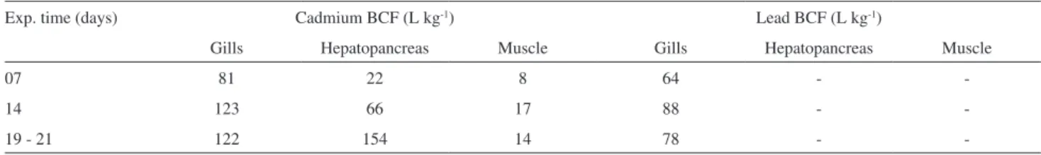
Bioconcentration factor (BCF) was calculated according to the equation 1, and the obtained values for tissues, for each one of the exposure periods, are shown in Table 4.
BCF = Cb / Cw (1)
Where: C b = average metal concentration in animals, µg kg-1; C
w = metal concentration in water, µg L -1.
For comparison, freshwater amphipods (Hyalella azteca) exposed for 5 days to a Cd concentration of 4.4 µg L-1 showed BCF of 31.803.33 Mussels and oysters
(bivalves) in natural environments with Pb concentrations of 0.64 µg L-1 and Cd concentrations of 0.08 µg L-1 have
shown bioaccumulation factors (when a chemical substance is absorbed in organism by all routes of exposure as occurs in the natural environment) of 220 e 216 for Pb and Cd, respectively. Crabs in these conditions shown bioaccumulation factors of 145 for Pb and 525 for Cd.34
Bioconcentration factors obtained in the present study were lower in comparison to those obtained for crabs naturally exposed to Cd and Pb, possibly due to conditions of higher concentrations in exposures.
Cadmium-binding biomolecules concentrations in tissues
The total Cd, inorganic cadmium (Cd(II)) and organic Cd (possibly Cd-metallothioneins) concentrations obtained from gills and hepatopancreas of exposed T. luviatilis are shown in Table 5. Gills and hepatopancreas were chosen for analysis once they are probably responsible for absorption and storage of Cd, respectively. As only Cd was detected in all tissues and it is expected low interaction between Pb and metallothioneins, the fractionation study focused only the irst element. The sum of Cd(II) and Cd-P concentrations for each animal represents values near 100% of total cadmium, determined directly on the extracts, which indicates that all Cd species existing on extracts were determined.
With the exception of animal L, it can be observed that total cadmium (CdT) concentrations in hepatopancreas Table 4. Bioconcentration factor (BCF) values for gills, hepatopancreas and muscle according to the exposure period
Exp. time (days) Cadmium BCF (L kg-1) Lead BCF (L kg-1)
Gills Hepatopancreas Muscle Gills Hepatopancreas Muscle
07 81 22 8 64 -
-14 123 66 17 88 -
-19 - 21 122 154 14 78 -
-Table 5. Total cadmium (CdT), inorganic cadmium (Cd(II)) and organic cadmium (Cd-P) concentrations (wet mass, mg kg-1) in cytosols of hepatopancreas and gills of T. luviatilis exposed to 200 µg L-1 of cadmium. The limit of detection (3 σ, wet mass) found with the procedure described in bioassay 2 (using at least 0.09 g of sample) was 4 mg kg-1
Animal Exp. time (days)
Sex CdT Cd(II) Cd-P
Hepatopancreas Gills Hepatopancreas Gills Hepatopancreas Gills Average SD Average SD Average SD Average SD Average SD Average SD
A 9 a M 18.77 0.56 < 4 - < 4 - < 4 0.47 11.28 0.33 < 4
-B 13 a M 30.44 0.72 < 4 - 4.20 0.83 < 4 2.22 25.24 0.43 < 4
-C 14 a F 25.50 2.79 6.93 0.01 25.55 4.06 5.40 0.58 9.03 1.04 < 4
-D 14 a M 14.74 0.26 - - < 4 - - - 12.14 0.48 -
-E 14 a M 12.51 0.21 7.59 0.30 < 4 - 7.88 0.22 11.75 0.48 < 4
-F 15 a M 21.70 1.01 < 4 - 22.30 2.00 < 4 0.79 4.74 0.58 < 4
-G 20 a M 13.00 0.19 < 4 - 4.60 0.68 < 4 3.28 8.45 1.11 < 4
-H 21 M 36.33 0.17 8.09 0.62 21.15 0.65 7.15 0.51 12.64 5.70 4.61 1.01
I 21 M 29.42 0.45 6.92 1.13 8.34 0.28 4.03 0.42 20.90 0.76 < 4
-J 21 M 53.28 0.29 6.59 0.13 20.45 1.39 8.91 1.26 34.48 1.73 < 4
-K 21 F 15.09 0.63 11.50 0.12 < 4 - 12.07 0.74 11.95 1.27 < 4
-L 21 F 9.02 0.03 13.82 0.22 < 4 - 12.36 0.55 8.15 0.69 < 4
are always higher than the ones obtained in gills, indicating an storage function for this tissue, as seen in previous results.
Higher levels of cadmium in hepatopancreas show that the biggest ratio of the metal is stored in the organ.11,14,16,29
In most crustaceans, hepatopancreas has central function to metabolize, to store and to detoxiicate many metals. Elevated concentrations in this organ (when compared to other tissues) probably are associated to its function and to its high amounts of metallothioneins.14,16,28
Friedman statistic test results showed no signiicant differences of Cd(II) and Cd-P concentrations in hepatopancreas (p = 0.1317), although it seems to be a trend towards higher values of the organic form (bound to proteins, possibly metallothioneins). On the other hand, in gills, there were found signiicant differences between the inorganic and organic forms (Friedman, p = 0.0348) evidencing that Cd in this tissue was found mainly as Cd(II), the “free” form.
Thus, the obtained results (Cd(II) and Cd-P) and CdT concentrations obtained for both tissues shows that cadmium is possibly absorbed through gills and rapidly transferred to hepatopancreas (at least, during the maximum period of exposure), where it is stored.
Conclusions
After exposure, Pb was found in detectable concentrations only in gills. It was not observed signiicant increase in Pb concentrations in gills towards periods of exposure (7, 14 and 21 days). Cadmium was found in all tissues after exposures (7-21 days). Considerable increases in cadmium concentration in gills and hepatopancreas indicate that the crab T. luviatilis may be a potential bioindicator species. In addition, results of Cd-P determination in tissues suggest that this metal possibly is absorbed through gills, followed by its transferring and storage in hepatopancreas.
Acknowledgments
The authors thank FAPESP (Fundação de Amparo à Pesquisa do Estado de São Paulo) and CNPq (Conselho Nacional de Desenvolvimento Cientíico e Tecnológico) for inancial support.
References
1. Bu-Olayan, A.-H.; Subrahmanyam, M. N. V.; Environ. Monit. Assess.1998, 53, 297
2. Mariño-Balsa, J. C.; Poza, E.; Vázquez, E.; Beiras, R.; Arch.
Environ. Contam. Toxicol. 2000, 39, 345.
3. Marsden, L. D; Rainbow, P. S.; J. Exp. Mar. Biol. Ecol. 2004,
300, 373.
4. Reinecke, A. J.; Snyman R. G.; Nel, A. J.; Water, Air, Soil Pollut.
2003, 145, 395.
5. López-Greco L. S.; Sánchez M. V.; Nicoloso, G. L.; Medesani, D. A.; Rodríguez, E. M.; Arch. Environ. Contam. Toxicol.2001,
41, 333.
6. Labunska, I.; Stringer, R. E.; Brigden, K.; Poluição porMetais e Compostos Orgânicos Associada à Unidade da Bayer em Belford
Roxo, Rio de Janeiro, Brasil; Exeter: Reino Unido, 2000.
7. Bjerregaard, P.; Depledge, M. H.; Mar. Biol.2002, 141, 741. 8. Du Preez, H. H.; Steenkamp, V. E.; Schoonbee, H. J.; Sci. Total
Environ.1993, 469.
9. Martín-Díaz, M. L.; Villena-Lincoln, A.; Bamber, S.; Blasco, J.; DelVall, T.; Chemosphere2005, 58, 615.
10. Sanders, M. J.; Du Preez, H. H.; Van Vuren, J. H.; Ecotoxicol.
Environ. Saf.1998, 41, 203.
11. Schuwerack, P.-M. M.; Lewis, J. W.; Jones, P.; Ecotoxicology
2001, 10, 159.
12. Wang, W.-X.; Rainbow, P. S.; Ecotoxicol. Environ. Saf.2005,
61, 145.
13. Ahearn, G. A.; Mandal, P. K.; Mandal, A.; J. Comp Physiol.,
B: Biochem Syst, Environ. Physiol.2004, 174, 439.
14. Rainbow, P. S.; Estuar. Coast. Shelf Sci.1997, 44, 169. 15. Rainbow, P. S.; Environ. Pollut.2002, 120, 497.
16. Amiard, J.-C.; Amiard-Triquet, C.; Barka, S, Pellerin, J.; Rainbow, P. S.; Aquat. Toxicol.2006, 76, 160.
17. Pourang, N.; Dennis, J. H.; Ghourchian, H.; Ecotoxicology
2004, 13, 519.
18. Wenli, M.; Lan, W.; Yongji, H.; Yao, Y.; Environ. Toxicol. 2008,
23, 393.
19. Pan, L.; Zhang, H.; Comp. Biochem. Physiol., Part C: Toxicol.
Pharmacol.2006, 144, 67.
20. Jui-Pin, W.; Hon-Cheng, C.; Comp. Biochem. Physiol., Part C:
Toxicol. Pharmacol.2005, 140, 383.
21. Silvestre, F.; Duchêne, C.; Trausch, G.; Devos, P.; Comp.
Biochem. Physiol., Part C: Toxicol. Pharmacol.2005, 140, 39.
22. Pourang, N.; Dennis, J. H.; Environ. Int. 2005, 31,325. 23. Menegario, A. A.; Tonello, P. S.; Biscaro, P. A.; Brossi-Garcia,
A. L.; Microchim. Acta. 2007, 159, 247.
24. High, K. A.; Azani, R.; Fazekas, A. F.; Chee, Z. A.; Blais, J. S.;
Anal. Chem.1992, 64, 3197.
25. Muñoz, J.; Baena, J. R.; Gallego, M.; Valcárcel, M.; J. Anal.
At. Spectrom2002, 17, 716.
26. Lobinski, R.; Chassaigne, H.; Szpunar, J.; Talanta1998, 46, 271. 27. DeForest, D. K; Brix, K. V; Adams, D. W.; Aquat. Toxicol. 2007,
84, 236.
28. Turoczy, N. J.; Mitchell, B. D.; Levings, A. H.; Rajendram, V. S.; Environ. Int.2001, 27, 327.
29. Sastre, M. P.; Reyes, P.; Ramos, H.; Romero, R.; Rivera, J.;
30. Rusanowski, P. C.; Gardner, L. A.; Jewett, S. C.; King, C. A.;
Proceedings of the International Symposium on King and
Tanner Crabs, Alaska, 1989. http://seagrant.uaf.edu/bookstore/
pubs/AK-SG-90-04.html.
31. Chen, M.-H; Chen, C.-Y; Chou, H.-Y; Wen, T.-C.; Mar. Pollut. Bull.2005, 50, 463.
32. Celik, M.; Kucukgulmez, A.; Yanar, Y.; Cikrikci, M.; Fresenius Environ. Bull. 2006, 15, 349.
33. Shuhaimi-Othman, M.; Pascoe, D.; Ecotoxicol. Environ. Saf.
2007, 66, 29.
34. Young-Tack, K.; Chan-Won, L.; Microchem. J.2001, 70, 255.
Submitted: February 11, 2010
Published online: September 21, 2010