Computational Analysis of Magnetic Resonance
Images of the Upper Airways: Algorithms and
Applications
Jessica Condesso Delmoral
Dissertation submitted in fulfillment of the requirements for the conclusion of the Integrated Master in Bioengineering – Branch of Biomedical Engineering, FEUP
Supervisor:
Prof. Dr. João Manuel R. S. Tavares
Prof. Associado do Departamento de Engenharia Mecânica, FEUP
Co-Supervisor:
Prof. Dra. Sandra Rua Ventura
Professora Adjunta da Área Técnico-Científica da Radiologia, ESTSP-IPP
iii
Abstract
The human upper airways anatomy consists of the jaw, tongue, pharynx, larynx, palate, nasal cavities, nostrils, lips, and adjacent facial structures.
The interplay and connective movement between all the anatomical structures present in this region is complex, and basic physiological functions such as muscle activation patterns associated with chewing, swallowing, and speech production are not well understood.
Specifically, one of the least studied organs in this region is the tongue, in which the tasks of imaging and quantification of its anatomy are of great relevance for further study and analysis of the anatomic and physiological mechanisms that govern it. Furthermore, new insight can be given on other applications such as surgical planning, post-operative rehabilitation and the study of new adaptations acquired upon possible changes in function of pathological origin, for example in the presence of tongue cancer, surgical intervention or aging. Magnetic Resonance imaging (MRI) is the state of the art methodology for visualization and study of soft tissues, since it provides the best image contrast of soft tissues such as the muscular tissue of the tongue.
Under the scope of the Computational Vision field, an area that has over recent years allowed the development of new tools of analysis that can be applied to medical images, this dissertation aims to present computational algorithms for object detection and segmentation in images, suitable for application on objects such as the tongue.
The proposed methodology includes a set of algorithms developed for human tongue image processing, in order to study morphology through the building of an Active Shape Model, that captures the shape variability of the anatomy during production of various European Portuguese sounds. The developed model allowed to simulate realistically the tongues shape capturing its variability in the production of different sounds. Subsequently, this model also allowed the building of a semi-automatic detection and segmentation algorithm of this structure. The study was carried out using midsagittal plane images, since this plane is the most representative in the depicting the overall tongue shape variability, which constitutes to be especially advantageous for speech assessment purposes. The suggested model made it possible to obtain a realistic segmentation of the tongue as well as efficiently perform segmentation in new images. Furthermore, the use of such image analysis techniques allows quantitative measures
with higher precision and are particularly advantageous when speech therapists and imaging specialists need to analyze a large volume of data.
In conclusion, the identification and analysis of human structures are complex tasks, since their shapes are not constant and vary through time. However, techniques of Computer Vision and objects modeling can assist in their achievement as is demonstrated throughout this dissertation.
v
Resumo
A anatomia das vias aéreas superiores humanas é contituída pela mandíbula, língua, faringe, boca, fossas nasais, narinas, lábios e estruturas faciais adjacentes.
Os mecanismos de interação combinada que se associam ao movimento conexivos entre todas as estruturas anatómicas presentes nesta região são complexos, e a sua funcionalidade inclui actividades fisiológicas básicas, tais como padrões de ativação muscular associados com a mastigação, deglutição, e produção da fala.
Um dos órgãos menos estudados nesta região é a língua, e portanto as tarefas de imagiologia e quantificação da sua anatomia são de grande relevância para o estudo e análise mais aprofundado dos mecanismos anatómicos e fisiológicos que a regem. Mais ainda, este estudo poderia produzir novo conhecimento passível de ser utilizado em outras aplicações, tais como o planeamento cirúrgico, reabilitação em pós-operatório e estudo de adaptações compensatórias na produção da fala, adquiridas pela presença de cancro da língua, após intervenção cirúrgica ou envelhecimento.
No âmbito da área de Visão Computacional, uma área que tem nos últimos anos permitido o desenvolvimento de novas ferramentas de análise que podem ser aplicadas em imagens médicas, esta dissertação tem como objetivo apresentar algoritmos computacionais para deteção e segmentação de objetos em imagens, adequado para aplicações em órgãos deformáveis tais como a língua. Para o estudo de tecidos moles, o estado da arte referente às técnicas de aquisição imagiólogica, a Ressonância Magnética (RM), uma vez que proporciona o melhor contraste de imagem de tecidos moles, tais como o tecido muscular da língua.
A metodologia proposta inclui um conjunto de algoritmos desenvolvidos para processamento de imagem da língua humana, para o estudo morfológico através da construção de um Modelo de Forma Activa, que capta a variabilidade anatómica desta estrutura durante a produção de vários sons do Português Europeu. O modelo desenvolvidos permitiu simular de forma realista a língua na sua variabilidade de forma durante a produção dos diferentes sons. Seguidamente, este modelo permite ainda, uma produção de um algoritmo de deteção semi-automática desta estrutura. O estudo foi realizado utilizando imagens do plano sagital médio, constituindo o plano mais representativo da variabilidade de forma da língua global, tornando-se este estudo especialmente vantajoso para fins de avaliação dos mecanismos de produção da fala. O modelo sugerido tornou possível obter uma segmentação realista da língua, bem como
executar eficientemente a segmentação da mesma em novas imagens. Mais ainda, o uso de tais técnicas de análise de imagem pode permitir a obtenção de medições quantitativas, com uma precisão mais elevada e são particularmente vantajosos para a análise por especialistas em imagem ou em produção da fala, no sentido da análise de grandes volumes de dados.
Em conclusão, a identificação e análise de estruturas humanas são tarefas complexas, uma vez que as suas formas não são constantes e variam ao longo do tempo. No entanto, as técnicas de visão computacional e modelagem de objetos podem ajudar na sua realização como é demonstrado ao longo desta Dissertação.
vii
Acknowledgments
Firstly, beginning by the direct participants in this work I would like to thank Professor Dr. João Tavares for all the availability, patience and counseling provided that guided me in this work, as well as, my co-supervisor, Professor Dr. Sandra Rua Ventura, for the availability and help to direct the purpose of this work and allowing me to achieve the objectives successfully. And I would furthermore like to thank the colleagues that accompanied me during the process of making this dissertation and of the long hours in front of the computer. Also, a special thanks goes to those that tried to keep me motivated, who helped me not to forget to relax and take a break to laugh.
Secondly, I would like to thank everyone that crossed my path, and marked it somehow, during the five years that have past. To all my friends with whom I have the best memories, with whom I grew and from whom I save the best moments. And also to the more recent ‘nerd’ friends. You have all played your part.
I would like to thank especially my oldest friends, which are the ones that accompanied me during the process of growing up to this point in our lives. Thank you for being there, Carolina, since 1998 and Rita, since an unknown year between 1992 and 1998.
Finally I would like to thank with all my heart my family, and my parents for accompanying me and supporting me until this point, thank you for always pulling me to be better, and to grow.
ix
“We are just an advanced breed of monkeys on a minor planet of a very average star. But we understand the Universe and that makes us something very special“
xi
Contents
... 1 INTRODUCTION ... 1 1.1. MOTIVATION ... 2 1.2. OBJECTIVES ... 3 1.3. REPORT ORGANIZATION ... 4 ... 5FUNDAMENTALS AND RELATED WORKS ... 5
2.1. HUMAN AERODIGESTIVE TRACT ANATOMY ... 5
2.2. MAGNETIC RESONANCE IMAGING IN THE CONTEXT OF AERODIGESTIVE ORGANS ... 16
2.3. UPPER AIRWAY IMAGING AND COMPUTATIONAL ANALYSIS ... 20
2.5. CONCLUSION ... 29
... 31
STATISTICAL MODELING OF THE TONGUE ... 31
3.1. IMAGE DATASET ... 32
3.2. METHODOLOGY ... 33
3.3. SHAPE MODEL ... 34
3.4. PROFILE MODEL ... 40
3.5. RESULTS AND DISCUSSION ... 47
3.6. CONCLUSIONS ... 62
... 65
ACTIVE SHAPE MODELING AND SEGMENTATION OF THE TONGUE ... 65
4.1. SEARCH ALGORITHM ... 67
4.2. MODEL INITIALIZATION ... 68
4.3. IMAGE FEATURE SEARCH ... 69
4.4. IMPOSING SHAPE CONSTRAINTS ... 70
4.6. RESULTS AND DISCUSSION ... 71
4.7. CONCLUSION ... 77
... 79
CONCLUSION AND FUTURE WORK ... 79
5.1. CONCLUSION ... 79
5.2. FUTURE WORK ... 80
xiii
List of Figures
Figure 1 - MR midsagittal image (slice) indicating the vocal tracts structures (Ventura et
al. (2011)). ... 6
Figure 2 - Side view of the skull. The styloid process is just posterior to the mandible (Georgia Highlands College, 2013) ... 7
Figure 3 - Tongues attachments and neighboring structures in a sagittal anatomical view (Gray (1918)). ... 8
Figure 4 - Extrinsic muscle of the tongue with styloglossus visible at center top (in red) (Gray, 1918). ... 8
Figure 5 - Muscles of the tongue (Takemoto (2001)): GG - genioglossus, T - transversus, V - verticalis, HG - hyoglossus, IL - inferior longitudinalis, S - superior longitudinalis, PG - palatoglossus, SG - Styloglossus. ... 11
Figure 6 - Tongue contour extracted from midsagittal images, during production of vocalic sounds present in Portuguese language (Ventura et al., 2008). ... 14
Figure 7 - Abd-El-Malek (1955) illustration of the preparatory stage of mastication (a), throwing stage of mastication (b), guarding stage of mastication (c), initial stage of deglutition (d). ... 16
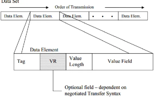
Figure 8 - DICOM data set structure consists of several data elements. ... 24
Figure 9 - Active Shape model structure scheme. ... 32
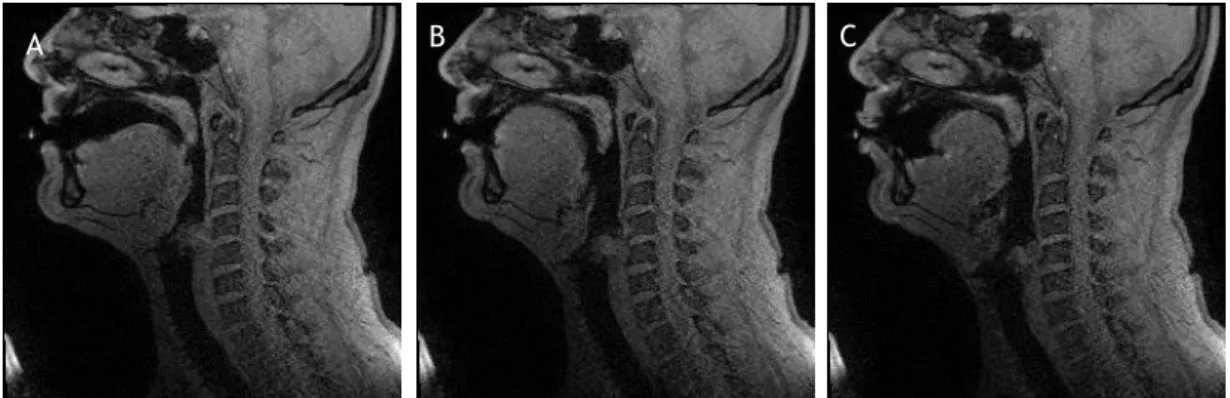
Figure 10 - Examples of images from the 3.0T image dataset used, of imaging of the oral sounds a (A), i (B) and u (C). ... 33
Figure 11 –Statistical shape model building scheme. ... 35
Figure 12 - Generalized Procrustes Analysis algorithm outline. ... 38
Figure 13 - Nonlocal Means Algorithm outline. ... 44
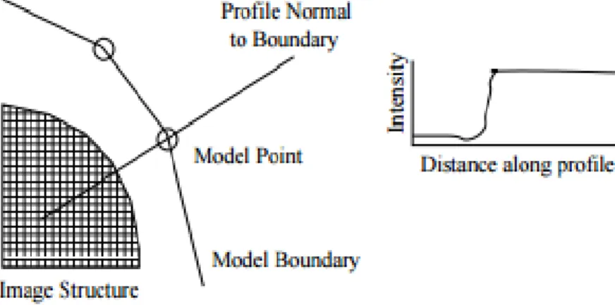
Figure 14 - The profile model takes upon the normals to the boundaries of the shape, at a given landmark (Cootes & Taylor., 2004). ... 45
Figure 15 - Example shape defined by vertexes A, B and C. ... 46
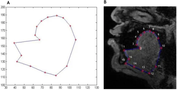
Figure 16 - Initial landmark map defined by hand representing the landmark connectivity (A), and in the image referential (B). ... 48
Figure 17 - Raw shapes (A) from dataset (blue) and initial mean shape (red), and aligned, origin centered shapes through Generalized Procrustes Analysis (B), with final shape dataset (blue) and final mean shape (red). Images plotted in image referential. ... 49
Figure 18 - Interpolated landmark map, with interpolation factor 4, depicting the original landmark constellation points in green and the interpolated points in red. ... 49
Figure 19 - Shape variance decay as the number of eigenvalues increases. ... 51 Figure 20 - Representation of the first six modes of variation plotted the model mean shape (blue) and the mean shape deformed by the model eigenvalues (red). ... 52
Figure 21 - Example image of female (top row) and male (down row) subject before and after non-local means denoising, with h parameter set to 0.1. . Error! Bookmark not defined.
Figure 22 - Non-local means denoising results, respective the original image (A), and denoised images with h parameter set to 0.05 (B), 0.1 (C) and 0.5 (D). ... 54
Figure 23 - NLM denoised example image and binarized image subtraction image result. 56 Figure 24 - A one-dimensional profile of each of the 16 initial hand-labeled landmarks of an example image of the training dataset. The blue line is the shape boundary. The red line are the whiskers, orthogonal to the boundary. ... 57
Figure 25 – Mean intensity profiles of landmarks 1 through 3 in the initial landmark constellation, corresponding to each labeled number in interpolation constellation, correspond to the tongues frenulum (anterior-posterior ends) and the tongues tip. ... 57
Figure 26- Unprocessed (left column) and processed (right column) image profiles examples, of each of the anterior, top, posterior and lower bounds (from top to bottom rows, respectively). ... 58
Figure 27 – Mean intensity profile of landmark 10, correspondent to the tongues root.
... Error! Bookmark not defined.
Figure 28 – Mean intensity profiles of landmarks 4 through 9 in the initial landmark constellation, corresponding to each labeled number in interpolation constellation, correspond to the tongues dorsum, or upper-posterior boundary. ... 60
Figure 29 - Mean profiles of landmarks 11 through 16 in the initial landmark constellation, corresponding to each labeled number in interpolation constellation, correspond to the tongues dorsum, or lower and sub-frenulum-anterior boundary. ... 61
Figure 30 - Gradient examples of each of the anterior, upper, posterior and lower walls of the tongue. ... 62
Figure 31 - Schematic model representing the final workflow of the Active Shape Model implemented, comprising the methods detailed in Chapter 3 and the present Chapter. ... 66
Figure 32 - Search model algorithm outline. ... 67 Figure 33 - Points selected in the manual initialization of the model. ... 69 Figure 34 - An image pyramid. The first level [128x128]px is a half of the resolution of the image above it [256x256]px. ... 71
Figure 35 - Segmentation process with the initial position of the shape model built overlapped (A) and the results after the 22nd (B) and 25th (C) iterations of the of the segmentation process by the active shape model. In D the produced shape (red) is overlapped with the hand-labeled shape (blue). ... Error! Bookmark not defined.
Figure 36 - Segmentation process with the initial position of the shape model built overlapped (A) and the results after the 2nd (B) and 12th (C) iterations. ASM using 17 pixels long profiles. ... 75
Figure 37 - Segmentation process with the initial position of the shape model built overlapped in image (A) and the results after the 2nd (B) and 12th (C) iterations of the of the segmentation process by the active shape model. In D the produced shape (red) is overlapped with the hand-labeled shape in the original image (blue)... 76
xvii
List of Tables
Table 1 - Muscles of tongue movement (Seikel et al. (2009))... 10 Table 2 - First seventeen modes of variation of the model obtained and their retained percentages, describing 99.9% of shape variation. ... 50
Table 3 – Mean peak signal to noise ratio (PSNR) and mean square error (MSE) of denoised images with NLM algorithm using different h parameters. ... 56
Table 4 - Mean and standard deviation (mean ± std) errors of the shapes segmented by the deformable models built in each test image with 7 pixels long profiles. ... 74
Table 5 - Mean and standard deviation (mean ± std) errors of the shapes segmented by the deformable model built in each test image with 17 pixels long profiles. ... 74
xix
Acronyms
1D One Dimensional 2D Two Dimensional 3D Three Dimensional AAM Active Appearance Model ACR American College of Radiology ASM Active Shape Model
CAD Computer-Aided Diagnosis CT Computed Tomography
DICOM Digital Imaging and Communications in Medicine DTI Diffusion Tensor Imaging
EPG Electropalatography FID Free Induction Decay GG Genioglossus muscle GTF Game-theoretic Framework MI Mutual Information
MRI Magnetic Resonance Imaging MRS Magnetic Resonance Spectroscopy
NEMA National Electrical Manufacturers Association NMCs Neuromuscular compartments
PACS Picture Archiving and Communication System PET Positron Emission Tomography
PDM Point Distribution Model RF Radiofrequency
RW Random Walker
SPECT Single-photon emission computed tomography SSM Statistical Shape Model
T Tesla
TR Repetition Time TE Echo Time US Ultrasound
1
Introduction
The tongue constitutes a unique anatomical structure among all the organs integrating the human body.
It is a specialized organ located in the oral cavity, which plays an important role in mastication and swallowing (for digestion process), taste and speech production. Breathing and swallowing processes are closely interrelated in their central control at brainstem and are highly coordinated. Many muscles and structures of the aerodigestive tract have dual roles in these processes, namely the tongue.
The ability to produce of fast and precise movements during the production of vocalic and consonant sounds, and doing so that there is an extensive variety of languages, each with its characteristic sounds, makes the study of the tongue of great interest and importance. The muscle components of the tongue have the unique purpose of contracting in order to deform the body of the tongue itself, and not simply function as most skeletal muscles in the human body. These act as a force generating organ for the movement and stabilization of attached body structures.
In speech production the tongue deforms to modulate the flow and acoustic resonances of air through the vocal tract. The transport of the bolus through and around the appropriate surfaces through tongue movements, followed by its propulsion into the esophagus is the purpose of mastication and swallowing tasks, respectively.
The functions of speech production and swallowing, can affect particularly the survival and quality of life. Therefore, for this process to occur, the tongue needs to be able to execute a sequence of organized and integrated motor events, mediated by neuro-motor stimulus, which can only be feasible if the anatomical and physiological integrity of this structure is preserved. All of these functions are controlled by highly evolved neuromuscular systems under both voluntary and involuntary control.
The purpose of this dissertation is to establish a semi-automatic segmentation tool of analysis for further understanding of how the tongue changes shape in response to muscular contraction, given that researchers have remarked that our knowledge of the tongue is extremely limited.
1.1. Motivation
The combined organs and tissues of the respiratory tract and the upper part of the digestive tract, called as aerodigestive tract, in all of the associated functionalities, represent a vital mean of survival for humans. Speech production is one of the processes secured by all these organs that constitute this tract, being the vocal tract one of the most important and complex structures.
One of the most important structures of the upper airways, or also referred to, the aerodigestive tract, is the tongue, an organ controlled by complex neuromuscular mechanisms, capable of high deformations of its shape to conquer the physiological tasks in which it intervenes, in the modulation of the upper airway properties. The study of the full detailed anatomy if this organ has recently gained significant relevance, and the comprehension level towards the study of the complex system of tongue conformation during the various functions, and have proven to play a key role in its correct execution, where speech impairments, respiratory disturbances (e.g. obstructive sleep apnea), as well as other pathologic consequences need to be studied in further depth.
Magnetic Resonance Imaging (MRI) is an imaging technique first discovered in 1952 by Felix Bloch (University of Stanford) and Edward Purcell (University of Harvard), for which they received the Nobel Prize in Physics. This technique revolutionized medical imaging, having been only comparable to the invention of the X-Ray by Wilhelm Conrad Roentgens, having been first applied to medical purposes in the 1970’s decade (Rinck, 2001).
Emerging researches are being carried out addressing the study of the functional, mechanical and dynamic properties, whereas it is well established that targeting specifically the tongue is a matter of high relevance.
Currently, there are no tools or exams that allow the complete characterization or evaluation of tongue motion and its modulation of the upper airways, by a non-invasive way.
The study in a Computational Vision point of view is therefore, of high importance in this field, and the objective is the creation of Computer Aided-Diagnosis (CAD) tools of modelling and quantification. Many are the advantages that derive from tongue segmentation, but its extent goes from the adequacy of imaging acquaintance through MRI to the two-dimensional and three dimensional analysis needed to understand its conformation and dynamics. Accordingly, the diagnostics and surgery planning related to the structures included in the upper airways holds a gap that can be fulfilled through the development of Computer Aided Diagnostics tool. Furthermore, the pertinence of the study of the tongue, is in practical
appliance expressed by speech therapists and imaging specialists, that in order to perform qualitative studies of the tongue, proceed to manual segmentations, done pixel by pixel, which obviously stands as highly time-consuming and subject to human error. The understanding of the mechanisms that govern the tongue, need therefore to be studied through qualitative and quantitative analysis with adequate precision, and it is accordingly advantageous to be possible to do so, in adequately large volumes of data for study validation.
1.2. Objectives
For the extraction of the tongue shape from MRI images, three key aspects must be considered:
• MRI images are usually very noisy, since this type of image is acquired through fourier transform reconstruction of the retrieved magnetic signals, that due to the presence of different tissues in each scan, are bound to present random noise;
• The tongues shape is highly deforming and cannot easily be represented by a parametric model;
• The study of 2D midsagittal tongue anatomies, would allow the performance of statistical studies of speech mechanisms;
• These studies would furthermore, allow the statistical analysis of the mechanisms acquired in pathological subjects comparatively to the normal deformation mechanisms observed in healthy subjects;
• The development of implementations with a certain automaticity, would be determinant to add value to the state of the art methodologies available.
Having the previous problem key points in mind, for the development of this work the main goals are:
• Development of the potential properties of magnetic resonance images for the analysis of the aerodigestive tract as to the 2D conformation and motion during speech production;
• Description of Landmark-based geometric morphometrics;
• Development of a semi-automatic segmentation process of the aerodigestive tract structure, specifically the tongue;
• Development of a computational analysis of the properties of the structures, namely the tongue;
• Demonstrate the viability of the segmentation results through quality measurements analysis;
• Demonstrate the viability of this analysis for the application as a Computer-Aided Diagnosis System (CAD system).
In order to establish the most adequate methodology to achieve the cited results, this dissertation included an analysis of the medical problems, focused on the tongue, that need to be tackled in the sense of defining the type of information that is pertinent to be retrieved, the methodologies reported in the literature based on Computational Vision, and the modelling techniques of identification of soft tissues in MRI images. This study also involved the selection of the appropriate platform of implementation.
1.3. Report Organization
The comprehensive analysis of human tongues anatomy and functionality will be addressed over Magnetic Resonance imaging, and the various stages of image analysis addressed, cover a wide spectrum of fields.
Chapter 2 presents an overview on the tongues full anatomy and functionality as well as the imaging technique used to acquire images of the complete aerodigestive tract. Regarding applications and previous works of computational analysis based in images the modelling and segmentation methods described in previous works are presented. An overview of the state of the art of tongue segmentation studies and techniques, from the very initial reports with poor description of the anatomy, which over the years was never very thoroughly described, and the perception that the complexity of its study has not been widely addressed. Only in recent years the developments of Computational Vision allowed that the studies address this organ with careful attention and the complexity of such anatomy as one of the most complex in the human body.
Chapter 3 introduces the developed methodology, to the modelling of the tongue, describing the standard Active Shape Model building, that is based on a Statistical Shape Model and a Profile Model, that characterize the tongues shape and boundary intensities, respectively, based on a set of training images, the tongues shape and intensity distribution based on landmark labeling.
Chapter 4 provides a thorough explanation of the developed segmentation methodology based on the building of an Active Shape Model, to segment the shape of the in new images, using the statistical models presented in the previous chapter, in new images.
5
Fundamentals and Related Works
2.1. Human Aerodigestive Tract Anatomy
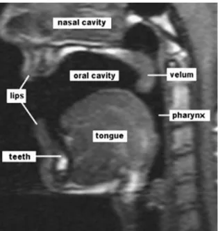
The human aerodigestive tract is regulated by many complex mechanisms and organs that sustain important functions such as mastication and swallowing (fundamental for the digestive process), taste, respiration and speech production. The importance of tongues functionality for said abilities implies actions of (1) positioning of food in the whole vocal cavity, (2) along with the buccinator muscle maintaining food in position for the mastication tasks, (3) propelling of the food to the palate and posteriorly into the pharynx initiating deglutition, (4) change its conformation in order to alter the sounds produced during speech production. In addition, humans have taste receptors including in the upper surface of the tongue and the epiglottis. The anatomical structure of the vocal tract (Figure1) is well established, being the tongue a central organ of this system, which plays a crucial role for the correct functioning of the referred tasks. The development of the anatomical structure of the human vocal tract, continues to change after birth. Specifically, the position of the tongue changes gradually, whereas the newborn tongue is initially flat, positioned almost entirely in the oral cavity, and later, as it descends into the pharynx, acquires a posterior rounded contour, carrying the larynx down with it. Suprapharyngeal horizontal and vertical proportions undergo comparative growth that reaches maturity by the age of 6-8 years old (Lieberman et al., 2001). This is confirmed by Vorperian et al., (2009) based on a longitudinal study of 605 subjects using MRI and CT images.
However, the anatomical study of this structure has been simply forgotten, since the actual knowledge and role in the execution of the referred tasks has only been attempted to be understood in very recent turn of investigations, being also aided by the application widening of the available imaging technics towards the characterization of this organ. In the literature, reported references that confer some extent of attention to the tongues anatomy are very
scarce. For instance, a gross anatomy of the tongue is, in very early anatomic discoveries, to be found in full human anatomy works (Gray (1918), Salter (1852)).
2.1.1.
Anatomy of the tongue
The human tongue is an organ composed primarily by skeletal muscle and located in the oral cavity, occupying a major portion of its volume. It is attached to the oral cavity through its posterior structures, namely via tendons, and other neighboring muscles as well as to its pavement through the lingual frenulum fold.
The tongue is attached to the support structure of bones of this region, specifically to the mandible, the hyoid bone and the styloid process of the skull. The styloid process and bone structure of the skull is shown in Figure 2, and the bone attachments of the tongue are depicted in Figure 3.
The posterior connection of the tongue is made by an attachment to the hyoid bone, which is suspended in the larynx structure, by muscles and cartilaginous tissue. Anteriorly, the tongue connects to the posterior aspect of the mandibular symphysis. The tongues base is connected by fascia to the supralaryngeal muscle that lies immediately inferior to the tongue and forms the muscular floor of the mouth, the mylohyoid.
The tongues structure is composed by a complex arrangement of muscles whereas, the muscles can be grouped in two categories: intrinsic muscles, those that are actually part of the tongue, have no bone insertions and are responsible for shape changing, flattening and up-lifting abilities, and extrinsic muscles, those that are connected to the main structure and attached to bone, responsible for protrusion and retraction, lateral movement and shape modification abilities (Seeley et al., 2008).
Figure 1 - MR midsagittal image (slice) indicating the vocal tracts structures (Ventura et al. (2011)).
The extrinsic muscles are genioglossus, hyoglossus, styloglossus, and palatoglossus. The remaining muscles, transversus, verticalis, superior longitudinalis, and inferior longitudinalis, are intrinsic to the tongue.
A groove, named terminal groove, divides the tongue into two portions. The anterior portion relatively to the groove corresponds to 2/3 of the surface of the tongue being covered with taste buds, with taste receptor cells. The posterior third portion is, in contrast, deprived of taste buds, having only some taste terminal receptors on its surface, being occupied by little glands and a big agglomerate of lymphoid tissue belonging to the lingual amygdalae.
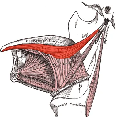
The musculature of the tongue has been described as being composed by eight paired muscles, as illustrated in Figure 4.
Genioglossus
Genioglossus constitutes the main volumetric portion of the tongue posteriorly, having a fan or wedge-shape. It is fixated through a musculo-tendinous origin from the inner surface of the symphysis menti, continuing from root to tip. Its muscular anterior fibers are arranged in a curved antero-dorsal direction that culminates in the anterior fibers of the inferior longitudinal, hyoglossus, and styloglossus muscles. Its posterior fibers run horizontally and backwards to the
Figure 2 - Side view of the skull. The styloid process is just posterior to the mandible (Georgia Highlands College, 2013)
Figure 4 - Extrinsic muscle of the tongue with styloglossus visible at center top (in red) (Gray, 1918).
Figure 3 - Tongues attachments and neighboring structures in a sagittal anatomical view (Gray (1918)).
root of the tongue towards the anterior surface of the hyoid bone and anterior surface of the base of the epiglottis. Also, intermediate bundles of fibers diverge with different degrees of obliquity between the two mentioned portions. In parasagittal plane, it becomes possible to identify its orientation.
Hyoglossus
Hyoglossus radiates in a fan-shaped manner in its upper portion, having a quadrangular conformation in base. Anatomically, towards the other tongue muscles, it is positioned medially, between the inferior longitudinal and genioglossus muscles. It arises from the body of the hyoid bone and interdigitates at its origin with superficial and deep fibers of the geniohyoid. Fiber orientation in the posterior portion of the muscle consists in an antero-posterior radiation. The anterior fibers run and terminate in an approximately longitudinal direction towards the tip of the tongue. The posterior portion lies therefore, under cover of styloglossus, terminating in a fusion to its fibers.
Styloglossus
Styloglossus departs from an insertion in the anterior and lateral surface of the styloid process, close to its apex, continuing in a descending and forward direction into the tongue. Its deep fibers interdigitate with the body muscle of the tongue. After inserting into the tongue, the fibers divide into two bundles. An anterior bundle continues anteriorly along the inferior surface of the inferior longitudinalis, laterally to the hyoglossus, finalizing in the tip of the tongue. A posterior bundle penetrates de hyoglossus and courses medially into the lingual septum.
Transversus
Transversus is part of the bulk of the tongue, along with the Verticalis. It is located between the superior longitudinal muscle, dorsally, the genioglossus and inferior longitudinal muscles, ventrally. The more superficial muscle fibers take a dorsal direction, and the deepest ones are disposed in a ventral direction.
Verticalis
Verticalis is the other muscle that constitutes the thickness of the tongue, being in a tight joint surface with the Transversus muscle. Verticalis fibers are generally vertical, spreading at its superior and inferior portions. The Genioglossus, transversus, and verticalis partially overlap with one another.
Table 1 - Muscles of tongue movement (Seikel et al. (2009)).
Elevate tongue tip Superior longitudinal muscles
Depress tongue tip Inferior longitudinal muscles
Deviate tongue tip Left and right superior and inferior longitudinal muscles for left and right deviation, respectively
Relax lateral margin
Posterior genioglossus for protrusion; superior longitudinal for tip elevation; transverse intrinsic for pulling sides medially
Narrow tongue Transverse intrinsic
Deep central groove Genioglossus for depression of the tongue body; vertical intrinsic for depression of central dorsum
Broad central groove
Moderate genioglossus for depression of the tongue body; vertical intrinsic for depression of dorsum; superior longitudinal for elevation of margins
Protrude tongue
Posterior genioglossus for advancement of body; vertical muscles to narrow tongue; superior and inferior longitudinal to balance and point the tongue
Retract tongue
Anterior genioglossus for retraction of the tongue into oral cavity; superior and inferior longitudinal for shortening of tongue; Styloglossus for retraction of tongue into pharyngeal cavity.
Elevate posterior tongue Palatoglossus for elevation of sides; transverse intrinsic to bunch tongue.
Depress tongue body
Genioglossus for depression of medial tongue; hyoglossus and chondroglossus for depression of sides if hyoid is fixed by infrahyoid muscles.
Superior Longitudinalis
Superior longitudinalis consists of a thin stratum muscle. Its fibers are directed longitudinally along the lamina propria, although this directionality is not clearly defined, being reported with disagreement in Anatomy bibliography. The muscle has a gradual reduction in thickness as it reaches the Styloglossus, hyoglossus and inferior longitudinal muscles, laterally in the tongue.
Inferior Longitudinalis
Inferior longitudinal is a narrow muscle that extends between the paramedian septum and the medial lamella of the lateral septum. It arises medially with the genioglossus muscle, having lateral attachment from the body of the hyoid bone. It is positioned medially with the
hyoglossus muscle. In the middle body of the muscle, it blends with the genioglossus hyoglossus, and Styloglossus muscles forming the tip of the tongue.
The whole description of the musculature existent in the tongue is based on the findings reported in (Abd-el-Malek, 1939). Takemoto, (2001) was able to describe and illustrate his findings on the relative positioning, especially well for the extrinsic muscles, stating the
Figure 5 - Muscles of the tongue (Takemoto (2001)): GG - genioglossus, T - transversus, V - verticalis, HG - hyoglossus, IL - inferior longitudinalis, S - superior longitudinalis, PG - palatoglossus, SG - Styloglossus.
difficulties of distinction between the genioglossus, transversus and verticalis, and produced a three-dimensional tongue model based on impressions from his tongue dissections, depicted in Figure 5. Also, muscle tongue movement has been established for each constitutive muscle of the tongue, as indicated in Table 1.
Despite the unclear definition of the myoarchitecture of anatomical fiber orientation and 3D arrangement of the tongue, in the last ten years a new interest has been taken by the scientific community in the comprehensive analysis of this structure. To answer these disparities, the detailed study of the tongue, specifically of the lingual myoarchitecture has been collected with new recordings through diffusion tensor magnetic resonance imaging, or diffusion tensor imaging (DTI). This technique is very attractive for these types of studies since it enables fiber orientation imaging and analysis in vivo. Gilbert and Napadow, (2005) report imaging three human tongues statically, and Shinagawa et al., 2008 reports imaging from single sections of in vivo human tongues during rest and protrusion movement. Many techniques such as electropalatography (EPG), X-ray imaging, ultrasound, and cine-MRI imaging have been reported in the study of lingual function (Shinagawa et al., 2008). However, the characterization and anatomy of the tongue are not well understood, in contrast to other neighboring structures such as, for instance, the hard palate. Other attempts of imaging the surface during movement and/or oral functions, new analysis of the activation of the tongue muscle fibers for deformation of its body, and a clear understanding of these mechanisms in
vivo has only been in recent years considered a matter of deserving attention.
2.1.2.
Neurophysiological control of the tongue
Neurophysiology is an advanced field that addresses the understanding of the mechanisms that govern the motor control system, especially at the level of last-order muscular output. Since the tongue is purely a muscular structure, the understanding of its complexity may address the neural complex mechanisms of activation that rule its functionality. This analysis is of preponderant importance since the neural control on tongue movement is crucial to the function of rhythmic tasks of respiration and swallowing, whereas disruptions of these mechanisms have even been associated with the highest mortality reported among the pathological problems that may arise (Sawczuk and Mosier, 2001).
The neuromotor system is based on the activation of motor units. These consist of single motor neurons and an assortment of muscle fibers onto which it is connected. Through this connection, synapses occur, through electrical potential signals that are sent along the specific motor neurons innervating the muscle fiber bundles that need to be activated, producing a simultaneous contraction of said fibers. Motor units are organized in motor pools activated in a systematic stimulation, by the central nervous system.
Tongue muscle movement, contractile properties and generator-produced rhythmic modulation derive all from the innervation of the hypoglossal motor neuron complex. The motor neurons are clustered in the hypoglossal nucleus, part of the brainstem, from which departs
the hypoglossal nerve, the twelfth cranial nerve XII. The system of motor neurons that innervate this group of muscles is astonishing, evidencing the remarkable complexity of such an important organ in all its functions. Although the actual number of neurons that intervene in this structure is reported with high disparity, placing, for instance, the total number of myelinated fibers in 9,900 (Atsumi and Miyatake, 1987). In contrast, other muscles of higher dimensions, including biceps or rectus femoris, for instance, are innervated by an average of 441.5 and 609 motor units, respectively (Hamilton et al., 2004).
Electromyographic studies have, on the other hand, been more recently carried out in order to comprehend the complete muscle activity involved. Recent studies report that the genioglossus is the primary upper airway dilator muscle, and its internal motion activation is inhomogeneous. The neuronal control has been vastly studied in the last ten years, and punctual conclusions have been established relatively to the phases of control of the Hypoglossus. EMG findings reveal that inspiratory neuronal activity begins approximately 250ms before the inspiratory process begins, whereas, during inspiration neuronal stimulus increases, and during expiration tonus level is maintained (Cheng et al., 2008).
Although this basic neuronal source is established, the tongue is very uniquely characterized by a complex mechanism of activation that is not yet known, whereas the highest difficulty of the comprehensive process is straightly related to its anatomical complexity. In fact, the human tongue is not only of higher complexity relatively to other mammals, but its anatomical nerve activation and gross neuroanatomy are also lacking. The most extensively studied muscle among tongue muscles is the Genioglossus, responsible for protrusion and depression motion, which has been demonstrated to take part in most tongue movements carried out.
It is hypothesized, in the literature, although it has not been directly reported, that neural control of the tongue may be done, as reported in other mammals for skeletal muscles control, by means of tissue composed of neuromuscular compartments (NMCs), that are morphologically and functionally activated by distinct neuromotor pools, defined as “smallest portion of a muscle to receive exclusive innervations by a set of motoneurons” (English et al., 1993). In (Mu & Sanders, 2000) is demonstrated a compartmental organization of the canine tongue, specifically the innervation present in the genioglossus, where it is reported the presence of two compartments, with fibers horizontal and an obliquely oriented, as well as the branches subdivision departing from the main genioglossus nucleus.
This mechanism is reported to base neuromuscular control of shoulder muscles (Wickham & Brown (2012), Lucas-Osma & Collazos-Castro (2009)); however, even in said anatomically simpler muscles NMCs boundaries are not completely defined.
Unfortunately, no careful anatomical data is found in the literature describing the neuronal organization of the human tongue, compartmental or non-compartmental wise.
2.1.3.
Speech Production, Respiration and
Swallowing
Speech production, respiration and swallowing are the three main activities that are carried out by the aerodigestive organs, with determinant aid of tongue motion.
Among these functions, speech production is the area that has been more extensively studied by the scientific community, due to its multidisciplinary character. The human phonetic apparatus may be divided in organs responsible for sound production and organs of speech articulation. Sound production or phonation, is achieved through the vibration of the vocal folds into the airstream of the airway, a process named voicing, following their fixation into specific position that modulates the aerodynamics of airstream passage. The vocal tract acts as an acoustic filter for a source signal generated in the vocal folds within the larynx, whereas the process of speech production implies the complement of simple phonation with the execution of an extremely well-organized and integrated sequence of movements of the speech articulator organs (lips, mandible, tongue and palatal velum), shaping the resonant cavities of the vocal tract and consequently altering the resulting acoustic output (Seikel et al., 2009). Tongue deformation is directly related to vocalic sounds as well as palatal, velar and pharyngeal consonants sound production. Many are the studies that model tongue conformation, during production of specific sounds present in various languages worldwide, as shown in Figure 6 for vocalic sounds of Portuguese language.
Moreover, the cross-sectional area along the vocal tract, in its supralaryngeal section determines formant frequencies, whereas records of studies addressing the human tongue deformation during speech production, exist from over 150 years (Lieberman, 2012). The analysis of the resonance cavities involved in phonation is, as obviously understood by the scientific community that has undergone an extensive amount of research relevance to the study of speech production anatomy and mechanism, in this sense of extreme importance. In addition, more importantly for the understanding of how the mechanisms allow the diversity of
Figure 6 - Tongue contour extracted from midsagittal images, during production of vocalic sounds present in Portuguese language (Ventura et al., 2008).
phonation capacity and how disturbances of pathological origin or otherwise to the structures involved may affect their functionality.
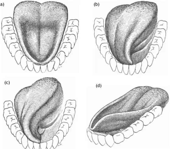
Swallowing consists in the passage of a bolus of food through the mouth to the pharynx, and into the esophagus that will trigger a swallowing reflex as it passes into these regions. To this process be succeeds the larynx must elevate, and the epiglottis (attached to the root of the tongue) drops down to cover the aditus, avoiding choking or pulmonary aspiration can occur. Food bolus building was illustrated and explained in (Abd-El-Malek, 1955). His observation of subjects masticating nuts, gelatin and chewing gum led to the description of the following steps:
a) Preparatory stage – acquires a pouch-like form, to collect the food on its dorsum; b) Throwing-stage - a twisting movement towards one side to deposit the bolus onto the molars;
c) Guarding stage - tongue twists even more, making contact with the upper and lower teeth, in order to keep the bolus between the molars during mastication;
d) Bolus building – after several chewing movements the cheeks move medially and the tongue moves side to side, mixing the bolus with saliva and coating it with mucus.
e) Swallowing - the tip of the tongue is raised and pressed against the posterior surface of the front teeth and the anterior part of the hard palate, as to close the mouth and pharynx;
These stages are illustrated in Figure 7.
Muscle activation during this process is automatic, and important processes regard studying swallowing to assess the stiffness of the tongues surface, or the force that the tongue is able to exert on the hard palate.
In humans, respiratory airway activity involves important tasks of patency maintenance. Substantial studies suggest that this function is provided by the tongues genioglossus muscle (GG). Airway patency is a matter of extreme importance, and delicate to control, since the human pharynx has no rigid support except at its extreme upper and lower ends where it is anchored to bone (upper extremity to hyoid bone) and cartilage (part of the larynx). Therefore, the airway depends on 20 skeletal muscles that dilate and keep the oropharynx open (Dempsey et al., 2010).
During respiration, tongue deformation has been analyzed through tagged MRI, a technique that arose later as a modality of MR imaging, allowing quantification of physiological motion. Expiratory and inspiratory tasks create pressure differences in the airway and muscle tonus of the involved structures that define its need to be able to maintain the adequate compliance. Inspiration tasks generate a negative inspiratory pressure that manifests at epiglottis level, that has been directly correlated with neuronal firing of the genioglossus (Pillar et al., 2001). In Cheng et al. (2008) is reported that the muscle movements activated throughout the respiratory cycle. Genioglossus muscle analysis indicated posterior movement during expiration as opposed to an anterior movement during inspiration, and over the geniohyioid. Geniohyioid has presented very little movement during respiration.
2.2. Magnetic Resonance Imaging in the
context of Aerodigestive Organs
Since the development of novel imaging techniques of the tissues that the in vivo anatomy of living organisms has been made possible.
Magnetic resonance imaging (MRI) is a diagnostic method that uses strong magnetic fields and radiofrequency (RF) waves to form images of the human body. This technique allows a non-invasive imaging method that presents a wide range of potential clinical applications.
MRI is therefore, nowadays a well-established imaging method used by physicians in the evaluation and characterization of soft tissues. The technique presents major advantages compared to conventional imaging methods: uses non-ionizing radiation, allows greater soft tissue contrast and also enables an analysis of the three-dimensional structures surrounding the upper airway. Analysis of images from MRI, relatively to other imaging techniques is characterized for being more informative in terms of output extent of data that can be
(a) (b)
(c)
(d)
Figure 7 - Abd-El-Malek (1955) illustration of the preparatory stage of mastication (a), throwing stage of mastication (b), guarding stage of mastication (c), initial stage of deglutition (d).
retrieved, allowing an analysis of the outputs to be oriented to the monitoring of the respiratory airway during sleep and the structures that play a determinant role in the study of normal functioning upper airway, relatively to the imaging of pathological aerodigestive tract. It allows therefore, the addition of tremendous value to screening, diagnostic, surgical planning and follow-up of patients, for a variety of pathologies developed in these organs. A particular case where this imaging technique is advantageous and necessary, precisely for the appearance of pathological scenarios during the developmental process in the aerodigestive tract, is when it is applied to children, to whom the usage of non-ionizing radiation is preferable. Despite the advantages presented, the use of MRI is not quite as common as it was idealized, being the main reason related to the high cost of the imaging technique.
In this chapter, the physical principles in which this technique is based are described, as well as the variable aspects that affect its quality and adequacy, in order to better understand the adaptability and potential in the application of imaging the human tongue.
2.2.1.
Basic Principles in Magnetic Resonance
Imaging
The rotational movement of protons presented in the 1H atoms nucleus – spins – implies that each of them is associated with magnetic dipolar moment (m.d.m). The most abundant atoms present in tissues are 1H atoms, with spin =1/2, being more sensitive to magnetic fields applied in Magnetic Resonance (MR). When a magnetic field is applied to the spins, these go from a state of null magnetization, to a state of magnetization where the m.d.m’s tend to align themselves with the orientation of the referred field, in a given volume element, assuming a magnetization value different from zero.
This alignment is done in its majority according to a parallel direction related to the field; however, a part of these spins does not respect this behavior and its movement is named precession movement that occurs with a given frequency, called Larmor frequency (Rinck, 2001). An external pulse applied in form of oscillations of the magnetic field in the range of radiofrequencies at Larmor frequency of those spins, forces them to enter in phase precession, which originates a signal of image in RM.
The phenomenon explained in terms of physical behavior, can be examined considering, where the magnetization vector is in the Z axis, and the precession phenomenon makes the spins rotate around that axis of magnetization with a deflection angle in the vertical plane containing said axis. Therefore, into an MR equipment, a given coil is positioned in the xy plane that detects a variable electromagnetic field, producing an oscillatory signal, which corresponds to the MR image signal. This method, consequently, intends to detect the energy released by the phenomenon of Relaxation, which occurs when the radiofrequency (RF) pulse ends and the spins start to relax to the minimum energy state.
2.2.2.
Relaxation Times
There are two types of relaxation of tissues, longitudinal or spin-lattice relaxation (T1 weighted time), made through the Z component of magnetization 𝑀𝑧 after the application of magnetization in the xy plane, and transversal relaxation or spin-spin relaxation (T2 weighted time), that occurs by the additional effect of dephasing of magnetization induced by interactions between spins of neighbor protons, that when subjected to magnetic fields with slight differences, rotate at corresponding Larmor frequencies. This process of continuous loss of phase coherence, becomes gradually more prominent with time. The magnetization then implies that T2 relaxation time is always less than T1, and that the timeline of the process starts at a magnetization in xy plane that then tends to zero, followed by an increase in the longitudinal magnetization until equilibrium is achieved, in axis Z. T1 relaxation results from the interaction with the mesh of atoms in the tissue, and is characterized by a rate of magnetization Mz vector through time given by:
𝑀𝑧 (𝑡) = 𝑀(0). (1 −
𝑒
−𝑇1𝑡) (1)This equation describes a profile, where the recovery tends to a thermodynamic equilibrium state, for which Eq. (1) given t=T1 is [1-(1/e1)], meaning that T1 characteristic time is the time
where the longitudinal magnetization recovers 63% of its equilibrium value (Rinck, 2001).
2.2.3.
K-space
Spatial encoding of the image is another part of the mechanism, of acquisition that includes: - Slice selection – implies the positioning of a gradient in the perpendicular direction to the cut to be retrieved (in the Z plane for an axial slice), the position of slice is selected by the frequency of the pulse, and the thickness by its bandwidth.
- Frequency encoding – applying a first signal according to a specific direction, the signal emitted by the different elements of volume, are characterized by different frequencies.
- Phase encoding – applying a second signal according to a determined direction, the different elements of volume according to that direction will be characterized by different phases.
Consequently, for an axial acquisition, the slice selection is done in the Z plane, the axis X and Y are responsible for the frequency and phase encoding. The two magnetic fields distributed, make for each orientation of the phase encoding gradient Gy correspondent to a line (y position), and the frequency encoding gradient dictates each columns value (x position) of that line, and in this way the (x,y) positions are stored in a matrix called K-space. Each combination is afterwards mapped in the image reconstruction to its position, and the amplitude into the corresponding intensity, by applying the Fourier transform to the 2D distribution (A. Bernstein et al., 2004).
The design of appropriate gradients, is preponderant so that k-space samples can be acquired and then inverse Fourier transformed to obtain an image of the magnetization M(x; y). K-space must be sufficiently sampled according to the Nyquist criterion to avoid object domain aliasing. The extent of k-space coverage determines the images resolution.
2.2.4.
Contrast and tissue signal in RM
Contrast in MRI is due to the occurrence of specific relaxation phenomena in the different tissues, where it depends on the different times of relaxation T1 and T2, as well as different proton densities, which are characteristic and intrinsic of each type of tissue. The different tissues contain large numbers of chemical components that contribute to the measured magnetic resonance signal, and this composition characterizes each type.
Image acquisition in MRI is made through specific sequences of pulses, of RF and orientation of the phenomena of relaxation where, given the dependence on time of these phenomena, contrast can be adjusted and chosen by applying specific combinations of temporal parameters of acquisition. In the conventional MRI acquisition, these phenomena will also be influenced by the technical factors of medical acquisition, or biologically extrinsic factors. These include the magnetic field strength and homogeneity, and are crucially determined by the pulse sequence contrast influencing components TR, TE, TI and FA.
The main objective since the discovery of this technique relies in combining these parameters in order to emphasize certain contrast determining factors, or determining relaxation phenomena among others, or even a set of different factors.
2.2.4.1. TR, TE and Pulse Sequences
Pulse sequences of acquisition consist in a sequence of signals sent to the tissues, by MR machines. The pulse sequence consists in repeated RF pulses that cause a free induction decay (FID) characterized by a specific initial amplitude, mediated by the pulse sequence parameters. The two time parameters that determine this method are TR (repetition time) and TE (echo time) of the pulse sequences. TR is the time interval between two successive RF pulses, and TE is the time at which the echo signal, the signal produced by induction of the spinning protons, reaches the detector of the machine and is measured. TR can therefore determine the degree of relaxation of protons back into alignment of the magnetic field, whereas specific rates of relaxation of the tissues will imply having TR times shorter than what is needed for a full relaxation decrease the signal retrieved from the analyzed tissues.
2.2.5.
Limitations
and
determinant
considerations
The growing interest in the tongues function over all its functionalities of taste, swallowing and speech production tasks has given rise to the importance of imaging the aerodigestive tract and its structures with the best imaging technique available; whereas for the correct imaging of such complex structures, a good contrast between tissues is fundamental to allow the differentiation of the different structures at its correct boundaries.
These factors are of extreme importance for the development of the dissertation work proposed here.
Therefore, the rigorous imaging of the structures at study is determinant for the correct function of the following computational tasks of retrieval of the target structural.
In spite of the image quality conditionings referred above, MRI technique is considered as the best, a non-invasive, accurate method imaging modality available for the imaging of the muscular organ under study.
2.3. Upper airway imaging and computational
analysis
Computational processing and analysis of medical images is a novel field that has gained a promising and relevant importance over the years, presenting astonishing developments in the areas of computer aided diagnostics, improving imaging technics, and imaging analysis processing of aspects that cannot be visualized and/or retrieved by plain image observation.
Volumetric imaging techniques can be used to reconstruct three-dimensional structures from serial two-dimensional images. This section provides a conceptual overview of those techniques by illustrating the reconstruction of the aerodigestive organs.
Segmentation of the target anatomical structures from MRI is still a challenging process. There are various reported methods of segmentation of static MR images/volumes (Balafar et al., 2010). Their applications to the particular segmentation of tongue, is reported in a scarce number of instances, highlighting the need of further studying this organ and the development of the adequate tools accordingly.
The imaging study of the tongue is a very underdeveloped field that has limited the improvement of anatomical and functional characterization of this organ. The recent development of Computer Vision and Machine Learning fields of Image Analysis in recent years have provided the availability of new tools of image computer analysis regarding 3D volume segmentation and reconstruction.
The first imaging reports of the tongue are made through ultrasound (US) imaging (Sonies, 1981), and subsequent applications towards the analysis of swallowing and articulation tasks
using snakes in (Unser and Stone, 1992), and using scale space filtering for edge detection in (Kelch and Wein, 1993). The main applied studies that address specifically this structure are extensively reported in speech studies. Therefore, US imaging presented the best imaging characteristics for a dynamic acquisition of multiple frames during speech production exercises. First tongue 3D modelling and reconstruction were reported in (Watkin and Rubin, 1989), that describes a trigonometric transformation of the 2D coordinates into a volume, and latter, more advanced segmentation methods were described by Akgul et al. (1998) and more recently for segmented 2D motion analysis applying Markov random fields in Tang et al. (2012).
Although the demonstrated applicability of US to tongue modelling, further study of its anatomy implies that a higher contrast and resolution imaging technique, such as MRI, prevails as more adequate in the intended study of the tongue.
The first reports of tongue anatomy imaging through MRI were reported in (Lufkin et al., 1983).
The analysis of tongue anatomy and physiology has been reported in studies using both static volumetric MRI, standard imaging modality for 3D imaging, Cine-MRI and even tagged-MRI imaging (another imaging modality that has been extensively used for temporal characterization of the tongues anatomy). Reported dynamic acquisition image analysis studies reinforce the necessity of a proper segmentation in 3D studies to the evaluation of the dynamic processes it is responsible for, such as swallowing and speech production (Lee et al., 2014). Other studies pretend to reinforce the study of the biomechanical modelling of this structure, and therefore, select a high resolution imaging modality such as static volumetric MRI (Harandi et al., 2014).
The emerging interest in the study of the tongues deformation and functionality has established that the requirement for an automated method of image analysis of this kind of anatomic data is expected to gain a rapid eminent relevance (Woo et al., 2012).
Reported studies on segmentation of the tongue, focus of the segmentation of static and dynamic acquisitions. Dynamic acquisition reveals to have obvious relevance in the study of tongue motion characterization. The processing needed is common since the format is usually based on 2D image segmentation. Vasconcelos et al. use statistical models to segment the tongues shape during the production of different sounds, in order to study speech production (Vasconcelos, Ventura, Tavares, & Freitas, 2009).
Stone et al. (2010) is one of the first reports that focuses on the strict tongue segmentation, and establishes the relevance of this study for motion patterns during speech production. In this 2D study, the images were to simply be registered through a landmark based transformation algorithm and aligned, following principal component analysis for the motion study.
The processes reported are usually divided into various basic phases: 1) Resolution wise pre-processing, 2) Segmentation, 3) Registration, 4) 3D Volume reconstruction.
In Lee et al. (2014) is reported an isotropic volume super-resolution reconstruction from dynamic tagged-MRI images. The images were subjected to a super-resolution volume reconstruction, in order to address inter-slice resolution. It was attempted to surpass the
limitation, extensively mentioned throughout this report, of long acquisition time, through the acquisition of three images with 6.0 mm thickness, which obviously affected the resolution in the through-plane direction. An up-sampling in the through-plane direction was developed using a fifth-order B-spline interpolation. Registration, for inter-slice alignment is reported in various studies (Lee et al. 2014, Woo et al. 2012), where the application of the Mutual information (MI) similarity measure is reported for registration of sagittal with axial and coronal volumetric image stacks. After the registration, a final intensity correction is made using a local intensity matching algorithm, following the application of the Random walker (RW) segmentation algorithm.
The Random walker algorithm, for segmentation of 3D super-resolution volumes was also cited in the literature for similar purposes, due to its attractive features in Woo et al. (2012).
Tagged-MRI is not adequate, regarding preponderant implications on volume reconstruction, to be used in these studies since the image quality is very low to when compared to static volumetric MRI.
A mesh modelling approach is reported in Harandi et al. (2014) whereas the registration technique departs from an initial source model of the tongue to whose vertices are applied external forces forcing it towards the target boundaries through a process dictated by local intensity profile registration and positions computed through normalized cross-correlation and finalized by shape matching. The advantage of this approach is that it allows user input to automatically correct the mesh nodes positioning.
The most recent study published attempted to go further in the investigation of functional behavior, and describes a novel method of segmentation of individual tongue muscles (Ibragimov et al., 2015), specifically genioglossus and inferior longitudinalis. In their work, it was implemented an adaptation to muscle segmentation of the game-theoretic framework (GTF) algorithm, based on land-mark-based segmentation.
2.3.1.
MRI 3D volumes image segmentation
techniques
Computer-aided modelling of the aerodigestive organs is beneficial for 3D visualization, and for the understanding of the associated physiology. Medical imaging is retrieved in a universal format, organized according to a predefined standard.
The studies that address image segmentation of the tongue are limited and therefore, an overview of this list of presented in the following points.