https://doi.org/10.1590/0004-282X20180070
ARTICLE
Spontaneous intracranial hypotension and
its complications
Hipotensão intracraniana espontânea e complicações
Marília Maria Vasconcelos Girão1, Rachid Marwan Pinheiro Sousa1, Mayani Costa Ribeiro2, Tânia Aparecida
Marchiori de Oliveira Cardoso2, Marcondes Cavalcante França Júnior2, Fabiano Reis1
Headache is a common complaint with a wide range of
differential diagnoses. Spontaneous intracranial hypoten
-sion (SIH) is an uncommon cause of novel onset persistent headaches, typically one that occurs or worsens upon stand
-ing and is relieved by ly-ing down, and is often an underdiag -nosed condition1,2,3.
Intracranial hypotension has an estimated incidence of 5/100,000 and typically results from a cerebrospinal fluid (CSF) leak, which leads to a decline in CSF volume. The leak can be explained by structural weakness of the dura mater, associated with minimal or no history of trauma. It is known that decrease in CSF volume, rather than CSF pressure decrease, is the core pathogenic factor. The disease is slightly more common in women and can occur in all age groups, with a typical age of incidence around 40 years1,2,4,5,6,7,8.
Delay or failure to diagnose SIH may lead to life-threat
-ening complications such as dural venous sinus thrombosis, subdural hematoma and subarachnoid hemorrhage3.
Imaging exams, particularly magnetic resonance imaging (MRI), play an important role in detecting such abnormali
-ties. Other complications described include bibrachial amy
-otrophy, superficial siderosis, syringomyelia, cranial nerve palsies and rebound intracranial hypertension1,4,5.
We report a series of patients with SIH, some of whom had complications detected by MRI of the brain.
METHODS
We reviewed the medical records and MRI studies of nine patients with SIH and describe the complications observed
1Universidade Estadual de Campinas, Faculdade de Medicina, Departamento de Radiologia, Campinas SP, Brasil;
2Universidade Estadual de Campinas, Faculdade de Medicina, Departamento de Neurologia, Campinas SP, Brasil.
Correspondence: Fabiano Reis; Rua Tessália Vieira de Camargo, 126; 13083-887 Campinas SP, Brasil; E-mail: [email protected]
Conflict of interest: There is no conflict of interest to declare.
Received 14 January 2018; Received in final form 06 March 2018; Accepted 21 April 2018.
ABSTRACT
Spontaneous intracranial hypotension (SIH) is a syndrome that was unknown until the advent of magnetic resonance imaging (MRI). It is a cause of orthostatic headache, which remains underdiagnosed and, rarely, can result in several complications including dural venous sinus thrombosis, subdural hematoma and subarachnoid hemorrhage. Some of these complications are potentially life-threatening and should be recognized promptly, mainly by imaging studies. We reviewed the MRI of nine patients with SIH and describe the complications observed in three of these patients. Two of them had subdural hematoma and one had a dural venous sinus thrombosis detected by computed tomography and MRI. We concluded that MRI findings are of great importance in the diagnosis of SIH and its complications, which often influence the clinical-surgical treatment of the patient.
Keywords: intracranial hypotension; subdural hematoma; sinus thrombosis, intracranial; magnetic resonance.
RESUMO
Hipotensão Intracraniana Espontânea (HIE) é uma síndrome desconhecida até o advento das imagens de Ressonância Magnética (RM). É uma causa de cefaleia ortostática que permanece subdiagnosticada e raramente resulta em complicações, como trombose de seios venosos durais, hematoma subdural e hemorragia subaracnoidea. Algumas dessas complicações são potencialmente ameaçadoras à vida e devem ser prontamente reconhecidas pelos estudos de imagem. Nós revisamos as RM de 9 pacientes com HIE e descrevemos as complicações observadas em 3 casos. Dois deles tiveram hematoma subdural e um teve trombose de seio venoso dural detectados por tomografia computadorizada e RM. Concluímos que achados de RM são de grande importância no diagnóstico de HIE e suas complicações, frequentemente influenciando o tratamento clínico-cirúrgico do paciente.
in three of these patients, evaluated by neurologists at the University of Campinas, São Paulo, Brazil, in 2016. The patients had given their informed consent. All patients met previously-reported diagnostic criteria for spontaneous spinal CSF leaks and intracranial hypotension. All of them underwent at least one brain MRI. All radiological images were reviewed by at least one board-certified neuroradiolo
-gist. We also reviewed the literature on possible complica
-tions of SIH.
Patient 1
A 49-year-old male truck driver, with a history of diabe
-tes mellitus type 2 and previous tension-type headaches,
reported a two-week history of headache that started when he was carrying out his usual activities. It was a severe holocranial headache (10/10intensity), maximum at the onset, which improved with lying down and wors
-ened with orthostasis/sitting up, associated with nausea, photo/phonophobia and rotational vertigo. He also had a history of a minimal head contusion on an aluminum plate a week before the onset of the symptoms, and took acetyl
-salicylic acid in the first two days of headache, without any improvement.
Computed tomography (CT) of the brain without con
-trast at admission showed chronic right and left subacute subdural hematomas, decrease in size of the ventricles and enlargement of the pituitary. He was discharged with symptomatic treatment.
The patient came back three days later without having had pain relief. He was evaluated by neurologists and had an MRI, which showed typical findings of SIH: subdural fluid col
-lections (hygromas), diffuse pachymeningeal enhancement, decrease in the size of the ventricles, enlargement of the pitu
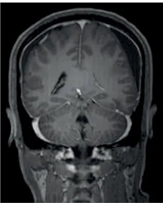
-itary, and cerebral venous sinus engorgement. There were also bilateral subdural hematomas (thickness of 1.0 cm on the left and 0.5 cm on the right), creating a discrete midline shift (Figure 1).
The diagnosis of a spontaneous CSF leak was confirmed by radionuclide cisternography (lumbosacral spontaneous dural fistula between L4-L5 on the right and bilateral L5-S1). He was treated by clinical measures (lying down, analgesics, hydration, pure coffee twice a day) and an epidural blood patch. A subdural hematoma drainage procedure was per
-formed as the symptoms did not improve. The patient was discharged with improvement of the headache and without neurological deficits.
Patient 2
A 35-year-old unemployed man with hypertension, presented at the emergency department with a history of three months of bilateral frontotemporal progressive head
-ache, 7/10 intensity, which worsened with orthostasis/sit
-ting up and was associated with nausea, vomi-ting and otalgia, with slight improvement with the use of NSAIDs.
He denied any history of trauma and reported that he had stopped working because he could not sit at work due to worsening of the headache. With the hypothesis of SIH, he underwent a CT scan of the brain, which showed a decrease in size of the ventricles and cisterns and a left frontoparietal laminar subdural hematoma. A few days later, he underwent an MRI to complement the investi
-gation, which showed a reduction in the size of the ven
-tricles, diffuse pachymeningeal thickening and enhance
-ment, volumetric increase of the pituitary, engorgement of the venous sinuses, bilateral subdural effusions and bilat
-eral frontotemporal laminar (thickness less than 0.5 cm) subdural hematomas (Figure 2).
The diagnosis of spontaneous CSF leak was confirmed by neuroaxis MRI (enlargement of the posterior epidural space with thin liquid in the epidural fat, and apparent foci of discontinuity in the dura mater at the lower tho
-racic levels, notably on the left), and radionuclide cister
-nography (altered CSF dynamics, without progression of the radiotracer from the base cisterns to the cerebral convexity even after 24 hours, and thoracolumbar spon
-taneous dural fistulas between T8-T12 bilaterally, notably T8-T9 on the left). He was treated with the same clinical measures as Patient 1, with improvement, and needed an epidural blood patch. There was no need for hematoma drainage and the patient was discharged with improve
-ment of symptoms.
Figure 1. Coronal contrast-enhanced MR image with diffuse
Patient 3
A 35-year-old female patient, unemployed (she was a pharmacist), with a history of bariatric surgery, use of oral contraceptives and ex-smoker, presented at the emergency department with a two-week history of severe (8/10) holo
-cranial headache (in the occipital region), with a progressive
pattern and a clear correlation with the orthostatic position,
which improved with lying down. It was associated with nau
-sea, vomiting, photophobia, phonophobia, hypoacusis and was barely responsive to common analgesics.
An MRI was performed and showed signs of intracranial hypotension (engorgement of the pituitary, sagging of the brain
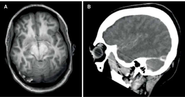
with herniation of the cerebellar tonsil, homogeneous and diffuse pachymeningeal enhancement, posterior fossa with reduced size, diffuse venous sinus engorgement, subdural effusions) and right transverse sinus thrombosis (Figure 3A). A CT angiography also confirmed the right transverse sinus thrombosis (Figure 3B).
The diagnosis of a CSF leak was made by cysternography, which showed a spontaneous dural leak at the cervical and thoracic levels, more evident between C7 and T1.
The patient’s oral contraceptive was discontinued and she was discharged with anticoagulation and significant improvement of headache after clinical treatment (lying down, hyper-hydration and caffeine).
A
B
Figure 2. Coronal T2-weighted MR image (A) showing hypointense left frontotemporal laminar subdural hematoma; (B)
Contrast-enhanced sagittal T1-weighted MR image showing diffuse pachymeningeal thickening and enhancement, enlargement of the pituitary gland and engorgement of the venous sinuses.
A
B
Figure 3. Axial T1-weighted MRI (A) demonstrating hyperintensity in the right transverse sinus; (B) sagittal CT angiography
DISCUSSION
Headache is a common complaint with a wide range of
differential diagnoses. Spontaneous intracranial hypotension is an uncommon cause, which remains underdiagnosed and should be considered in cases of orthostatic headache1-5.
In this context, it is important to perform imaging of the brain, and that the radiologist suspect the diagnosis. Tomographic examination can be used for screening, espe
-cially in the emergency department, and ventricular collapse, descent of the cerebellar tonsils1,2 and even pachymeningeal
enhancement can be observed if an iodinated contrast is used. Hemorrhagic complications are easily detected in a CT and venous sinus thrombosis can also be seen in this method. However, in general, the accuracy of MRI is much higher than that of CT, and a complementary MRI should be performed in patients with a high clinical suspicion, even when a CT scan does not reveal major alterations. For example, small ventri
-cles may not draw much attention in a young patient’s CT, and pachymeningeal enhancement areas are not as well defined as they are in the MRI. When a CT raises suspicion of an SIH, it is also important to perform an MRI with gadolinium, which is better able to depict typical findings and complications. Some classic findings of SIHs seen in MRIs are diffuse pachymenin
-geal enhancement, decrease in the size of the ventricles, sub
-dural fluid collections, enlargement of the pituitary, flattening of the pons against the clivus, cerebral venous sinus engorge
-ment and sagging of the brain1,2,4,5,6,9,10,11.
However, there are some pitfalls to avoid, such as the downward displacement of the cerebellar tonsils into the spi
-nal ca-nal, which may mimic a Chiari malformation (pseudo-Chiari malformation), subdural fluid collections mimicking a primary subdural hematoma, pachymeningeal enhancement that may be mistaken for an infectious or neoplastic disease, and pituitary hyperemia mimicking a pituitary tumor, such
as an adenoma8,11,12.
Complications associated with SIH are rare, but poten
-tially serious, and early radiological diagnosis favors proper management. Complications described include subdural hematoma, cerebral venous and venous sinus thrombosis, bibrachial amyotrophy, superficial siderosis and syringomy
-elia.3,4,5 We have reviewed the literature on these conditions.
Subdural fluid collections in SIH range from simple thin hygromas to massive subdural hematomas. Subdural fluid collections are common in SIH, occurring in about 50% of patients. Mostly, they represent hygromas and are bilateral, thin, usually found over the cerebral convexities, and do not cause significant mass effect1,2,5,13,14. However, subdural hema
-tomas with varying degrees of mass effect (seen in Patient 1), sometimes shifting the midline, are not uncommon and have been described in about 20% of patients with SIH, this being the most common complication of this entity. Subdural hematomas are typically bilateral but asymmetrical and the maximal thickness usually ranges from 1 cm to 3 cm4,13.
The decrease of CSF volume can lead to compensatory enlargement of the subdural/subarachnoid resulting in sub
-dural hygromas; on the other hand, sub-dural hematomas are probably caused by bleeding from enlarged veins in the sub
-dural zone or tearing of bridging veins13,14.
It is important to point out that a careful anamnesis should include risk factors for hemorrhagic events, such as acetylsalicylic acid (seen in Patient 1) or anticoagulant use, or history of hemophilia, which, in addition to SIH, increase the risk of these complications.
The heterogeneous appearance and attenuation of the subdural hematoma in the imaging indicates that recurrent bleeding within the subdural space is common in untreated SIH. Studies on this entity have shown no significant differ
-ences in symptomatology, age, sex or location of the CSF leak among patients with or without subdural hematomas as a complication of SIH13.
Resolution of subdural hygromas (and also the typical MRI imaging features of SIH) can be demonstrated within days of successful treatment of the CSF leak. For subdural hematomas with mass effect, however, resolution occurs more slowly and may take up to a few months13.
The main treatment of the collection of subdural fluid is the management of the underlying CSF leak, almost always with
-out the need for neurosurgical intervention. Besides, a craniot
-omy may increase sagging of the brain. If subdural hematomas are evacuated but the CSF leak is not treated, there is a high risk of persistent or recurrent subdural hematomas4,13.
Cerebral venous thrombosis is found in about 2% of the population with SIH15.
The Virchow’s triad comprises three categories of fac
-tors that contribute to venous thrombosis: venous stasis, hypercoagulability (hyperviscosity) and vessel damage16,17.
Spontaneous intracranial hypotension is a risk factor for cere
-bral venous thrombosis according to the following mecha
-nisms that make up the triad: venous engorgement due to the loss of CSF (Monro-Kellie doctrine)18 results in the slowing
of venous blood flow (venous stasis); the loss of CSF reduces absorption of CSF into the cerebral venous sinuses leading to an increase of blood viscosity (hypercoagulability); and sinking of the brain due to the loss of buoyancy may reflect in damage of cerebral veins and sinuses (vessel damage)15,19.
As previously mentioned, a thorough anamnesis is very important, and should include other risk factors for throm
-botic events, such as a previous history of thrombosis, use of contraceptives with estrogen (seen in Patient 3), pregnancy, puerperium, and thrombogenic disorders.
As concern imaging findings, a CT scan can show a spon
Central venous thrombosis is a serious complication, potentially life threatening, with a patient fatality rate of up to 5% and it may also complicate a cerebral venous infarc
-tion, which may occur in up to 40% of the patients, according to some studies. Successful treatment combines anticoagula
-tion and the treatment of the spinal CSF leak15,19.
Bibrachial amyotrophy is characterized by weakness and atrophy of a few contiguous cervical myotomes (segmental amy
-otrophy) and it is associated with an extra-arachnoid encapsu
-lated fluid collection that causes chronic pressure on the ventral aspect of the cord. These extra-arachnoid fluid collections may be associated with a CSF leak. This may be mistaken for a motor neu
-ron disease, and sensory symptoms are minimal or absent4,21.
Superficial siderosis, in the context of SIH, is a result of hemosiderin deposition in the subpial layers of the brain
and spinal cord and may be caused by repeated hemorrhage into the subarachnoid space. This may happen due to bleed
-ing from friable vessels of a dural defect or due to exudation from engorged vessels ( frequently seen in CSF hypovolemia). Impaired hearing and ataxia may be seen as neurologic man
-ifestations, especially when the superficial siderosis occurs in the posterior fossa and cranial nerves, and cerebellar involve
-ment occurs4,21,22.
Syringomyelia is a rare complication associated with sig
-nificant downward displacement of the cerebellar tonsils. It may vary from very small to quite extensive4.
It is important that the radiologist identifies not only the classic imaging findings related to SIH, but also the possible associated complications, which often influence the clinical or surgical treatment of the patient.
References
1. Schievink WI. Spontaneous spinal cerebrospinal fluid leaks. Cephalalgia. 2008 Dec;28(12):1345-56. https://doi.org/10.1111/j.1468-2982.2008.01776.x
2. Schievink WI. Spontaneous spinal cerebrospinal fluid leaks and intracranial hypotension. JAMA. 2006 May;295(19):2286-96. https://doi.org/10.1001/jama.295.19.2286
3. Ade S, Moonis M. Intracranial hypotension with multiple complications: an unusual case report. Case Rep Neurol Med. 2013;2013:913465. https://doi.org/10.1155/2013/913465 4. Mokri B. Spontaneous intracranial hypotension. Continuum
(Minneap Minn). 2015 Aug;21(4 Headache):1086-108. https://doi.org/10.1212/CON.0000000000000193
5. Couch JR. Spontaneous intracranial hypotension: the syndrome and its complications. Curr Treat Options Neurol. 2008 Jan;10(1):3-11. https://doi.org/10.1007/s11940-008-0001-5
6. Hoffmann J, Goadsby PJ. Update on intracranial hypertension and hypotension. Curr Opin Neurol. 2013 Jun;26(3):240-7. https://doi.org/10.1097/WCO.0b013e328360eccc
7. Understanding AP. Understanding and managing spontaneous intracranial hypotension. Can J Neurol Sci. 2013;40(2):139-40. https://doi.org/10.1017/S0317167100013640
8. Schievink WI. Spontaneous spinal cerebrospinal fluid leaks: a review. Neurosurg Focus. 2000 Jul;9(1):e8. https://doi.org/10.3171/foc.2000.9.1.8
9. Lin WC, Lirng JF, Fuh JL, Wang SJ, Chang FC, Ho CF et al. MR findings of spontaneous intracranial hypotension. Acta Radiol. 2002 May;43(3):249-55. https://doi.org/10.1034/j.1600-0455.2002.430304.x
10. Tosaka M, Sato N, Fujimaki H, Tanaka Y, Kagoshima K, Takahashi A et al. Diffuse pachymeningeal hyperintensity and subdural effusion/hematoma detected by fluid-attenuated inversion recovery MR imaging in patients with spontaneous intracranial hypotension. AJNR Am J Neuroradiol. 2008 Jun;29(6):1164-70. https://doi.org/10.3174/ajnr.A1041
11. Leung GK, Ho J, Pu JK. Pituitary enlargement in spontaneous intracranial hypotension—a diagnostic pitfall. Acta Neurochir (Wien). 2011 Dec;153(12):2445-6. https://doi.org/10.1007/s00701-011-1099-x
12. Schievink WI. Misdiagnosis of spontaneous intracranial hypotension. Arch Neurol. 2003 Dec;60(12):1713-8. https://doi.org/10.1001/archneur.60.12.1713
13. Schievink WI, Maya MM, Moser FG, Tourje J. Spectrum of subdural fluid collections in spontaneous intracranial hypotension. J Neurosurg. 2005 Oct;103(4):608-13. https://doi.org/10.3171/jns.2005.103.4.0608
14. Noronha RJ, Sharrack B, Hadjivassiliou M, Romanowski CA. Subdural haematoma: a potentially serious consequence of spontaneous intracranial hypotension. J Neurol Neurosurg Psychiatry. 2003 Jun;74(6):752-5. https://doi.org/10.1136/jnnp.74.6.752
15. Schievink WI, Maya MM. Cerebral venous thrombosis in spontaneous intracranial hypotension. Headache. 2008 Nov-Dec;48(10):1511-9. https://doi.org/10.1111/j.1526-4610.2008.01251.x
16. Brotman DJ, Deitcher SR, Lip GY, Matzdorff AC. Virchow’s triad revisited. South Med J. 2004 Feb;97(2):213-4. https://doi.org/10.1097/01.SMJ.0000105663.01648.25 17. Kumar DR, Hanlin E, Glurich I, Mazza JJ, Yale SH. Virchow’s
contribution to the understanding of thrombosis and cellular biology. Clin Med Res. 2010 Dec;8(3-4):168-72. https://doi.org/10.3121/cmr.2009.866
18. Mokri B. The Monro-Kellie hypothesis: applications in CSF volume depletion. Neurology. 2001 Jun;56(12):1746-8. https://doi.org/10.1212/WNL.56.12.1746
19. Park JH, Yoon SH. New concept of cerebrospinal fluid dynamics in cerebral venous sinus thrombosis. Med Hypotheses. 2008;70(1):143-7. https://doi.org/10.1016/j.mehy.2002008;70(1):143-7.03.036
20. Lee EJ. The empty delta sign. Radiology. 2002 Sep;224(3):788-9. https://doi.org/10.1148/radiol.2243990978
21. Deluca GC, Boes CJ, Krueger BR, Mokri B, Kumar N. Ventral intraspinal fluid-filled collection secondary to CSF leak presenting as bibrachial amyotrophy. Neurology. 2011 Apr;76(16):1439-40. https://doi.org/10.1212/WNL.0b013e3182166e6f