Arq Neuropsiquiatr 2003;61(2-A):274-276
POSTERIOR FOSSA GANGLIOCYTOMA WITH
FACIAL NERVE INVASION
Case report
Andrei Koerbel
1, Daniel Monte-Serrat Prevedello
2, Cláudio Esteves Tatsui
2,
Luciano Pellegrino
3, Ricardo Alexandre Hanel
1, Luiz Fernando Bleggi-Torres
4,
João Cândido Araújo
5ABSTRACT - A 5 year-old boy with a cerebellar gangliocytoma with a peripheral right facial paresis and ataxia is presented. His MRI showed a heterogenous, diffuse lesion, isointense on T1 and hyperintense on T2-weigthed sequences, involving the right cerebellar hemisphere with direct extension into the right facial nerve. The present case is the first description of a gangliocytoma with direct facial nerve invasion, as demonstrated for the facial nerve paresis and supported by MRI and surgical inspection.
KEY WORDS: gangliocytoma, facial paresis, facial nerve, CNS tumor.
Gangliocitoma de fossa posterior com invasão de nervo facial: relato de caso Gangliocitoma de fossa posterior com invasão de nervo facial: relato de casoGangliocitoma de fossa posterior com invasão de nervo facial: relato de caso Gangliocitoma de fossa posterior com invasão de nervo facial: relato de caso Gangliocitoma de fossa posterior com invasão de nervo facial: relato de caso
RESUMO - Um menino de 5 anos de idade com gangliocitoma cerebelar manifestando paralisia facial periférica e ataxia é apresentado. O estudo de ressonância magnética (RM) mostrou lesão difusa e heterogênea isointensa em T1 e hiperintensa em sequências ponderadas em T2, envolvendo o hemisfério cerebelar direito com extensão direta ao nervo facial direito. O presente caso é a primeira descrição de gangliocitoma com invasão direta do nervo facial, tal como demonstrado por paralisia facial periférica a direita e sustentado por RM e inspeção cirúrgica.
PALAVRAS-CHAVE: gangliocitoma, paralisia facial, nervo facial, tumor de SNC.
Divisions of Neurosurgery and Pathology of Hospital Nossa Senhora das Graças, Curitiba PR, Brazil: 1Neurosurgeon; 2Resident in
Neurosurgery; 3Medical Student; 4Pathologist; 5Neurosurgeon Coordinator of the Residency Program in Neurosurgery.
Received 22 August 2002, received in final form 24 October 20002. Accepted 7 November 2002
Dr. Andrei Koerbel - Rua Alcides Munhoz 433 - 80810-040 Curitiba PR - Brasil. FAX: 055 41-3350191. E-mail: akoerbel@hotmail.com
A case of conventional gangliocytoma involving
the cerebellum, brainstem and superior cervical
spi-nal cord with right facial nerve invasion is presented.
Gangliocytomas are rare, benign, well differentiated,
slowly growing neuroepithelial tumors composed of
neoplastic, mature ganglion cells
1. Cranial nerve
in-vasion by the lesion is extremely rare, with only one
case of trigeminal nerve involvement previously
re-ported in the literature
2. To our knowledge the
pre-sent case is the first one showing facial nerve invasion.
CASE
A 5 year-old boy was referred to our institution for evaluation of peripheral right facial paresis and ataxia. He started with slowly progressive right facial weakness one year before admission. Three months later, his mother no-ticed gait disturbance and difficulty with his right arm. Two months before admission, he started with vomiting
and headache. His neurological examination revealed peri-pheral right facial paresis, ataxia, and right sided dysmetria. A CT scan showed obstructive hydrocephalus caused by a diffuse right sided cerebellar lesion, hypodense in nature, with heterogenous contrast enhancement. He was submit-ted to a ventriculoperitoneal (VP) shunt, with relief of syn-toms due to intracranial hypertension. A cranial MRI per-formed three days after VP shunt showed a heterogenous, diffuse lesion, isointense on T1-weigthed images, hype-rintense on T2-weigthed sequences, at the right cerebellar hemisphere involving the superior part of cerebellar vermis, right middle cerebellar peduncle, pons and medulla oblongata (Fig 1). The lesion extented inferiorly to upper cervical spinal cord. A direct extension into the right vestibulo-cohclear-facial complex was also detected from brainstem through the internal acoustic meatus (Fig 2).
Arq Neuropsiquiatr 2003;61(2-A) 275
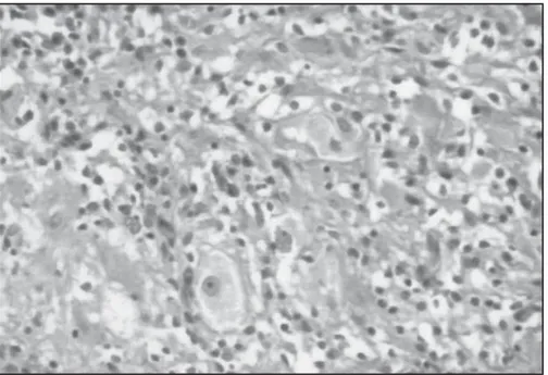
thickening of the facial nerve, from its central origin to-wards the periphery, entering the internal auditory canal. Histopathological examination was compatible with con-ventional gangliocytoma, showing diffuse proliferation of mature-looking nerve cells in a disorganized pattern. No necrosis and no mitosis were seen (Fig 3). One week after surgery, some improvement in the degree of ataxia and dysmetria was noted but facial paresis remained un-changed. At one-year follow-up, he showed a stable neu-rologic condition. MRI obtained at that time demonstrated no further progression of the lesion.
Fig 1. T2-weighted MRI image shows a hyperintense, heterogenous, diffuse lesion, at the right cerebellar hemisphere involving the su-perior part of cerebellar vermis, right middle cerebellar peduncle, pons and medulla oblongata.
Fig 2. T2-weighted MRI image shows direct extension into the right facial nerve, through the internal acoustic meatus; A. on a MRI coronal scan; B. on a MRI axial scan.
Fig 3. Histopathological examination compatible with conventional gangliocytoma, showing diffuse proliferation of mature-looking nerve cells in a disorganized pattern. No necrosis and no mitoses were seen (H&E x 400).
A
276 Arq Neuropsiquiatr 2003;61(2-A)
DISCUSSION
Neuronal and mixed neuronal-glial tumors are
ex-tremely rare, accounting for 0.1% to 0.5% of all brain
tumors
3. They are mostly seen in children and young
people
3, but the age of incidence ranges from 2
months to 80 years
1.
They include gangliocytoma,
dys-plastic gangliocytoma of the cerebellum
(Lhermite-Du-clos Disease), ganglioglioma, dysembryoplastic
neuro-epithelial tumor, central neurocytoma, cerebellar
lipo-neurocytoma and paraganglioma.
Some authors
consider ganglioneuroma as a synonym for
gangli-ocytoma. Such terminology is not accepted by the
World Health Organization classification of tumors and
its use should be avoided. De Arriba-Vilamor et al. state
that their pathogenesis is not fully understood,
consi-dering the possibility of being dysplasias or
malfor-mations rather than true neoplasias
4.
The most frequent site for gangliocytomas is the
temporal lobe, but they can arise anywhere in the
central nervous system, such as in the cerebellum,
brainstem, floor of third ventricle and spinal cord.
5Image findings are not specific. At MRI, the tumor
would emit a low-intensity signal on T1-weighted
sequences and a high-intensity signal on T2. Contrast
enhancement varies in intensity from none to
mar-ked, and it may be solid, heterogeneous, rim or
nodu-lar.
Differential diagnosis on imaging includes
astro-cytomas and oligodendrogliomas as well other
tu-mors of neuronal origin
6. Gangliocytomas are usually
radioresistant, therefore surgical removal is the only
option with a favorable prognosis,
even in those
ca-ses of partial resection.
There is only one description of cranial nerve
inva-sion by gangliocytoma in the literature, where Abe
et al report a case of trigeminal nerve involvement
2.
To our knowledge the present case is the first
des-cription of direct facial nerve invasion by
gang-liocytoma, as demonstrated clinically and supported
by MRI and surgical inspection.
REFERENCES
1. Nelson JS, Bruner JM, Wiestler OD, VandenBerg SR. Ganglioglioma and gangliocytoma. In Kleihues P, Cavenee WK (eds). Pathology and genetics of tumors of the nervoussystem. Lyon: IARCPress, 2000:96-98.
2. Abe T, Asano T, Manabe T, Matsura H, Furuta T, Taguchi K. Trigeminal ganglioneuroma. Brain Tumor Pathol1999;16:49-53.
3. Ebina K, Suzuki S, Takahashi T, Iwabuchi T, Takei Y. Gangliocytoma of the pineal body: a case report and review of the literature. Acta Neurochir (Wien)1985;74:134-140.
4. Arriba-Vilamor C, Martinez-Mata A, Espinosa-Mogro H, Rubio-Viguera V. Ganglion cell tumors. Rev Neurol(Barc) 1998;27:1008-1011.
5. Atlas SW. Magnetic resonance imaging of the brain and spine. New York: Raven Press, 1991:266-273.