Letters to the Editor
Radiol Bras. 2015 Set/Out;48(5):333–340
334
http://dx.doi.org/10.1590/0100-3984.2015.0046
Central nervous system involvement in sarcoidosis
Envolvimento do sistema nervoso central na sarcoidose
Dear Editor,
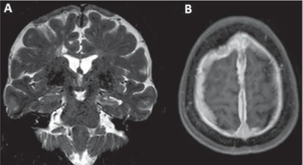
A 51-year-old female patient complained of mild frontotem-poral headache of insidious-onset for two years. One year ago, she had an episode of focal, tonic-clonic seizures (with right lower limb paresthesia) and was prescribed carbamazepine. Cerebrospinal fluid demonstrated increased protein levels and intrathecal im-munoglobulin (IgG) synthesis, suggesting an inflammatory com-ponent. Magnetic resonance imaging was performed (Figure 1). Sarcoidosis is a multisystem disease of unknown etiology characterized by noncaseating granulomatous inflammation(1).
There is a genetic predisposition, with T-lymphocyte receptor
activation by some unknown antigen. The disease affects prefer-entially the respiratory system(1). In the lungs, granulomas are
observed in the interstitial compartment, showing a perilymphatic distribution along the peribronchovascular sheaths, interlobular septa and pleural surface(1).
It is estimated that in about 5% to 15% of cases sarcoidosis affects the central nervous system. Rarely the patient presents with exclusively neurological manifestations like in the present case. Most commonly, neurosarcoidosis is observed in cases of dissemi-nated disease(2).
The clinical manifestations of neurosarcoidosis are pleomor-phic. Cranial nerve compromise, visual alterations, headache, weakness, paresis, paresthesia, psychiatric alterations and signs of meningeal irritation may be observed. Although rare, symp-extraluminal extension of the tumor(6,12). The prognosis is poor,
with mean survival time of approximately one year and a half after symptoms onset(8). Due to pulmonary artery occlusion and acute
symptoms, surgical resection is generally the treatment of choice(8).
In conclusion, the present case reinforces the important role of the imaging methods in the differentiation between pulmo-nary artery intimal sarcoma and chronic PTE. The relevant as-pects for this differentiation, such as contrast enhancement, dis-tention of the affect vessels and extraluminal extension, allow for a correct diagnosis, avoiding delay in the required surgical ap-proach.
REFERENCES
1. Yamanari MGI, Mansur MCD, Kay FU, et al. Bullet embolism of pul-monary artery: a case report. Radiol Bras. 2014;47:128–30. 2. Agnollitto PM, Barreto ARF, Barbieri RFP, et al. Rendu-Osler-Weber
syndrome: what radiologists should know. Literature review and three cases report. Radiol Bras. 2013;46:168–72.
3. Yamada AM, Melo ALKO, Lopes GP, et al. Bilateral breast swelling secondary to superior vena cava obstruction and subclavian vein throm-bosis. Radiol Bras. 2013;46:252–4.
4. Daud DF, Campos MMF, Fleury Neto LAP. Cardiac tamponade in an infant during contrast infusion through central venous catheter for chest computed tomography. Radiol Bras. 2013;46:385–6.
5. Eifer DA, Arsego FV, Torres FS. Unilateral pulmonary veins atresia: evaluation by computed tomography. Radiol Bras. 2013;46:376–8.
6. Chong S, Kim TS, Kim BT, et al. Pulmonary artery sarcoma mimick-ing pulmonary thromboembolism: integrated FDG PET/CT. AJR Am J Roentgenol. 2007;188:1691–3.
7. Grosse C, Grosse A. CT findings in diseases associated with pulmonary hypertension: a current review. Radiographics. 2010;30:1753–77. 8. Wong HH, Gounaris I, McCormack A, et al. Presentation and
manage-ment of pulmonary artery sarcoma. Clin Sarcoma Res. 2015;5:3. 9. Dornas AP, Campos FT, Rezende CJ, et al. Intimal sarcoma of the
pulmonary artery: a differential diagnosis of chronic pulmonary throm-boembolism. J Bras Pneumol. 2009;35:814–8.
10. Cheng HM, Chou ASB, Chiang KH, et al. Serial CT findings of pul-monary artery intimal sarcoma in 4 months: a case report. Chin J Radiol. 2009;34:35–8.
11. Wittram C, Maher MM, Yoo AJ, et al. CT angiography of pulmonary embolism: diagnostic criteria and causes of misdiagnosis. Radiographics. 2004;24:1219–38.
12. Yi CA, Lee KS, Choe YH, et al. Computed tomography in pulmonary artery sarcoma: distinguishing features from pulmonary embolic dis-ease. J Comput Assist Tomogr. 2004;28:34–9.
Marianna Nunes Batista1, Miriam Menna Barreto1,
Renata Fukamati Cavaguti1, Gláucia Zanetti1, Edson
Marchiori1
1. Universidade Federal do Rio de Janeiro (UFRJ), Rio de Janeiro, RJ, Brazil. Mailing Address: Dr. Edson Marchiori. Rua Thomaz Came-ron, 438, Valparaíso. Petrópolis, RJ, Brazil, 25685-120. E-mail: [email protected].
Figure 1. A: Coronal magnetic resonance imaging – T2-weighted sequence demonstrating diffuse pachymeningeal thickening most prominent at the high convexity and extending bilaterally toward the falx, with predominance of hyposignal in association with reduction in volume and hypersignal of the left hippocampus (mesial sclerosis). B:
Letters to the Editor
Radiol Bras. 2015 Set/Out;48(5):333–340
335
Vinicius Silles Machado1, Nivaldo Adolfo Silva Junior1,
Luciano Souza Queiroz1, Fabiano Reis1, Danilo dos Santos
Silva1, Flavia Fagundes Bueno1, Ana Carolina Coan1
1. Universidade Estadual de Campinas (Unicamp), Campinas, SP, Brazil. Mailing Address: Dr. Fabiano Reis. Faculdade de Ciências Médicas – Uni-versidade Estadual de Campinas, Departamento de Radiologia. Rua Tessá-lia Vieira de Camargo, 126, Cidade Universitária Zeferino Vaz. Caixa Postal: 6111. Campinas, SP, Brazil, 13083-887. E-mail: [email protected].
http://dx.doi.org/10.1590/0100-3984.2014.0113 toms of diabetes insipidus such as polydipsia and polyuria may also
occur due to the involvement of the hypothalamus and hypophy-sis. In cases of spinal cord involvement, weakness of lower limbs and other nonspecific signs of myelopathy are observed(3).
Although sarcoidosis may manifest in all the regions of the central nervous system, it is most commonly seen in the skull base, hypothalamus, pituitary and optic chiasm(4). At magnetic
reso-nance imaging, a common finding is intraparenchymal lesions with hypersignal on T2-weighted and FLAIR sequences, gener-ally multifocal, periventricular, subcortical or in the deep white matter. Such findings can hardly be differentiated from vasculitis or demyelinating diseases. Intraparenchymal lesions are gener-ally located near the areas with leptomeningeal involvement (with enhancement by paramagnetic contrast medium), and may be either single or multiple, possibly also involving cranial nerves(4).
Like in the present case, diffuse pachymeningeal thicken-ing may be observed, with hyposignal on T2-weighted, isosignal on T1-weighted sequences and contrast enhancement. Thus, differential diagnoses such as neurotuberculosis, dural lymphoma, meningioma en plaque, IgG4 deposition disease, pseudotumor, adenocarcinoma metastasis, Wegener’s granulomatosis, idio-pathic hypertrophic pachymeningitis might be considered, requir-ing biopsy to define the etiology. Simultaneous dural and leptom-eningeal involvement is rarely observed(4). In the present case, the
anatomopathological findings corresponded to typical noncaseating granulomas in the pachymeninges (Figure 2). Intracranial hy-potension is another differential diagnosis to be considered,
gen-erally presenting with diffuse pachymeningeal thickening, but with hypersignal on T2-weighted sequences (in the present case, hyposignal was observed on T2-weighted sequences).
A consensus is still to be reached on the treatment for sarcoi-dosis. In cases where the patient is symptomatic the treatment is initiated with high doses of corticosteroids, gradually reduced along the treatment up to complete withdrawal(3).
REFERENCES
1. Melo ASA, Marchiori E, Capone D. Tomographic and pathological find-ings in pulmonary sarcoidosis. Radiol Bras. 2011;44:220–4. 2. Spencer TS, Campellone JV, Maldonado I, et al. Clinical and magnetic
resonance imaging manifestations of neurosarcoidosis. Semin Arthritis Rheum. 2005;34:649–61.
3. Nozaki K, Judson MA. Neurosarcoidosis: clinical manifestations, diagno-sis and treatment. Presse Med. 2012;41(6 Pt 2):e331–48.
4. Christoforidis GA, Spickler EM, Recio MV, et al. MR of CNS sarcoidosis: correlation of imaging features to clinical symptoms and response to treat-ment. AJNR Am J Neuroradiol. 1999;20:655–69.
Femoral artery injury during aneurysm coiling
Lesão da artéria femoral durante embolização de um aneurisma
Dear Editor,
Endovascular artery reconstruction with low-profile stents, flow-diverters and flow-disrupting devices represent a significant progress in the endovascular therapy of intracranial aneurysms. Despite the improvement in technical expertise and developments in device technology, endovascular treatment still has inherent risks(1). In the literature, most reports are focused on neurological
complications during procedures(2), however, reports on access
vessel complications are scarce. Some of the well known access-related complications include: arterial pseudoaneurysms,
arterio-venous fistulae, hematomas, arterial dissection leading to acute vessel occlusion(3,4), intracavitary bleeding, and retroperitoneal
hematoma following femoral artery puncture(5). The authors
re-port the case of a large groin hematoma caused by a hypodermic needle connected with the black cable of the detachable coil power supply (Boston Scientific; Natick, MA, USA) and its endovascular management.
Local compression is the first line treatment for femoral ac-cess complications(6), but such strategy may fail when indicated
for patients under combined antiplatelet and anticoagulation regi-mens. Open surgery is effective in the treatment of groin compli-cations(7). However, the endovascular approach is a safe and