Universidade de Lisboa
Faculdade de Ciências
Departamento de Física
Development of a membrane based
detection of Candida albicans
Catarina Guerreiro Silva de Almeida
Orientador Interno: Professor Dr. Hugo Ferreira, Faculdade de Ciências da
Universidade de Lisboa, Lisboa, Portugal
Orientador Externo: Dr. Dorota Kwasny, Danmarks Tekniske Universitet, Lyngby,
Dinamarca
Mestrado Integrado em Engenharia Biomédica e Biofísica
Perfil de Engenharia Clínica e Instrumentação Médica
Dissertação
University of Lisbon
Faculty of Sciences
Physics Department
Development of a membrane based
detection method for Candida albicans
Catarina Guerreiro Silva de Almeida
Internal Supervisor: Professor Dr. Hugo Ferreira, Faculdade de Ciências da
Universidade de Lisboa, Lisboa, Portugal
External Supervisor: Dr. Dorota Kwasny, Technical University of Denmark, Lyngby,
Denmark
Integrated Master in Biomedical and Biophysics
Field of Clinical Engineering and Medical Instrumentation
Dissertation
Abstract
Candida albicans (C. albicans) is a yeast that can be found in 80% of the population. It is a normal constituent of the human flora and is commonly found in the gastrointestinal tract and in the female genital tract. In people with low immunity, such as HIV (human immunodeficiency virus) patients and radiotherapy patients, this common yeast can cause serious problems. In fact, Candidemia is one of the most common fungal infections in human and it is difficult to eradicate due to the resistance of this yeast.The detection systems available nowadays involve techniques such yeast culturing and PCR (polymerase chain reaction) that are in general time consuming. In order to reduce the time of detection and also to identify early stages of the infection (low concentrations of yeast) we propose a new system of filtration of the blood sample integrated with electrochemical gold electrodes functionalized with specific antibodies to detect C. albicans. The novel sensor consists of the gold electrodes directly fabricated on a filter membrane.
Electrochemical sensors are widely used in the biosensor field in order to study and detect reactions between different biological elements transforming these reactions into electrical signals. The first step of building a functional sensor is the development of the functionalization protocol. Here, we tested two different protocols of antibody immobilization. Protocol 1 consists of the attachment of a cross-linker on the tip of the antibody allowing for a better affinity to the surface and an oriented position of the antibody during the functionalization. Protocol 2 has the same purpose as Protocol 1, to allow a better affinity and orientation of the antibodies, but it is made with a different order. The attachment of a cross-linker is directly made on to the gold electrodes’ surface and, only after the preparation of the surface, the antibody is incubated to bind to the cross-liker. In order to compare both protocols, the C. albicans cells were marked with a fluorescent cell tracker and the number of cells in each sample was counted. The protocol with best results was Protocol 1.
After the selection of the best functionalization protocol, the developed membrane-based sensor capable of detecting Candida albicans yeast in culture broth is presented in this work. The yeast cells were captured on electrodes specifically functionalized with anti-C.albicans antibodies during 15 minutes incubation and detection is achieved by electrochemical impedance spectroscopy. The sensor allows for quick detection of the yeast cells at clinically relevant concentrations (150 CFU/ml).
Resumo
Candida é uma família de fungos, normalmente, presente na flora gastrointestinal, nos orgãos genitais, no sistema respiratório e na pele de pessoas saudáveis e, até determinada quantidade, não trazem nenhum risco. Apenas 17 espécies de Candida podem ser consideradas como patogénicas para o ser humano e, dentro deste grupo, Candida albicans é a espécie mais comum e pode ser facilmente encontrada em ambiente hospitalar, comida, solo e outros animais, sendo, por isso, a probabilidade de adquirir este fungo elevada.Candida albicans é unicelular e com forma ovóide, tendo entre 4 e 6 μm de diâmetro. Esta espécie reproduz-se produzindo blastoconídios (ou gêmulas) e quando o processo de divisão não está completo produz pseudo-hífas ou, dependendo das condições, pode mesmo produzir hífas. O nível de desenvolvimento de hífas promove a patogenicidade deste tipo de fungos. C. albicans cresce aerobicamente em ambientes com temperaturas entre 25 e 37ºC e formam colónias redondas e brancas após 48 - 72h de incubação em pratos de agar.
Como foi referido, C. albicans é geralmente inofensiva. No entanto, certas alterações no corpo, como variações de pH, podem provocar o crescimento excessivo deste fungo, potenciando a sua patogenicidade e tornando-se a causa de uma série de condições médicas incluindo infecções vaginais ou orais. Estas infecções podem ter também fontes exógenas, como uso de catéteres ou transmissão entre pessoas.
Quando a infecção por C. albicans, designada por Candidíase, atinge a corrente sanguínea facilmente se dissemina para outros orgãos internos. Este é o estágio de infecção mais grave pois o seu diagnóstico é difícil e, caso não seja feito em tempo útil, pode ter consequências graves. Este fungo pode atingir a corrente sanguínea quando o sistema imunitário do doente se encontra comprometido.
O uso excessivo de antibióticos, o crescimento do números de tratamentos invasivos aplicados (como quimioterapia e o transplante de órgãos) que comprometem o sistema imunológico dos doentes, juntamente com o aumento de portadores de HIV faz com que o grupo de risco seja elevado e tem provocado um aumento do número de casos de Candidíase Invasiva. As espécies de Candida são a causa de 70-80% de infecções do sangue causadas por fungos, e 45-65% destes casos são causados por Candida albicans. Para além dos custos médicos que estes casos representam, 40% deles levam os pacientes à morte, sendo as infecções causadas por Candida albicans consideradas um problema de saúde pública.
A principal razão para a elevada taxa de mortalidade referida deve-se ao facto deste tipo de infecções gerarem sintomas que são comuns a outros tipos de problemas, como febre ou outro tipo de sintomas dependendo do orgão atingido. Caso o organismo não responda ao tratamento utilizado, os órgãos do paciente podem parar de funcionar.
Outra razão que contribui para dificultar o tratamento eficiente deste tipo de infecções são as próprias técnicas de diagnóstico presentes no mercado. O uso de técnicas de cultura é o mais utilizado para diagnóstico de infecções por fungos. No entanto, esta técnica demora entre 4 a 16 dias até confirmação do resultado definitivo e, para além disso, é muito pouco sensível, sendo 50% dos resultados falsos negativos.
Outra técnica utilizada é ELISA (enzyme-linked immunosorbent assay), um teste imunoenzimático que permite a detecção de antigénios específicos (por exemplo, determinados anticorpos no plasma sanguíneo). Este teste é usado no diagnóstico de várias doenças e baseia-se na ligação do analito a ser detectado ao anticorpo específico desse analito e na posterior adição de um substracto que, caso essa ligação antigénio-anticorpo se verifique, muda de cor. Em adição, a densidade óptica deste produto dá informações sobre a concentração de antigénio presente. Esta técnica é altamente sensível mas bastante complexa e trabalhosa, demorando entre 2 a 3 dias para o resultado final.
Concluindo, os métodos de detecção utilizados actualmente, são, em geral, complexos e demorados. Sendo por isso necessária a investigação de métodos mais rápidos, sensíveis e fáceis de utilizar.
A área de biosensores está em grande desenvolvimento pelo seu potencial de redução da duração do processo de detecção e obtenção do resultado final, no entanto, ainda é necessário mais investigação para que eles sejam eficientes o suficiente para o seu uso na prática.
Um biosensor é um sistema que converte uma resposta biológica a um analito específico num sinal mensurável. Os sistemas de transdução destes sinais podem ser, por exemplo, piezoeléctricos, ópticos ou electroquímicos.
Os sensores electroquímicos são bastante utilizados na área de biosensores, inclusive com aplicações biomédicas com o objectivo de estudar as propriedades electroquímicas dos materiais e reações entre diferentes elementos biológicos. Um tipo de elementos biológicos associados aos sensores electroquímicos são anticorpos que funcionam como o elemento que permitem a identificação e detecção de diferentes tipos de antigénios, como fungos. Esta associação entre biossensores electroquímicos e anticorpos têm apresentado bons resultados em termos de sensibilidade e selectividade.
Assim, tendo em conta a necessidade existente de um novo sistema de detecção de Candida albicans mais eficiente, este projecto consistiu no desenvolvimento de um biosensor electroquímico baseado na funcionalização de eléctrodos de ouro aplicados na superfície de uma membrana com anticorpos específicos C. albicans e cuja a reacção entre anticorpos e células provoca uma alteração da impedância da superfície dos eléctrodos permitindo assim a detecção da presença de Candida albicans na amostra analisada.
A produção dos eléctrodos é feita sobre uma membrana pois a ideia do projecto final, que deverá continuar em desenvolvimento, é a produção de um sistema de filtragem que permita a separação dos vários elementos constituintes do sangue das células de C. albicans, de modo que o processo de tratamento da amostra e a sua detecção sejam feitos no mesmo dispositivo e em simultâneo.
O projecto foi dividido em três grandes etapas. A primeira etapa consistiu na utilização da técnica ELISA, por ser um método já bastante desenvolvido e utilizado, para analisar os anticorpos utilizados e ter esta experiência como referência para os resultados futuros. No entanto, embora seja uma técnica comum, é bastante difícil de optimizar. Ainda assim, foi possível concluir que tipo de anticorpos a utilizar nas etapas seguintes é anti-candida policlonal e qual o limite de detecção desta técnica, 1680 CFU/ml (CFU significa unidade formadora de colónias e corresponde ao número de colónias contabilizadas na amostra em análise).
A segunda etapa consistiu no estudo de qual a técnica de funcionalização mais eficiente a ser aplicada nas superfícies de ouro dos eléctrodos. Esta é uma das fases mais críticas no desenvolvimento de um sensor, pois é através da funcionalização adequada dos eléctrodos que é possível que a detecção ocorra. Assim, dependendo do processo de modificação da superfície, o sensor vai ser mais ou menos eficiente, quer a nível de selectividade, quer a nível de sensibilidade. Foram testados dois métodos diferentes, sendo que ambos utilizam a molécula LC-SPDP (succinimidyl 6-(3-[2-pyridyldithio]-propionamido)hexanoate) como cross-linker. No Protocolo 1, esta molécula foi previamente ancorada aos anticorpos e só depois eles foram aplicados à superfície. No Protocolo 2, o cross-linker foi primeiro aplicado à superfície dos eléctrodos e só depois os anticorpos foram utilizados para finalizar a modificação da superfície. Como método de avaliação da eficiência dos protocolos as células foram marcadas com um marcador de fluorescência, e após o tratamento das superfícies, incubação das células e lavagem para remoção das células que não foram capturadas pelos anticorpos, as superficies foram analisadas ao microscópio e o número de células à superfície foi contado. O Protocolo 1 verificou-se mais eficiente e, por isso foi o aplicado à última etapa do projecto.
A última etapa do projecto consistiu no desenvolvimento do sistema electroquímico de detecção de Candida albicans em meio de cultura. Para tal, foi aplicado o Protocolo 1 de funcionalização e analisados vários espectros de impedância obtidos nas várias experiências que foram efectuadas de forma a caracterizar o sensor e optimizar o processo de detecção. Foram estudadas várias fases, desde optimização do processo de limpeza da superfície dos eléctrodos sem a degradar; testadas várias concentrações de anticorpos na funcionalização dos eléctrodos e, por fim, foram realizados testes para estudar a sensibilidade e selectividade do sensor na detecção de C. albicans. Dentro das várias experiências efectuadas foram obtidos resultados bastante promissores tendo sido possível atingir níveis de detecção de concentrações relevantes para a aplicação em ambiente clínico, 150 CFU/ml.
Com os resultados obtidos durante este projecto, está a ser preparado em conjunto com a minha orientadora externa um artigo intitulado “Membrane based detection of Candida albicans using electrochemical impedance spectroscopy” e os dados foram ainda apresentados na 4th International Conference on Bio-Sensing Technology, que decorreu em Maio, na cidade de Lisboa.
Preface
This project is the master thesis and completion of the Biomedical and Biophysics Engineering course at the Faculty of Sciences, University of Lisbon (FCUL). The project was carried at Institute of Micro- and Nanotechnology at DTU from August 2014 to February 2015 and corresponds to 45 ECTS points. The project was conducted in the Nano Bio Integrated Systems (NaBIS) group and the work has been done under supervision of Post Doc Dorota Kwasny, Associate Professor Winnie Edith Svendsen and Professor Hugo Ferreira (FCUL).Selected results in this thesis were presented at the 4th International Conference on Bio-Sensing Technology, in Lisbon and will be subject of a scientific paper.
Catarina Almeida September 17th, 2015
Acknowledgments
The experience of these last seven months would not have been so meaningful if I did not have the right people around me and the next words are for them.First of all I would like to thank to Winnie E. Svendsen and Andrea Pfreundt for accepting me to be part of NaBIS group. It was with them this adventure started!
A really special thanks to my supervisor Dorota Kwasny for her support and encouragement, for always being available to help me and for trusting me to work independently. Her optimism and calm even when the future of the project was not the most promising was an important key to keep me motivated.
Thanks to NaBIS group for the good work atmosphere and inspirational meetings. Specially to Susan for being available for work discussions or random conversations when a break was needed.
Thanks to Professor Hugo for his encouragement e-mails and help during all the process.
Thanks to my great group of friends who helped me to feel at home even when I was miles far from them. A special thanks for those who came to visit me, you are amazing! Other special thanks to my new friends I made during this journey and helped on making this experience more special.
Finally thanks to all my family, specially to my parents for all the support they give me and for trusting me all the time that make all this possible and my sister Maria, because she is my sister and that says everything. Without you and your inspiration I would not be where I am today.
Thank you,
Catarina
Contents
ABSTRACT I RESUMO III PREFACE VIII ACKNOWLEDGMENTS X CONTENTS XI LISTOF FIGURES XIII LISTOF TABLES XIV LISTOF ACRONYMS XV1. INTRODUCTION 1
1.1. Motivation 1
1.1.1. Established methods in pathogen detection 2
1.1.2. Biosensors 6
1.2. Aim of the project 7
2. THEORETICAL BACKGROUND 8
2.1. Antibodies 8
2.2. ELISA 11
2.3. Surface immobilization of antibodies 13
2.4. Electrochemical Detection 17
2.4.1 Electrochemical sensors 17
2.4.2. Impedance definition 18
3. MATERIALSAND METHODS 22
3.1. ELISA 22
3.2. Surface Functionalization 23
3.2.1 Protocol 1 24
3.2.2 Protocol 2 24
3.3. Electrochemical Detection 25
3.3.1. Electrodes and holder fabrication 25
3.3.2. Electrochemical detection of Candida albicans 27
4.1. ELISA 29 4.1.1. Conclusions 30 4.2. Surface Functionalization 31 4.2.1 Conclusions 34 4.3. Electrochemical Detection 34 4.3.1. Electrodes Cleaning 34
4.3.2. Characterization of the immobilized antibody 38
4.3.3. Analysis of the selectivity and sensitivity 40
4.3.4. Conclusions 44
5. CONCLUSIONSAND FUTURE WORK 45
6. BIBLIOGRAPHY 49
APPENDICES 54
Appendix 1: Protocol for Candida albicans growth 54
List of Figures
1.1. C. albicans colonies in agar medium 1
2.1. Antibody structure 9
2.2. Scheme of Direct ELISA 11
2.3. Scheme of the different types of antibodies on Indirect ELISA 12
2.4. Scheme of Indirect ELISA (Sandwich) 12
2.5. Scheme of physical adsorption 13
2.6. Scheme of Avidin-biotin system 14
2.7. Scheme of self-assembled monolayers method 14
2.8. Scheme of Protocol 1 15
2.9. Scheme of Protocol 2 16
2.10. Nyquist plot from EIS 20
2.11 Bode plot from EIS 20
2.12. Randles circuit with a constante phase element 20
3.1. Picture of the gold surface with a drop of C.albicans solution during incubation 23
3.2. Structure of the membrane based electrodes 25
3.3. Setup for fabrication of the membrane electrodes 26
3.4. Holder for electrochemical measurements 26
4.1. ELISA results obtained from OD measurements 30
4.2. Microscopy picture of stained cells of Candida albicans 31
4.3. Number of counts depending on the concentration of antibody 32
4.4. Number of counts depending on the concentration of C. albicans 33
4.5. Study of cleaning time 35
4.6. Impedance spectra fitted with the simulation made using Randles circuit 36
4.7. Impedance spectra of 11 electrodes of the same membrane before cleaning 37
4.8. Impedance spectra of the same 11 electrodes after cleaning 37
4.9. Average Rct values before and after cleaning and after functionalization
of the electrodes 38
4.10. Variation of Rct values after incubation of different concentrations of
antibody 39
4.11. Nyquist plot of the same electrode after different steps on the experiment 41
4.12. Variation of Rct values after incubation of different concentrations
C. albicans and S. cerevisiae 42
4.13. A typical Nyquist plot for a good electrode 43
List of Tables
Table 1. Different studies for C. albicans detection 5
List of Acronyms
HIV - human immunodeficiency virus PCR - polymerase chain reaction
ELISA - enzyme-linked immunosorbent assay CFU - colony-forming unit
DNA - deoxyribonucleic acid LFD - lateral flow devices HRP - horseradish peroxidase
TMB - 3,3',5,5'-Tetramethylbenzidine PBS - phosphate buffered saline BSA - bovine serum albumin
EIS - electrochemical impedance spectroscopy
Sulfo-LC-SPDP - sulfosuccinimidyl 6-(3'-(2-pyridyldithio)propionamido)hexanoate CPE - constant phase element
Rct - charge transfer resistance SAM - self assembly monolayer AC - alternating current
1. Introduction
1.1. Motivation
Candida species are normally harmless constituents of the human flora and commonly found in the gastrointestinal tract, sexual organs, the respiratory system and on the skin of healthy people. Only 17 Candida species are known as human pathogens. Candida albicans is the most predominant species and it has been also found in hospital environments, food, soil, and in animals [1-3].
Candida albicans is unicellular and ovoid with 4-6 μm in diameter. This yeast multiplies by producing blastoconidia and when the division process is not completed it develops pseudohyphae or under certain conditions true hyphae. The development of hyphaes promote the pathogenicity of C. albicans. C. albicans grows aerobically at 25-37ºC and form creamy, white, smooth and flat colonies on agar plates as it can be visualized in Fig. 1.1. These colonies become visible after 48-72 hours of incubation [3].
1. Introduction
As it was referred, C. albicans is usually harmless but when an overgrowth occurs it can become pathogenic, causing a range of medical conditions including painful superficial infections, such as vaginitis in otherwise healthy women and severe surface infections of the mouth and esophagus. Candidiasis is mainly an endogenous infection produced by the overgrowth of human body’s own fungi due to some physiological alteration such as pH change. However, Candidiasis can also be acquired from exogenous sources like catheters or prosthetic devices, person-to-person transmission or vertical transmission [5].
When Candidiasis spreads to the bloodstream it is called Invasive Candidiasis. Once the fungus is in the bloodstream, it can spread to other organs causing infection. The spread of this yeast to the bloodstream occurs mainly in patients with impaired immune system.
The widespread use of antibiotics, the increase of invasive procedures (like chemotherapy, organ transplantation, the use of indwelling catheters) that compromise the defense mechanisms of the human host have further aggravated the problem of Invasive Candidiasis. In addition, the increase in the numbers on prevalence of HIV (human immunodeficiency virus) patients has dramatically raised the number of patients susceptible to opportunistic fungal infections. Invasive Candidiasis is the fourth most common cause of hospital-acquired infections and species of the genus Candida, in particular, are responsible for 70-80% of diagnosed fungal bloodstream infections in the United States. In particular, 45-65% of these cases are specially caused by Candida albicans. This problem costs around $1 billion to Medicare and the rate of mortality is 40% which make Candida infections a major public health concern [1,5].
The symptoms of this kind of infection are not specific which makes its diagnosis more difficult. Fever and chills that do not disappear after antibiotherapy are the most common symptoms. If the infection spreads to other organs or parts of the body such as kidneys, other symptoms may develop, depending on the site of infection. If the infection does not respond to the treatment, the patient’s organs may stop working [6].
1.1.1. Established methods in pathogen detection
Diagnosis of Invasive Candidiasis can be difficult and the oldest bacterial detection techniques, and still the “gold standard”, are culturing and plating. In general, these methods use selective liquid or solid culture media, to grow, isolate, and enumerate the target microorganism and simultaneously prevent the growth of other microorganisms present in the the sample to analyze [7,8]. The quantitative plate method is based on culturing dilutions of sample suspensions in the interior or on the surface of an agar layer in a Petri dish. Individual
1. Introduction
microorganisms or small groups of microorganisms will grow to form individual colonies that can be counted visually. The qualitative procedures are used when it is not necessary to know the amount of a microorganism present in a sample but only its presence or absence [9]. These techniques are very time-consuming. It can take from 4 to 16 days for confirmation of results and the rate of false negatives is high (50% of blood cultures can be false negative) [6, 7].
Polymerase chain reaction (PCR) is a nucleic acid amplification method developed in the 80’s and it is based on the isolation, amplification and quantification of a short DNA sequence including the targeted bacteria’s genetic material. PCR uses oligonucleotide primers which sequence is homologous to the ends of the genomic DNA region to be amplified. Conventional PCR relies on the amplification of the target gene in a thermocycler. The method is performed in repeated cycles of denaturation by heat of the extracted and purified DNA, followed by an extension phase using specific primers and a thermostable polymerization enzyme. The products of one cycle serve as the DNA template for the next cycle, doubling the number of target DNA copies in each cycle. The PCR products separation is made by a electrophoresis gel, followed by analysis of the resulting electrophoretic patterns. Comparing with other techniques like culturing it is less time-consuming but still takes from 5 to 48h to achieve a result. Many PCR tests have been validated and commercialized to make PCR a standard tool used by microbiology laboratories to detect pathogens. Aside from that, if a positive result for PCR is reached, this must be confirmed using culturing techniques because, as already referred, they cannot distinguish an ongoing infection. They often result in false positives as microorganisms’ DNA can be present in the blood stream following previous infections or after eating some products, such as cereal products or leftovers, that can have plenty of funghi [7, 9]. Moreover, they cannot distinguish ongoing infections, only the presence of specific yeast genes, which may remain in the bloodstream from previous infections or food products.
Immunology-based methods are based on the antibody-antigen interaction which has been used in the design of a variety of assays and formats. In some cases, the antigen–antibody complex formed is directly measurable or even visible. Incubation times are usually very short comparing with the techniques described above. Normally, the antibody is labeled with a fluorescent reagent or with an enzyme so that the antigen–antibody interaction may be visualized more easily when it occurs. Among the different immunology-based methods there are the lateral flow devices (LFD) and the enzyme-linked immunosorbent assay (ELISA) [7].
LFD are composed by a dipstick made of a porous membrane that contains colored latex beads or colloidal gold particles coated with detection antibodies targeted toward a specific microorganism. When the target organism is present, then it will bind with the particles and both move by capillary forces until they find and bind the immobilized capture antibodies
1. Introduction
forming a color line that is in the device window, indicating a positive result. It is a quick and easy technique, simple to interpret and can be completed within 10 min after culture enrichment [9]. However, this type of devices needs to be improved in terms of reproducibility and sensitivity because when the analyte is very low concentrated, a preliminary concentration step is obligatory. LFD allows mostly qualitative results and when a quantitative result is needed, it is necessary to introduce a labeled analyte, for example with a fluorescent label, which adds extra steps and increases the costs, since a more sophisticated read-out system has to be used [10].
ELISA is a biochemical technique that combines an immunoassay with an enzymatic assay and, currently, it is the most established technique being source of inspiration for many biosensor applications. Generally, this method consists of the immobilization of the antigen-specific antibody on a solid matrix that captures the antigen from enrichment cultures following by the addiction of a second antibody conjugated to an enzyme capable of generating a product detectable by a change in color or, in case of enzyme-linked fluorescence assay (ELFA) in fluorescence, which allows for indirect measurement using spectrophotometry (or fluorometry for ELFA) of the antigen present in the sample (microorganism or toxin). The results can be obtained in 2–3 days [7-11].
Immunoassay-based methods’ success depends on the specificity of the antibody and the limit of detection of these techniques is around 103-105 CFU/ml, which is high if an early detection
is aimed. Based on the literature [12] and on several studies in this area (Table 1), a limit of detection of 10 CFU/ml is one of the goals for this project.
Table 1. summarizes some of the several studies made about C. albicans detection in terms of limit of detection (the minimum concentration possible to detect) and time to perform the tests. The most common technique used is PCR and its variations. Besides, it is also common the combination of techniques, for example, application of ELISA to detect PCR products. It is possible to observe that there are already techniques with very low limit of detection in 8h or less. However they are the combination of techniques which usually brings difficulty to the protocols and the need of specialized people to work with these systems. This is the reason why a new system is needed, where it is necessary the less sample preparation possible using simple techniques.
1. Introduction
Technique
Limit of detection
(CFU/ml)
Time
Article
PCR 200 5h Tirodker, Urmila H.,
et. al, 2003 [13] ELISA (for detection of PCR
products)
4 8h Flahaut, M., et. al., 1998 [14]
Culturing <1 (but 50% of false positives)
4-16 days Meyer, W., [15]
PCR paired with mass spectrometry analysis (PCR-ESI/
MS)
20 No information Metzgar, D., et. al., 2013 [16]
Electrochemical detection of PCR products
10 No information Muir, A., et. al, 2011 [17]
Combination of nested and multiplex PCR
3 No information Gosiewski, T., et. al., 2011 [18]
Combination of immunomagnetic separation (IMS) with solid-phase cytometry (SPC) using viability
labeling
8 4h Vanhe, L., et. al.,
2010 [19]
MagNA Pure LC assay, Roche 1 6h Schmidt, K., et. al., 2001 [20]
1. Introduction
1.1.2. Biosensors
As described, currently, there are several methods for pathogen detection and, in general, they are very complex, involving different techniques as extraction and purification or separation. Most of them are also very time consuming.
Biosensing devices are under development due to their potential to shorten the waiting times between sample uptake and results. However, they still need to be comparable the conventional methods in terms of sensitivity, selectivity and costs [7].
A biosensor is an analytical device that converts a biological response to a target analyte into a measurable signal. The transducing system can be, for example, piezoelectric, optical, or electrochemical [7,11].
Piezoelectric biosensors are based on the measurement of changes in resonant frequency of a piezoelectric crystal resulting in mass changes on the crystal surface which is very often quartz. The quartz crystal is the critical component of the quartz crystal microbalance (QMC) because it reports the mass deposited on its electrodes quantitatively in which the mass changes its oscillation frequency. Piezoelectric immunosensors based on QCM are classified as direct immunosensors when the immobilising antigen or antibody is applied on the surface of a piezoelectric material. The intrinsic vibration frequency of the support is modulated with the immunochemical recognition reaction. After the antibody immobilization on the electrode surface, the interaction with its corresponding antigen changes the resonance frequency. Numerous piezoelectric immunosensors have been reported for the detection of various analytes from small molecules to macromolecules and also viruses and cells [7].
Optical biosensors have been developed for the rapid detection of contaminants, toxins, drugs and pathogenic bacteria, showing high selectivity and sensitivity, and they are based on several optical phenomena such as surface plasmon resonance changes, scattering and interferometry. Surface plasmon resonance measure changes in refractive index caused by structural alterations in a thin metal surface. Another type of optical immunosensor use a compact fiber-optic evanescent-wave sensing. In this system, the fluorescent signal is confined in the fiber system so the signal-to-noise ratio is greatly improved and the system can be operated in ambient light conditions [11]. There are other examples of simpler optical biosensors, which is the case of colorimetric biosensors which are based on the change of color when a specific reaction occurs.
Electrochemical biosensors are used in different fields to investigate the electrochemical properties of materials allowing the detection of certain interactions through current or
1. Introduction
potencial changes [21]. As the the system under development in this project is based on electrochemical sensors, this type of sensors will be described in detail in section 2.4.
1.2. Aim of the project
As it was referred above, the risk group referred is incredibly large and Candida albicans infections can be fatal if not detected early enough. The development of a system that enables aN early diagnosis of these infections is thus mandatory.Based on this need, the purpose of this project was to develop an ultrasensitive electrochemical biosensor to detect Candida albicans in the bloodstream using a membrane based sensing with integrated electrical impedance spectroscopy, which, theoretically, would allow an early detection of this infection.
The main part of this project was the development of the electrochemical sensor and its optimization to achieve the highest possible sensitivity on the detection of Candida albicans. The project is structured in three stages. First, the antibodies were tested with ELISA, a well developed method, in order to have a reference to compare the results from this new system and confirm if the purchased antibodies were working properly. Then, two different gold functionalization procedures were studied in order to optimize the specificity of the system and to choose the best method to apply on the electrochemical sensor. Finally, the chosen protocol was applied to functionalize the gold electrodes’ surface and several electrochemical measurements were made for optimizing Candida albicans detection.
2. Theoretical Background
2.1. Antibodies
In biosensing there are three main classes of biological recognition elements:
-
enzymes (in the specific case of bacterial and yeast detection. They are used more often as a label than as a biological recognition element)-
antibodies-
nucleic acidsIn this project the biological recognition elements consist of antibodies. Antibodies or immunoglobulins (Ig) are a group of glycoproteins secreted by specialized B lymphocytes, known as plasma cells, that constitute one of the most important specific defense mechanisms in vertebrate animals. They are recruited by the immune system to identify and neutralize foreign objects like bacteria and viruses [22].
The ability of antibodies to bind an antigen with a high degree of affinity and specificity has led to their wide use in a variety of scientific and medical disciplines. Their use in diagnostic assays and as therapeutics has had a profound impact on the improvement of health and welfare systems in both humans and animals. Today antibodies are used extensively for research purposes in many areas of biology, such as immunoprecipitation, histochemistry, enzyme linked immunosorbent assays (ELISA), diagnosis of disease, immunoturbidimetric methods, Western blots and Biochip technology [22,23].
All of the antibodies have a very similar Y shaped structure composed of four polypeptides. Each Y contains two identical copies of a heavy chain and two identical copies of a light chain which are different in their sequence and length. Each heavy and light chain are held together by disulfide and nonequivalent bonds. Fig. 2.1 is a schematic representation of the general antibody structure [24].
The amino acid sequence in the tips of the Y varies greatly among different antibodies. In this region the light and heavy chains associate to form an antigen-binding domain (Fab fragment), and the variability of this region gives the antibody its own specificity. The carboxy terminal regions of the two heavy chains fold together to form the Fc domain. The
2. Theoretical Background
two domains (Fab and Fc) are connected by a region known as hinge. This region imparts lateral and rotational movement to the antigen-binding domains, providing the antibody the ability to interact with a variety of antigen presentations [23].
Antibodies are divided into five major classes, IgM, IgG, IgA, IgD, and IgE, based on their constant region structure and immune response and they are distinguished by the type of heavy chain found in the molecule.
IgG, a monomer, is the predominant Ig class present in human serum. Produced as part of the secondary immune response to an antigen, this class of immunoglobulin constitutes approximately 75% of total serum Ig. Because of its relative abundance and excellent specificity toward antigens, IgG is the principle antibody used in immunological research and clinical diagnostics [22].
Antibodies (whatever their class or subclass) are produced and purified in two basic forms for use as reagents in immunoassays: polyclonal and monoclonal.
Polyclonal antibodies are produced as a response for the same antigen but with different specificities and epitope affinities. Typically, the immunological response to an antigen is heterogeneous, resulting in many different cell lines of B-lymphocytes (precursors of plasma
2. Theoretical Background
cells) producing antibodies to the same antigen. All of these cells originate from common stem cells, yet each develops the individual capacity to make an antibody that recognizes a particular determinant (epitope) of the same antigen. For production purposes these antibodies are generally purified directly from the serum of immunised animals were the antigen of interest stimulates the B-lymphocytes to produce a diverse range of immunoglobulin's specific to that antigen [23, 25].
Due to its selectivity, polyclonal antibodies are ideally suited for use in sandwich assays as second stage antigen detectors. Often polyclonal antibodies will be tagged with reporter molecules such as horseradish peroxidase (HRP) or alkaline phosphatase (AP) so that under specific conditions the antibodies presence can be detected by light or color changes [25].
Because an individual B-lymphocyte produces and secretes only one specific antibody molecule, clones of B-lymphocytes produce monoclonal antibodies. All antibodies secreted by a B-cell clone are identical, providing a source of homogeneous antibody having a single defined specificity. B-cells can be isolated easily from the spleen and lymph nodes of immunised animals; however, these cells have a limited life span and cannot be cultured directly to produce antibody in useful amounts due they limited division capacity [25].
For an antibody to be useful in research or industry, it must be readily available in large quantities. Due to the referred limitations, this would not be possible using B-cells cultured in vitro as they would eventually stop dividing and the population would die out. However, this restriction has been overcome with the development of hybridoma technology, wherein isolated B-lymphocytes in suspension are fused with myeloma cells from the same species (usually mouse) to create monoclonal hybrid cell lines that are virtually immortal while still retaining their antibody producing abilities. This technology allows scientists to extract and purify one antibody from the complex mixture of antibodies present in the in vivo polyclonal response. This cell line, once stabilised via single cell cloning, can be frozen and stored indefinitely under liquid nitrogen, allowing the antibody to be produced in vitro, in large quantities when required. Monoclonal antibodies can be raised against many targets. Specific antibody characteristics can be identified and selected e.g. sensitivity requirements and cross reactivity levels can be specified and monoclonal antibodies screened to identify any cell lines exhibiting the required characteristics. Monoclonal antibodies are especially useful as primary antibodies in applications that require single epitope specificity [22-26].
In conclusion, polyclonal antibodies are less selective comparing with monoclonal antibodies. However, polyclonal production is easier and cheaper and the work with monoclonal antibodies can be more demanding since small changes in the structure of an epitope (e.g., as consequence of genetic polymorphism, glycosylation, denaturation) can markedly affect the function of a monoclonal antibodies and they are also more susceptible to small pH and salt
2. Theoretical Background
concentration changes. The decision about which antibody should be used depends on the requirements of the device under development [24]. In this project both monoclonal and polyclonal antibodies against Candida albicans were initially tested. Due to project time schedule the polyclonal antibodies were primarily used throughout the development of the electrochemical sensor.
2.2. ELISA
In order to study the antibodies that were used during the project and to have a second method to compare the results to, a sandwich ELISA was performed.
As referred, ELISA is another basic technique based on the antibody-antigen interaction that results in color change and allows the detection and quantification of substances such as peptides, antibodies, hormones or organisms as yeast. ELISA is widely used because it allows the analysis of a large number of samples simultaneously. There are different formats of ELISA but the most common classifications are direct or indirect. All the formats have in common the coating of a solid surface, typically a 96-well plate:
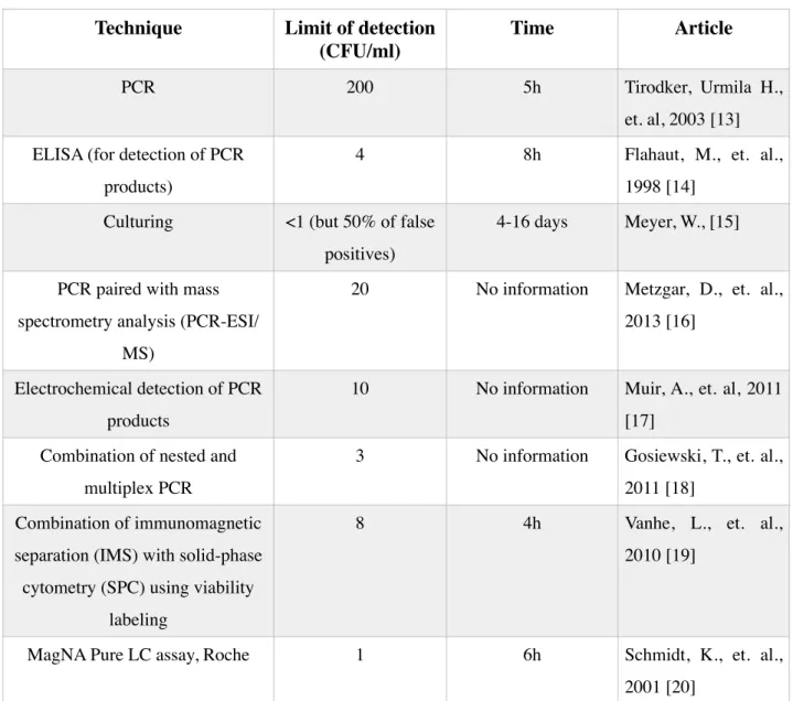
• Direct: in this method antigens from the sample are attached to a surface. Then, a further specific antibody is applied over the surface so it can bind to the antigen. This antibody is linked to an enzyme, and, in the final step, a substance containing the enzyme's substrate is added. The subsequent reaction produces a detectable signal, most commonly a color change in the substrate [27]. Fig. 3.1. represents how direct ELISA occurs.
2. Theoretical Background
• Indirect (sandwich): this approach allows for simultaneous processing of many small samples and is used to measure the amount of antigen between two layers of antibodies. It requires two antigen specific antibodies. Briefly, one antigen-specific antibody (named as capture or primary antibody) is coated onto a solid substrate to capture the antigen from the applied solution. Then the sample is added and any analyte present is bound by the immobilized antibody. A second labeled antibody (named as detection or secondary antibody) is then added and binds to the captured analyte. The label of the secondary antibody is often a HRP enzyme (horseradish peroxidase) that reacts with TMB susbtract (3,3’,5,5’-Tetramethylbenzidine) resulting in a change of color and allows the detection. After the substrate solution is added, a color change occurs and the optical density can be measured. Depending on the optical density value, it is possible to conclude about the quantity of analyte present in the sample. The capture and detection antibodies must be of different isotypes, if not from different species, so that the detection antibody will only detect the presence of the primary antibody and correctly indicate that the antigen has been captured in the well [28]. This was the format used and will be explained in more detail in the Materials and Methods section. Fig. 2.3. represents the distinction between capture and detection antibody. Fig. 2.4. represents how this method works.
12
Fig. 2.3. Schematics of the different types of antibodies on Indirect ELISA
2. Theoretical Background
Several ELISA assays were performed to test two different types of antibodies: monoclonal and polyclonal, as explained above, and different concentrations of C. albicans were analyzed. The general procedure was based on the one provided by the antibody supplier with modification in regards to concentration of antibodies and analytes and washing solutions. Incubation times were also tested.
2.3. Surface immobilization of antibodies
Surface immobilization of antibodies is crucial for biosensor sensitivity. The most important aspect is to identify a surface chemistry that allows optimal orientation (antigen-binding regions away from the surface) and a good degree of freedom for the antibody. The immobilization of antibodies on a substrate is made using different techniques:
-
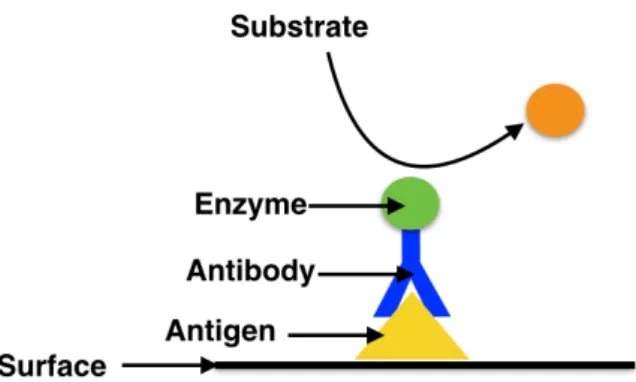
Physical adsorption: consists of the random attachment of antibodies on the surface. This is the simplest and quickest technique. However, as the correct orientation of the antibodies can not be controlled it is also the least reliable and most unstable method [7]. Fig 2.5. represents how physical adsorption occurs.-
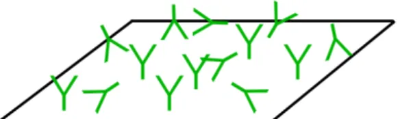
Avidin-biotin system: it is a very effective way to anchor biomolecules to an avidin coated surface. Since the biotin label is stable and small, it rarely interferes with the function of labeled molecules enabling the avidin-biotin interaction (that is the strongest known non-covalent interaction, Kd =10-15M, between a protein and a ligand) to be used for the development of
robust and highly sensitive assays. Although this method allows a very oriented disposition of the antibodies, it is dependent on the success of, at least, three bonding steps which is very demanding and brings more uncertainty to the success of the functionalization: first, it is necessary to label the antibody with the biotin group; then it is made the binding of the
Fig. 2.5. Schematic of physical adsorption. The antibodies attach to the surface without any
2. Theoretical Background
avidin unit to the surface (this can be done by direct adsorption which is not very stable as it was referred above); and finally the binding between avidin and biotin [7]. Fig 2.6. represents the avidin-biotin system.
-
Self-assemble monolayers (SAMs): SAMs of organic molecules are molecular assemblies formed spontaneously on surfaces by adsorption and are organized into more or less large ordered domains. This method consists of the attachment of a functional group that has a strong affinity to the substrate and anchors the molecule to it. Basically, a SAM is formed by a head group, a tail and a functional end group that will attach to the target. Common head groups include for example thiols. Due to the robustness of immunosensing devices based on SAMs, they have been applied in different systems and it was the chosen method for this project [9,11]. Fig 2.7. represents how the SAM method occurs.14
Fig. 2.6. Schematic of avidin-biotin system. It is obtained an oriented position of the antibodies
with antingen binding site free.
Fig. 2.7. Schematic of self-assembled monolayers method. The orientation of the antibodies are
not so controlled as the avidin-biotin system but its disposition is more organized than physical adsorption
2. Theoretical Background
The orientation and freedom-of-movement of immobilized antibodies is important to obtain a maximal functional sensor surface. To reach this goal several studies have shown that bio-functionalized surfaces can be made more active by coupling the biomolecules to linkers. This gives the protein an environment that is not restricted by being too close to the sensor surface but instead have more freedom to interact with the analyte. Furthermore, these methods help orientate the antibodies, so that the antigen-binding sites are freely available for analyte interaction [23].
Two different immobilization procedures were tested based on two scientific articles [30, 31]. Both protocols have in common the usage of a thiol-cleavable sulfonated succinimidyl 6-(3-[2-pyridyldithio]-propionamido)hexonoate (sulfo-LC-SPDP). Sulfo-LC-SPDP is a water-soluble, long-chain cross linker for amine-to-sulfhydryl conjugation via the N-hydroxysuccinimide (NHS) ester that reacts with lysine residues to form a stable amide bond. The other end of the spacer arm terminates with a pyridyldithiol reactive group that will react with sulfhydryl groups to form a reversible disulfide bond that is used for self assembly on the gold surfaces [32].
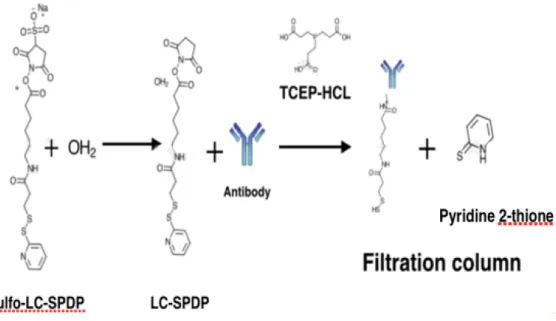
In the first method (Protocol 1) sulfo-LC-SPDP is initially attached to the primary amine groups on the antibodies, as it is represented in the Fig. 2.8., followed by immobilization of the entire complex to the gold surface [30].
Fig. 2.8. Schematics of Protocol 1. Sulfo-LC-SPDP when dissolved in water forms the molecule LC-SPDP
which will attach to the antibodies. TCEP-HCl (tris(2-carboxyethyl)phosphine hydrochloride) breaks dissulfide bonds and the thiol group of the cross-liker is free to bind to the surface. Any excess of sulfo-LC-SPDP and pyridine 2-thione molecules are eliminated after using a filtration column
2. Theoretical Background
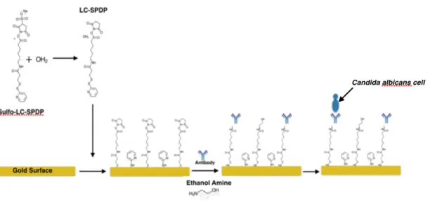
In the second method (Protocol 2) the linker molecule sulfo-LC-SPDP is first immobilized on the gold surface to form a uniform and aligned SAM due its long chain length that will permit the immobilization of the antibody to the gold surface. Afterwards, the surface is incubated with the antibodies in the presence of ethanolamine in order to form stable bonds between the antibodies and the linker. This long chain also helps to keep reactive succinimidyl group slightly away from the surface. Fig. 2.9. represents the process. [31]
16
Fig. 2.9. Schematics of Protocol 2. Gold surfaces are immersed into a solution of sulfo-LC-SPDP in water
at room temperature overnight. In water, sulfo-LC-SPDP divides in LC-SPDP and pyridine 2-thione and both attach to the gold surface. Pyridine 2-thione is, in this method, used as a spacer that avoids unspecific binding. The antibody is incubated to bind to LC-SPDP molecules and ethanolamine is used to wash the surface to eliminate excess of antibodies that did not attach.
2. Theoretical Background
2.4. Electrochemical Detection
2.4.1 Electrochemical sensors
Electrochemical sensors are widely used in the biosensor field, including biomedical applications, in order to investigate the electrochemical properties of materials, electrode processes and interfaces, allowing the analysis of reactions between different biological elements [21].
In the case of electrochemical immunosensors, antibodies or antigens are used as bio-recognition elements linked with electrochemical transducers. These sensors exhibit good sensitivity and selectivity and are completely label-free [8].
These devices are mainly based on the observation of current or potential changes due to interactions occurring at the sensor surface. During the catalysis of substrates by an enzyme conjugated to an antibody, electrochemical species such as electrons are consumed or generated and this produces an electrochemical signal (e.g., ions, and pH change or oxygen consumption) which can be measured by an electrochemical analyzer. There are three parameters that can be observed:
•
Current (amperometric): measures the current resulting from an electrochemical reaction at the electrode’s surface while keeping the potential constant.•
Potential (potentiometric): measures the electrochemical potential of a solution between two electrodes. The potential is then translated to a concentration of one of the analytes present in the solution.•
Impedance: measures changes in the resistance of a system. Impedimetric sensors are very sensitive to changes on the electrodes surfaces allowing the measurement and recognition of certain events.The detection of Candida albicans was made using electrochemical impedance spectroscopy (EIS). EIS based label-free biosensors are the most promising for detection of antigens due to simplicity in transducing the detection signal and high sensitivity in monitoring the changes occurring at the interface [21].
The basic components of an electrochemical sensor are a working (or sensing) electrode, a counter electrode and usually a reference electrode as well. The working electrode is the transduction element in the biochemical reaction where the desired reaction occurs, it is also known as the sensing or redox electrode. A reference electrode is kept at a distance from the
2. Theoretical Background
reaction site in order to maintain a known and stable potential. A counter-electrode establishes a connection to the electrolytic solution and carries the current flow away from the reference electrode. These electrodes should be conductive and chemically stable [33]. More details about the electrochemical sensors fabrication and design can be read in section 3.3.1.
2.4.2. Impedance definition
Electrical resistance is the ability of a circuit element to resist to the flow of electrical current and this characteristic is described by Ohm’s law that defines resistance in terms of the ratio between voltage, E, and current, I:
(2.1)
However, this relationship is limited to an ideal resistor and for that it has to follow the next requirements:
- it follows Ohm’s law at all current and voltage levels - its resistance value is independent of frequency
- AC current and voltage signals through a resistor are in phase with each other.
This simplification is not true for most of the electrical systems, as they exhibit much more complex behaviors. There is a more general concept of impedance, Z. Impedance is a measure of the ability of a circuit to resist the flow of electrical current. It is a complex electrical property consisting of a real part, the resistance, R, and an imaginary part, the reactance, X [34].
For measuring the electrochemical impedance, an alternating current (AC) potencial is applied to the electrochemical cell and then the current is measured through the cell. Considering that a sinusoidal potencial excitation is applied, the response to this potential is an AC current signal that can be analyzed as a sum of sinusoidal functions.
In order to ensure that the cell’s response is pseudo-linear, it is applied a small excitation signal. This means that the current response will be a sinusoid at the same frequency but shifted in phase [35, 36].
Electrochemical impedance is usually measured by applying a sinusoidal excitation signal with the form:
2. Theoretical Background
(2.2)
where Et is the potential at time t, E0 is the amplitude of the signal and w is the radial
frequency (rad/s), which can also be represented by:
(2.3)
where f is the frequency (Hz).
The response current, It, in a linear system, is shifted in phase (Φ) and has a different
amplitude than I0:
(2.4)
The ratio Et/It at a particular frequency is defined as the impedance (Z) of the electrochemical
cell. The system’s impedance is characterized by two values Z0 magnitude and φ phase shift:
After some mathematical manipulation the impedance can also be presented as a complex number:
(2.6)
There are two ways to represent the data of EIS: the Nyquist plot if the real part of the impedance (Z’) is plotted versus the imaginary part (-Z’’) or the Bode plot where the absolute impedance (|Z|) or the phase shift of the system is plotted versus the log of the frequencies range. Examples of both types of plots are represented in Fig. 2.10. and 2.11. and it is possible to see that Nyquist plot has one major shortcoming: the information about the frequency used to record a specific point is hidden [33, 36].
2. Theoretical Background
In EIS biosensors, the change in impedance after antigen binding to the bio-functionalized electrode surface that is modified with mono-layers and receptors (oligonucleotide, DNA, antibody, aptamer) is typically interpreted as detection. The electrode-cell system behavior is analyzed by fitting experimental impedance data to an equivalent electrical circuit model composed of common electrical elements such as resistors, capacitors, and inductors. EIS models usually consist of a number of elements in serial or/and in parallel combinations [35].
The Simplified Randles cell is one of most common electrochemical cell models and consists of a parallel combination of the constant phase elements (CPE) and charge transfer (Rct) and
Warburg (W) resistances connected in series with a solution resistance (Rsol). The equivalent
circuit for a simplified Randles Cell is shown in Fig. 2.12. Rsol depends on the concentration
of ions in the solution, temperature and the electrode geometry and it is connected in series as the signal goes through the electrolyte to reach the electrodes. The change in Rct upon antigen
binding is proportional to antigen concentration and thus provides quantitative information.
20
Fig. 2.11. Absolute impedance Bode plot from EIS [37] Fig. 2.10. Nyquist plot from EIS [37]
Fig. 2.12. Randles circuit with a constante phase element. It consists in the combination of a
constant phase element (CPE), the charge transfer resistance (Rct), the Warburg (W) resistance and
2. Theoretical Background
To understand the influence of coating the electrodes, the parallel combination of resistors and capacitors is used as they both contribute to the charge transfer reaction. When two conductive media such as electrode and electrolyte are separated by a non-conducting layer, a capacitor is formed and the capacitance between the metal electrode and ions in solution (CPE) can be modeled as a series combination of the surface modification capacitance and the double layer capacitance. These occurs, for example, when the electrode surface is covered by biomolecules and in the measurements there are often deviations from an ideal capacitor. This means that these measurements are imperfect semi-circles comparing with the result from an ideal capacitor. The capacitance CPE is often modeled by a constant phase element instead of a pure capacitance. To avoid the controversial discussion about the explanation of the non-ideal behavior of the system, very often the capacitor element is exchanged for an empirical value α. The charge transfer resistance (Rct) is a manifestation of two effects: (1) the energy
potential associated with the oxidation or reduction event at the electrode (i.e. the overpotential) along with (2) the energy barrier of the redox species reaching the electrode due to electrostatic repulsion or steric hinderance. Once the surface of the electrode is blocked by biomolecules it changes the charge transfer resistance Rct. The Warburg impedance (W),
only of physical significance in faradaic EIS, represents the delay arising from diffusion of the ions to the electrode surface. It is only appreciable at low frequencies, and results in an increase of the Warburg resistance and has a phase shift of −45° [37].
3. Materials and Methods
3.1. ELISA
• Phosphate buffered saline (PBS) (Sigma-Alridch);
• Antibodies (anti-candida albicans mouse monoclonal antibody (ab23368), anti-candida albicans rabbit policlonal antibody (ab53891) and anti-candida albicans rabbit polyclonal conjugated with HRP (ab20028)), were all purchased from Abcam;
• 96-wells Nunc plate (from Thermo Scientific); • Bovine serum albumin (BSA) (Sigma-Alridch);
• Kit TMB (3,3’,5,5’-tetramethylbenzine) and Stop Solution (Sigma-Alridch).
The 96-wells plate was coated with the capture antibody testing concentrationsof 0, 2, 6 and 10 μg/ml in PBS (pH 7.4). Then the plate was covered with a plastic adhesive and incubated overnight at 4°C.
The coating solution was removed and the plate was washed twice by filling the wells with 200 μl of PBS. The washing solutions were removed pipetting and the remaining drops were removed by patting the plate on a paper towel.
In order to block the remaining protein-binding sites in the coated wells, 200 μl of blocking buffer (1% BSA in PBS) were added, the plate was covered again with an plastic adhesive and incubated for 2h at room temperature.
100 μl of yeast broth with different dilutions of C. albicans (0, 13, 160, 1680 CFU/ml) were added to each well and incubated for 90 min at 37°C. After this incubation period, the samples were removed, the wells were washed twice by filling them with 200 μl PBS and 100 μl the HRP-secondary antibody diluted in blocking buffer was added to each well. The plate was thereafter covered with plastic adhesive and incubated for 2h at room temperature. The plate was washed four times with PBS to remove unbound detection antibody. Then TMB substrate solution was added to each well for color development (blue) and after 15 min of
3. Materials and Methods
incubation, the stop solution for TMB substrate was added (to form a yellow complex) and the optical density was read at 450 nm. A scheme of this method is presented in Fig. 2.4.
3.2. Surface Functionalization
• PBS (Sigma-Alridch);
• Anti-candida albicans rabbit policlonal antibody (ab53891); • Sulfo-LC-SPDP (Thermo-Scientific);
• TCEP-HCl (Sigma-Alridch); • Acetone (Sigma-Alridch); • Ethanol amine (Sigma-Alridch);
• VectaSpin Micro centrifuge tube (GE Healthcare Life Sciences). • CellTracker™ Blue CMAC (Life technologies)
For the functionalization of simple gold surfaces, small 8x8 mm2 gold squares were fabricated
by depositing on a silicon wafer of 10 nm of Cr followed by 100 nm of Au in Alcatel SCM 600 E-beam metal deposition system. These surfaces were used for the immobilization of antibodies and the success of the procedure was validated by microscopic visualization using yeast cells stained with a cell tracker fluorescent probe (CellTracker™ Blue CMAC).
3. Materials and Methods
3.2.1 Protocol 1
The first step in this protocol is the thiolation of the antibodies. For that, the stock antibody solution (4mg/ml) was diluted in PBS to a final concentration of 2 mg/ml and 100 μl of this were mixed with 100 μl of PBS and 50 μl of sulfo-LC-SPDP. The solution was incubated for 60 min at room temperature. In order to break dissulfide bonds, TCEP-HCl (5 mM in H2O)
was added and incubated for 15 min at room temperature [30].
Any excess of unbound antibodies, LC-SPDP and pyridine 2-thione was removed using a VectaSpin Micro centrifuge tube (GE Healthcare Life Sciences). In this way, any molecules smaller than antibodies bound to the linker were removed from the solution. 250 μl of antibody solution was introduced in the column and it was centrifuged for 1 min at 6000g; after this 500 μl PBS were added and the solution was again centrifuged during 1 min at 6000g. In order to elute the antibodies from the dialysis column and to keep the same initial concentration of antibodies (2 mg/ml) 200 μl of PBS were added and the column was centrifuged for 3 min at 2000g.
After the preparation of the antibody solution, the gold surfaces were washed in 50 mM KOH/25 mM H2O2 solution. This step allows the cleaning of the surfaces and render the
surfaces more hydrophilic [39].
The cleaning process was followed by the incubation of 5 μl of antibody with the linker solution at different concentrations (0, 5, 10, 50, 100 μg/ml) overnight at 4ºC. After washing with PBS (pH 7.4) 50 μl of stained yeast solution at different concentrations (0, 20, 2000, 2x10⁶, 2x10⁷ CFU/ml) was added and incubated for 30 minutes at room temperature. Finally
the gold surfaces were washed in PBS for 10 min.
3.2.2 Protocol 2
The cleaning process of the gold surfaces was the same as described above. Then the surfaces were immersed into a 0.5 mg/ml solution of sulfo-LC-SPDP in water at room temperature overnight. Next day, they were washed with water and acetone to remove any unbound sulfo-LC-SPDP followed by rinsing in MilliQ water and drying under nitrogen gas [31].
5 μl of antibody solution in PBS at various concentrations ((0, 5, 10, 50, 100 μg/ml) ) were incubated for 1.5h in a humidity chamber. Afterwards, the surfaces were washed with PBS (10 mM, pH 7.4) followed by washing with 1% ethanolamine solution in PBS for 10 min.
3. Materials and Methods
Similarly to Protocol 1, the surfaces were incubated with 50 μl of stained yeast solution at different concentrations (0, 20, 2000, 2x10⁶, 2x10⁷ CFU/ml) for 30 min at room temperature
and washed in PBS for 10 min.
Protocol 1 and 2 were tested and repeated 3 times each. Stained C. albicans were used for visualization of antibody-gold binding and 3 pictures for each sample were analyzed. After all the experiments, the cells in each picture were counted manually in order to calculate the average number of cells and the respective standard deviation.
3.3. Electrochemical Detection
3.3.1. Electrodes and holder fabrication
The design and fabrication of the membrane based electrodes used in this project are described in [38]. Polycarbonate (PC) membranes with 5 μm of pore size (Millipore, Darmstadt, Germany) were used. Each membrane is composed of 1 reference electrode, 1 counter electrode and 11 working electrodes, as presented in Fig. 3.2. These electrodes are fabricated with deposition of a 200 nm of gold layer using E-beam evaporation in an Alcatel SCM 600 E-beam metal deposition system.
3. Materials and Methods
For the deposition of gold to produce the electrodes, a 0.5 mm thick poly(methyl methacrylate) (PMMA) shadow mask and a second PMMA substrate with alignment marks were used. The placement of the membranes and the alignment of all the setup is made manually, respecting the alignment marks as illustrated in Fig. 3.3.
The holder for the electrochemical measurements is composed by two PMMA structures: one on the bottom and the other with an O-ring fitted on top. The membranes are clamped between these two PMMA parts. A custom made Polydimethylsiloxane (PDMS) O-ring is fitted in the top of the PMMA substrate. A custom designed PCB placed on top of the PMMA holder allows the electrical connections. Fig. 3.4. shows the holder used. The holder used for the several experiments was produced by my supervisor Dr. Dorota Kwasny.
26
Fig. 3.4. Holder for electrochemical experiments [38]
Fig. 3.3. Setup for fabrication of the membrane electrodes with a thin PMMA substrate with alignment marks for
the membranes on the bottom; in the middle membrane where the metal is going to be deposited and, on top, the shadow mask fabricated in thin PMMA using laser ablation [38].
![Fig. 1.1. C. albicans colonies on potato dextrose agar medium [4]](https://thumb-eu.123doks.com/thumbv2/123dok_br/19186561.947841/21.892.248.618.654.901/fig-c-albicans-colonies-potato-dextrose-agar-medium.webp)
![Fig. 2.1. Antibody structure (adapted from [26])](https://thumb-eu.123doks.com/thumbv2/123dok_br/19186561.947841/29.892.234.622.314.616/fig-antibody-structure-adapted-from.webp)
![Fig. 2.4. Schematics of Indirect ELISA procedure, adapted from [29]](https://thumb-eu.123doks.com/thumbv2/123dok_br/19186561.947841/32.892.166.723.850.1076/fig-schematics-indirect-elisa-procedure-adapted.webp)
![Fig. 2.11. Absolute impedance Bode plot from EIS [37]](https://thumb-eu.123doks.com/thumbv2/123dok_br/19186561.947841/40.892.112.842.148.380/fig-absolute-impedance-bode-plot-eis.webp)
