UNIVERSIDADE DE LISBOA
FACULDADE DE MEDICINA VETERINÁRIA
CLINICAL RELEVANCE OF SERUM CYTOTOXIC T-LYMPHOCYTE ASSOCIATED PROTEIN
4 (CTLA-4) AND ITS CORRELATION WITH THE PRO-INFLAMMATORY CYTOKINES
INTERLEUKIN 6 (IL-6) AND TUMOR NECROSIS FACTOR ALPHA (TNF-α) IN FELINE
MAMMARY CARCINOMA
ANA CATARINA FERNANDES URBANO
ORIENTADOR:
Doutor Fernando António da Costa Ferreira COORIENTADOR:
Doutor Jorge Manuel Jesus Correia
i
UNIVERSIDADE DE LISBOA
FACULDADE DE MEDICINA VETERINÁRIA
CLINICAL RELEVANCE OF SERUM CYTOTOXIC T-LYMPHOCYTE ASSOCIATED
PROTEIN 4 (CTLA-4) AND CORRELATION WITH THE PRO-INFLAMMATORY CYTOKINES
INTERLEUKIN 6 (IL-6) AND TUMOR NECROSIS FACTOR ALPHA (TNF-α) IN FELINE
MAMMARY CARCINOMA
ANA CATARINA FERNANDES URBANO
DISSERTAÇÃO DE MESTRADO INTEGRADO EM MEDICINA VETERINÁRIA
JÚRI
PRESIDENTE:
Doutora Maria da Conceição da Cunha e Vasconcelos Peleteiro
VOGAIS:
Doutor Fernando António da Costa Ferreira Doutora Solange Judite Roque Coelho Alves Gil Neves
ORIENTADOR:
Doutor Fernando António da Costa Ferreira COORIENTADOR:
Doutor Jorge Manuel Jesus Correia
ii
DECLARAÇÃO RELATIVA ÀS CONDIÇÕES DE REPRODUÇÃO DA TESE OU DISSERTAÇÃO
Nome: Ana Catarina Fernandes Urbano Título da Tese ou
Dissertação:
Clinical relevance of serum cytotoxic t-lymphocyte associated protein 4 (ctla-4) and its correlation with the pro-inflammatory cytokines interleukin 6 (IL-6) and tumor necrosis factor alpha (TNF-α) in feline mammary carcinoma Ano de conclusão (indicar o da data da realização das provas
públicas): 2019
Designação do curso de
Mestrado ou de Doutoramento: Mestrado Integrado em Medicina Veterinária Área científica em que melhor se enquadra (assinale uma):
Clínica Produção Animal e Segurança Alimentar
Morfologia e Função
Sanidade Animal
Declaro sobre compromisso de honra que a tese ou dissertação agora entregue corresponde à que foi aprovada pelo júri constituído pela Faculdade de Medicina Veterinária da ULISBOA.
Declaro que concedo à Faculdade de Medicina Veterinária e aos seus agentes uma licença não-exclusiva para arquivar e tornar acessível, nomeadamente através do seu repositório institucional, nas condições abaixo indicadas, a minha tese ou dissertação, no todo ou em parte, em suporte digital.
Declaro que autorizo a Faculdade de Medicina Veterinária a arquivar mais de uma cópia da tese ou dissertação e a, sem alterar o seu conteúdo, converter o documento entregue, para qualquer formato de ficheiro, meio ou suporte, para efeitos de preservação e acesso.
Retenho todos os direitos de autor relativos à tese ou dissertação, e o direito de a usar em trabalhos futuros (como artigos ou livros).
Concordo que a minha tese ou dissertação seja colocada no repositório da Faculdade de Medicina Veterinária com o seguinte estatuto (assinale um):
1. Disponibilização imediata do conjunto do trabalho para acesso mundial;
2. Disponibilização do conjunto do trabalho para acesso exclusivo na Faculdade de Medicina Veterinária durante o período de 6 meses, 12 meses, sendo que após o tempo assinalado autorizo o acesso mundial*;
* Indique o motivo do embargo (OBRIGATÓRIO)
Os resultados serão alvo de publicação científica
Nos exemplares das dissertações de mestrado ou teses de doutoramento entregues para a prestação de provas na Universidade e dos quais é obrigatoriamente enviado um exemplar para depósito na Biblioteca da Faculdade de Medicina Veterinária da Universidade de Lisboa deve constar uma das seguintes declarações (incluir apenas uma das três):
1. É AUTORIZADA A REPRODUÇÃO PARCIAL DESTA TESE/TRABALHO (indicar, caso tal seja necessário, nº máximo de páginas, ilustrações, gráficos, etc.) APENAS PARA EFEITOS DE INVESTIGAÇÃO, MEDIANTE DECLARAÇÃO ESCRITA DO INTERESSADO, QUE A TAL SE COMPROMETE.
Faculdade de Medicina Veterinária da Universidade de Lisboa, 22 de Novembro de 2019
iii
Agradecimentos
Ao Professor Fernando Ferreira, por me ter proporcionado este estágio, por ter confiado nas minhas capacidades e pela partilha do entusiamo pela investigação.
À Doutora Maria João Soares, porque esta tese também lhe pertence. Pelo enorme esforço investido na criação do biobanco sem o qual este estudo não teria sido possível, e pela amabilidade, disponibilidade e ajuda prestada.
Às colegas do grupo de trabalho de oncologia, pela partilha do gabinete, pelas trocas de informação, ideias e boa disposição.
Ao Professor Jorge Correia, pela boa disposição, disponibilidade e entusiasmo na partilha de conhecimentos.
À Professora Manuela Oliveira e aos colegas do grupo de trabalho de microbiologia, pelo uso do leitor de placas, e mais concretamente à Dra. Eva pela extrema amabilidade e paciência.
A todos os docentes, médicos e enfermeiros que contribuíram para a minha formação ao longo da licenciatura e mestrado, por todos os conhecimentos, dedicação e contributo para o meu crescimento pessoal e educacional.
Às minhas colegas de curso, Sofia Andrade, Marion Carreira, Cátia Rojão, Raquel Santos e Dídia Gomes, que me acompanharam neste percurso, pela camaradagem, pela energia e pelo grande apoio nos momentos mais difíceis. Amizade é pouco para descrever o que sinto por vocês.
Ao Eduardo, por ter começado este percurso comigo e acreditar sempre.
À minha mãe, por todo amor e dedicação e ao meu pai, por ter sido quem foi, a minha enciclopédia para a vida, parte integral de mim.
À minha família, sempre presente; aos meus irmãos Paulo e João, que apesar de longe está sempre perto do meu coração; à tia Clita, à tia Lena e ao tio Zé, pelo carinho e palavras amigas.
Ao Kristof, por me ter mostrado um mundo maior que o meu e por me encorajar todos os dias a ir mais longe.
iv
RELEVÂNCIA CLÍNICA DA PROTEÍNA 4 ASSOCIADA AO LINFÓCITO T CITOTÓXICO 4 (CTLA-4) E SUA CORRELAÇÃO COM AS CITOQUINAS PRÓ-INFLAMATÓRIAS INTERLEUCINA 6 (IL-6) E FACTOR DE
NECROSE TUMORAL ALFA (TNF-α) NO CARCINOMA MAMÁRIO FELINO
Resumo
A associação entre a expressão da CTLA-4 e o prognóstico no cancro tem sido amplamente investigada, valorizando o papel da inflamação e do microambiente tumoral (TME), do qual mediadores inflamatórios como as citoquinas são uma importante componente não celular. Até à data, não existem estudos sobre reguladores de checkpoint imunológico em gatos com carcinoma mamário, nem foram avaliados perfis de citoquinas. Assim, foram investigados pela primeira vez, os perfis séricos da CTLA-4 e das citoquinas pró-inflamatórias IL-6 e TNF-α em 57 gatas com carcinoma mamário e verificada a existência de associações entre os níveis séricos da CTLA-4 e das referidas citoquinas. Os resultados obtidos demonstram que os níveis de CTLA-4 estão aumentados no soro das gatas com carcinoma mamário, quando comparadas com animais saudáveis (P=0.022). Foi também demonstrada uma correlação forte com os níveis séricos do TNF-α (R=0.88, P<0.001) e da IL-6 (R=0.72, P<0.001), reforçando o papel imunomodulatório deste regulador. Adicionalmente foi encontrada uma associação significativa entre os níveis séricos elevados da CTLA-4, e várias características clinicopatológicas menos agressivas: tumores mais pequenos (P<0.001), estadiamento precoce, (P=0.002), ausência de necrose tumoral (P<0.001), sem envolvimento dos linfonodos (P=0.007), sem invasão linfática (P=0.006), com positividade para os receptores hormonais (P=0.007), subtipo não-TN (P=0.041), subtipo não-basal (P<0.001), e baixo índice Ki67 (P=0.001). Os resultados obtidos ainda revelaram uma associação com subtipos específicos de cancro da mama, nomeadamente o HER-2 positivo com sobre-expressão da CTLA-4 (P<0.001) e do TNF-α (P=0.00CTLA-4) e o luminal A com sobre-expressão da IL-6 (P=0.020). Não foi possível confirmar a associação entre os níveis séricos da CTLA-4 e das citoquinas e o tempo de sobrevivência, devido ao tamanho reduzido da amostra. No entanto, os resultados obtidos sugerem um efeito protetor dependente da concentração da CTLA-4 e IL-6 séricos, como evidenciado pelos tempos medianos de sobrevivência mais altos nos grupos CTLA-4high (28 vs 22 meses para o grupo CTLA-4low) e IL-6high (28 vs 19 meses para o grupo IL-6low). Em contraste, o TNF-α parece ser um fator de prognóstico negativo, como sugere o tempo mediano de sobrevivência mais baixo no grupo TNF-αhigh (16.5 vs 23.5 meses para o grupo TNF-αlow). Permanece a questão de como o CTLA-4 influencia ou é influenciado pelas citoquinas pró-inflamatórias. A avaliação da expressão tumoral da CTLA-4, dos subtipos de linfócitos T, e dos perfis de macrófagos associados ao tumor e células supressoras da linha mieloide no microambiente tumoral, são aspetos importantes a avaliar em estudos futuros. Palavras chave: carcinoma mamário felino, proteína 2 associada ao linfócito t citotóxico, citoquinas pró-inflamatórias, biomarcadores séricos, oncologia comparada
v
CLINICAL RELEVANCE OF SERUM CYTOTOXIC T-LYMPHOCYTE ASSOCIATED PROTEIN 4 (CTLA-4) AND CORRELATION WITH THE PRO-INFLAMMATORY CYTOKINES INTERLEUKIN 6 (IL-6) AND TUMOR
NECROSIS FACTOR ALPHA (TNF-Α) IN FELINE MAMMARY CARCINOMA
Abstract
The association between CTLA-4 expression and cancer prognosis has been extensively investigated in recent years, pointing to the link with inflammation, and highlighting the role of the tumor microenvironment (TME), of which inflammatory mediators like cytokines are an important non-cellular component. To the best of our knowledge, no studies on immune checkpoint regulators had been conducted on cats with mammary carcinoma before, nor had cytokine profiles been previously assessed. Thus, we investigated the serum profiles of CTLA-4 and pro-inflammatory cytokines IL-6 and TNF-α in 57 female cats with mammary carcinoma and checked for associations between CTLA-4 and cytokine serum levels. Our results clearly demonstrate that serum CTLA-4 levels are increased in cats with mammary carcinoma when compared to healthy animals (P=0.022). Furthermore, we show a strong positive correlation with TNF-α (R=0.88, P<0.001) and IL-6 levels (R=0.72, P<0.001), advancing the concept of an immunomodulatory role for this regulator in breast cancer pathogenesis. We also show a statistically significant association between higher levels of serum CTLA-4 and less aggressive clinicopathological features: smaller tumors (P<0.001), lower stage (P=0.002), absence of necrosis (P<0.001), no lymph node involvement (P=0.007), no lymphatic vessel invasion (P=0.006), positive hormone receptor status (P=0.007), non-TN status (P=0.041), non-basal status (P<0.001) and low Ki67 index (P=0.001). Our findings further expand this concept by indicating an association with specific breast cancer subtypes, namely, HER-2 positive with CTLA-4 (P<0.001) and TNF-α (P=0.004) and luminal A-like with IL-6 (P=0.020). We could not confirm an association between serum CTLA-4 and cytokines levels and survival due to the small sample size. Nevertheless, our findings suggest a potentially concentration-dependent protective role for serum CTLA-4 and IL-6, as evidenced by higher median survival times in the CTLA-4high (28 vs 22 months for the CTLA-4low group) and IL-6high (28 vs 19 months for the IL-6low group) groups. Conversely, TNF-α seems to be a negative prognostic factor, as shown by the lower median survival in the TNF-αhigh group (16.5 vs 23.5 months for the TNF-αlow group). An intriguing question that remains is how serum CTLA-4 influences or is influenced by the pro-inflammatory cytokines. Assessment of CTLA-4 tumor expression, T-lymphocyte subtypes, and tumor associated macrophages and myeloid derived suppressor cell profiles in the microenvironment, are important features to evaluate in future studies.
Keywords: feline mammary carcinoma, cytotoxic t-lymphocyte associated protein 4, pro-inflammatory cytokines, serum biomarkers, comparative oncology
vi
Table of Contents
List of Figures ... viii
List of Tables ... x
List of Abbreviations ... xi
1. Introduction ... 1
1.1. Feline mammary carcinoma as a model for human breast cancer ... 1
1.2. Inflammation and tumor development ... 3
1.3. CTLA-4: a key immune checkpoint regulator ... 6
1.4. Cellular components of the TME: the role of Tregs, TAMs and MDSCs ... 11
1.5. Soluble mediators of cancer related inflammation: TNF-α and IL-6 ... 15
2. Retrospective Study ... 17
2.1. Objective ... 17
2.2. Materials and Methods ... 18
2.2.1. Study Population ... 18
2.2.2. Measurement of serum CTLA-4 and cytokine levels ... 18
2.2.3. Statistical Analysis ... 20
2.3. Results ... 21
2.3.1. Serum CTLA-4 levels ... 21
2.3.2. Prognostic value of serum CTLA-4 for overall and disease-free survival ... 23
2.3.3. Serum TNF-α and IL-6 levels ... 24
2.3.4. Prognostic value of TNF-α and IL-6 for overall and disease-free survival ... 27
2.3.5. Correlation between CTLA-4 and serum TNF-α and IL-6 levels ... 29
2.4. Discussion ... 30
2.4.1. Serum CTLA-4 is increased in cats with mammary carcinoma and correlates with less aggressive clinicopathological features and positive HER-2 status ... 30
2.4.2. Higher serum CTLA-4 tends to correlate with improved survival in cats with mammary carcinoma ... 31
2.4.3. Serum TNF-α levels are increased in cats with mammary carcinoma and correlate with less aggressive clinicopathological features and positive HER-2 status ... 32
2.4.4. Higher serum TNF-α tends to correlate with worse survival in cats with mammary carcinoma…… ... 33
2.4.5. Serum IL-6 levels are increased in cats with mammary carcinoma and correlate with lymph-node negative status and a luminal A-like subtype ... 33
2.4.6. Higher serum IL-6 tends to correlate with improved survival in cats with mammary carcinoma…… ... 34
vii
2.4.7. Serum CTLA-4 levels correlate with TNF-α and IL-6 levels in cats with mammary
carcinoma……. ... 35
2.5. Conclusions ... 35
3. References ... 37
ANNEX I – Results of the Statistical Analysis ... 46
viii
List of Figures
Figure 1. Molecular pathways connecting inflammation and tumor development ... 4 Figure 2. T-lymphocyte activation and inhibition by the immunoglobulin superfamily receptors cluster of differentiation 28 (CD28) and cytotoxic T-lymphocyte associated protein 4 (CTLA-4) ... 6 Figure 3. Alignment of feline (Fe) CTLA-4 amino acid sequence with homologues from bovine (Bo), human (Hu) and mouse (Mu) species ... 7 Figure 4. Generation of full-length, soluble and ligand-independent cytotoxic T-lymphocyte associated protein 4 (CTLA-4) mRNA ... 8 Figure 5. Possible mechanisms of immune regulation by soluble cytotoxic T-lymphocyte associated protein 4 (sCTLA-4). ... 9 Figure 6. Functions of regulatory T-lymphocytes (Tregs) in the tumor microenvironment .. 12 Figure 7. Contrasting functions of immune cells in the tumor microenvironment ... 14 Figure 8. Signal transduction pathways and major biological responses of tumor necrosis factor alpha (TNF-α) and interleukin 6 (IL-6) ... 17 Figure 9. Sandwich enzyme-linked immunosorbent assay (ELISA) technique ... 20 Figure 10. Box plot analysis of serum cytotoxic T-lymphocyte associated protein 4 (CTLA-4) levels in healthy cats and cats with mammary carcinoma ... 21
Figure 11. Box plot analysis of serum cytotoxic T-lymphocyte associated protein 4 (CTLA-4) levels and their association with clinicopathological parameters ... 22 Figure 12. Box plot analysis of serum cytotoxic T-lymphocyte associated protein 4 (CTLA-4) levels and their association with immunohistochemical parameters ... 23 Figure 13. Kaplan–Meier survival curve for serum cytotoxic T-lymphocyte associated protein 4 (CTLA-4) high and low groups ... 24 Figure 14. Box plot analysis of serum tumor necrosis factor alpha (TNF-α) levels in healthy cats and cats with mammary carcinoma ... 25 Figure 15. Box plot analysis of serum tumor necrosis factor alpha (TNF-α) levels and their association with clinicopathological parameters ... 25 Figure 16. Box plot analysis of serum tumor necrosis factor alpha (TNF-α) levels and their association with immunohistochemical parameters ... 26 Figure 17. Box plot analysis of serum interleukin 6 (IL-6) levels in healthy cats and cats with mammary carcinoma ... 26 Figure 18. Box plot analysis of serum interleukin 6 (IL-6) levels and their association with clinicopathological criteria ... 27 Figure 19. Box plot analysis of serum interleukin 6 (IL-6) levels and their association with immunohistochemical parameters ... 27
ix
Figure 20. Receiver operator curve (ROC) analysis of sensitivity versus specificity for tumor necrosis factor alpha (TNF-α) and interleukin 6 (IL-6) ... 28 Figure 21. Kaplan–Meier survival curve for tumor necrosis factor alpha (TNF-α) and interleukin 6 (IL-6) high and low groups ... 28 Figure 22. Pearson correlation of sCTLA-4 and serum IL-6 and TNF-α levels ... 30
x
List of Tables
Table 1. Clinicopathological characteristics of female cats with mammary carcinoma ... 18 Table 2. Median survival times for cytotoxic T-lymphocyte associated protein 4 (CTLA-4) high and low groups ... 24 Table 3. Median survival times for tumor necrosis factor alpha (TNF-α) high and low groups ... 29
xi
List of Abbreviations
AKT – Protein kinase B APC – Antigen presenting cell Arg1 – Arginase 1
BRCA – Breast cancer susceptibility gene CCL22 – C-C motif chemokine 22
CD – Cluster of diferentiation CK5/6 – Cytokeratin 5/6 COX-2 – Cyclooxygenase 2
cSMAC – Central supramolecular activation cluster CTLA-4 – Cytotoxic T-lymphocyte associated protein 4 DC – Dendritic cell
DFS – Disease free survival EGF – Epidermal growth factor ER – Estrogen receptor
gp130 – Glycoprotein 130
HER-2 – Epidermal growth factor receptor 2 HRP – Horse radish peroxidase
IDO – Indoleamine 2,3-dioxygenase IFN – Interferon
IL – Interleukin
IL-6Rα – Interleukin 6 receptor alpha subunit iNOS – Inducible nitric oxide synthase JAK – Janus kinase
mCTLA-4 – Membrane bound cytotoxic T-lymphocyte associated protein 4 MDSC – Myeloid derived suppressor cell
NFκB – Nuclear factor kappa B NK – Natural killer cell
NO – Nitric oxide OS – Overall survival
PDCD4 – Programmed cell death protein 4 PIP3 – Phosphatidylinisitol (3,4,5)-trisphosphate PR – Progesterone receptor
PTEN – Phosphatase and tensin homolog RONS – Reactive oxygen and nitrogen species ROS – Reactive oxygen species
xii
sCTLA-4 – Soluble cytotoxic T-lymphocyte associated protein 4 SOCS3 – Suppressor of cytokine signaling 3
STAT – Signal transducer and activator of transcription TAM – Tumor associated macrophage
TCR – T-cell receptor
TGF – Transforming growth factor Th1 – Type 1 T-helper cells Th17 – Type 17 T-helper cells Th2 – Type 2 T-helper cells
TIL – Tumor infiltrating lymphocytes TLR – Toll-like receptor
TMB – 3,3’,5,5’-Tetramethylbenzidine TME – Tumor microenvironment TN – Triple negative
TNBC – Triple negative breast cancer TNF – Tumor necrosis factor
TNFR – Tumor necrosis factor receptor TP53 – Tumor protein p53
TRADD – Tumor necrosis factor receptor type-1 associated protein TRAF – Tumor necrosis factor associated factor
Treg – Regulatory T-lymphocyte
1
1. Introduction
1.1. Feline mammary carcinoma as a model for human breast cancer
Cats have proved to be valuable models for various non-neoplastic (Narfström et al. 2013) and neoplastic diseases (De Maria et al. 2005). Compared with traditional mouse models, they demonstrate more features in common with humans: they share many anatomical and physical similarities, have longer life spans, greater size, a genetically more heterogeneous background, and are exposed to the same environmental risk factors (Cannon 2015). They continue to grow in popularity as pets and experience increasingly higher levels of medical surveillance, making them especially useful models for the study of spontaneous disease and better candidates for use in clinical trials (De Vico and Maiolino 2008).
Several spontaneous feline tumors are currently considered relevant for human cancer studies, including injection-site sarcoma, oral squamous cell carcinoma, lymphoma and mammary carcinoma (De Vico and Maiolino 2008; Cannon 2015; Thomas 2015). Mammary gland tumors are frequently reported as the third most common tumor type affecting female cats and the most common type of cancer in women. Recently they were identified as the most common type in a 10-year retrospective study of feline tumors conducted in Portugal (Garcês et al. 2019). Feline mammary carcinomas are the most representative lesion within this group (Zappulli et al. 2015). Mean age of development is 10-11 years, similar to that described for human breast cancer after adjusting for age, and all breeds may be affected, although evidence points to a hereditary predisposition in the Siamese (Zappulli et al. 2005; Cannon 2015). Similarly, a hereditary predisposition has been observed in women, often associated with mutations at the BRCA1 and BRCA2 genes. Women carrying these mutations are significantly younger at time of diagnosis. Interestingly, the mean age of presentation in Siamese cats is also lower than in other breeds (Zappulli et al. 2005; Cannon 2015).
Feline mammary carcinoma occurs either as single or multiple nodules frequently showing ulceration associated with extensive tumoral necrosis, probably owing to their generally advanced stage at time of diagnosis (Zappulli et al. 2005). They are highly infiltrative and metastasizing, showing a metastatic pattern similar to that described in women (regional lymph nodes, lungs, pleura and liver) (Zappulli et al. 2005).
The influence of steroid hormones on the development of breast cancer is well-known in women and there is evidence of a similar involvement in cats. Intact females have a significantly higher risk of developing disease, as do those exposed to regular and prolonged administration of progestagens (Zappulli et al. 2005). Most cats however tend to have estrogen
2
growth factor receptor 2 (HER-2) overexpression is also well documented in human breast cancer often associated with poor prognosis (De Maria et al. 2005). Increased HER-2 expression has been documented in a significant proportion of feline mammary carcinomas, although there is variation among studies as to the degree of overexpression (De Maria et al. 2005; Santos et al. 2013; Cannon 2015). Regardless, taken together with the high level of homology between the feline HER-2 gene transcript and the human sequence this qualifies cats as suitable models (De Maria et al. 2005).
Additional molecular analyses distinguish several other subtypes of breast cancer: luminal A, expressing the luminal epithelial markers (CK7, CK8, CK18, CK19) and with high expression of ER markers and lower expression of proliferation markers; luminal B, expressing the luminal epithelial markers, with lower expression of ER markers and higher expression of proliferation markers; basal-like, negative for hormone receptors (ER, PR, HER-2) and expressing basal markers (CK5, CK6, CK14, CK17, SMA, calponin, vimentin, and p63); HER-2 positive tumors overexpressing the HER-HER-2 receptor; and normal-like, negative to all markers (Goldschmidt et al. 2016). Similar subtypes were identified in cats in a 3-year follow-up study conducted at this institution (Soares et al. 2016b) which identified a higher prevalence of luminal B and triple-negative subtypes, associated with a worse prognosis. Triple-negative breast cancer (TNBC) is also associated with poor prognosis in humans and is especially challenging to treat because of the lack of specific targets. The findings of Soares and colleagues suggest that the cat might also be a suitable model for this highly aggressive subtype.
Studies like the ones conducted by De Maria (2005) and Soares (2016b) reflect a rising interest in the use of immunohistochemical prognostic markers in veterinary oncology. Tissue-based biomarkers, however, often require highly invasive procedures to obtain and may be difficult to include in routine clinical practice. These limitations make biomarkers from liquid biopsies especially valuable. Samples can be collected all through the disease course or before and after specific treatments, to monitor disease progression and predict patient response to therapy (Chakrabarti et al. 2019). Several markers have emerged over the years, some of which have been extensively studied. Serum HER-2 is one such. It can be used to evaluate HER-2 status (i.e. diagnosis), and several studies indicate a role for predicting prognosis and response to treatment (Lüftner et al. 2003). HER-2 serum levels have also been investigated in cats and showed significant association with HER-2 in tissue samples (Soares et al. 2016a) making cats promissing candidates for use in the study of novel serum markers.
The use of cats as a model for human cancers, however, also presents some challenges. Most cat owners are still unwilling to consider enrolling their animals in clinical trials and their use as pre-clinical models for assessment of new drugs is hampered by differences in drug metabolism and pharmacokinetics (Cannon 2015). Cats are known, for
3
example, to have reduced glucuronidation capacity in comparison to humans, and several chemotherapeutic drugs, including cisplatin, 5-fluorouracil, doxorubicin and ifosfamide have
differing toxicities in cats (reviewed in Cannon 2015). Clinical follow-up data is also not always
available and some predictors of clinical behavior, namely overall survival (OS) and disease-free survival (DFS; time from surgery to the development of recurrences and/or metastases) rely on information obtained retrospectively through methods that are often imprecise (Zappulli et al. 2015).
When considering the benefits of cats as models in biomedical research it is also important to consider the ethical costs of involving these animals in experiments. The use of novel therapies in the treatment of spontaneous diseases in companion animals might seem more ethically acceptable than in experimentally induced pathologies in animal models. However, the animals enrolled in experimental trials should still be considered as veterinary patients and their management should involve a close partnership between the owners, veterinary practitioners and the veterinarians in the research institutions. To guarantee
reliability of results, increase enrolment and, above all, ensure the patient’s well-being, an
approach based on information, commitment, responsibility and care must be taken (De Vico and Maiolino 2008).
Despite these challenges, feline models remain an exciting prospect in the field of comparative oncology and contribute a diverse range of opportunities to the “One Health” concept, which capitalizes on the integration of biomedical research efforts to achieve better
health care for humans and animals. Investigating cancer in cats may additionally generate
new insights into aspects of tumor biology that are less accessible in other species (Thomas 2015), such as the role of viruses in malignant transformation and the relationship between inflammation and tumor development.
1.2. Inflammation and tumor development
Inflammation is a well-established risk factor for several cancers. It can contribute to tumor initiation by inducing genetic and epigenetic changes such as point mutations, DNA methylation, and post translational modifications of genes that regulate critical pathways related to cell homeostasis (Hussain and Harris 2007). Injection site sarcoma (ISS), a well-recognized phenomenon in cats particularly associated with vaccine administration, is thought to result from chronic inflammation due to an inappropriate and excessive response to injection or trauma, which causes proliferation and malignant transformation of fibroblasts (Cannon 2015). In Kaposi’s sarcoma virus infections, inflammation is also essential for tumor development. Other types of alterations concurring to tumor progression, such as activation of oncogenes and inactivation of tumor suppressors may also trigger the inflammatory cascade,
4
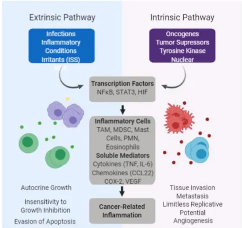
leading to changes in the cellular microenvironment which favor selection and expansion of cells with growth or survival advantages (called the “intrinsic” pathway, Figure 1). Examples of these adaptive changes include increased expression of antioxidant enzymes, matrix metalloproteinases and growth factor receptors, increased anaerobic respiration and de novo synthesis of angiogenic factors (Federico et al. 2007). During tumor promotion, these initiated cells produce inflammatory mediators such as reactive oxygen and nitrogen species (RONS), cytokines, prostaglandins, growth factors and specific microRNAs (Schetter et al. 2009). These, in turn, induce cell proliferation and recruit inflammatory cells, increasing the production of RONS and leading to further DNA damage and reduced DNA repair, perpetuating the cycle (Coussens and Werb 2010).
Figure 1. Molecular pathways connecting inflammation and tumor development. ISS –
Injection site sarcoma; ROS – Reactive oxygen species; NFκB – Nuclear factor kappa B; STAT – Signal transducers activator of transcription; HIF – Hypoxia inducible factor; TAM – Tumor associated macrophages; MDSC – Myeloid-derived suppressor cells; PMN – Polymorphic nuclear cells; COX-2 – cyclooxygenase 2; VEGF - vascular endothelial growth factor. Redrawn from the original in Denardo, 2017. Created with BioRender.
Key players in cancer related inflammation include transcription factors such as nuclear factor κB (NFκB), signal transducer activator of transcription (STAT)-3, and primary inflammatory cytokines such as interleukin (IL)-1β, IL-6, and tumor necrosis factor (TNF)-α
5
(Del Prete et al. 2011). NFκB induces the expression of inflammatory cytokines, such as IL-6, adhesion molecules, cyclooxygenase-2 (COX-2), and inducible nitric oxide synthase (iNOS), generating a strong pro-inflammatory microenvironment. It also promotes cell survival and proliferation through the activation of genes regulating cell cycle progression and apoptosis and other pro-tumorigenic changes, including stimulation of angiogenesis by activating vascular endothelial growth factor (VEGF) (Schetter et al. 2009). NFκB activation can follow sensing of microbes or tissue damage by the toll-like receptor (TLR)-MyD88 pathway, inflammatory cytokines TNF-α and IL-1β or can be the result of cell-autonomous genetic alterations in cancer cells (Del Prete et al. 2011). STAT3 is a critical regulator of cytokine, chemokine and growth factor expression. It’s persistent activation in tumor cells, either through increased production of positive effectors such as IL-6 or decreased expression of negative regulators such as suppressor of cytokine signaling 3 (SOCS3), in turn activates STAT3 in stromal cells, inducing and maintaining an inflammatory microenvironment (Chang et al. 2015). Activated STAT3 also increases tumor cell proliferation, survival and invasion, while suppressing anti-tumor immunity by promoting pro-tumorigenic pathways like NFκB. Both transcription factors have been shown to play a role in human TNBC, a useful clinical example of the connection between oncogenes and inflammation. In TNBC somatic mutations leading to the inactivation of the tumor suppressor genes tumor protein p53 (TP53) and phosphatase and tensin homolog (PTEN) were implicated in the SOCS3-mediated activation of an IL-6/STAT3/NF κB inflammatory loop (Kim et al. 2014). Mutations in TP53 and PTEN have also been reported in feline mammary carcinoma (Mayr et al. 2000; Ressel et al. 2009; Adega et al. 2016) and considering the high level of sequence homology between the human and feline TP53 and PTEN genes it is reasonable to assume that the mechanisms of tumorigenesis may be similar in the two species. In fact, aberrant activation of the phosphatidylinositol 3,4,5-triphosphate (PIP3)/protein kinase B (AKT)/PTEN pathway, another pathway widely implicated as a driver of tumor development and progression in human breast cancer, was recently shown to be correlated with tumor malignancy, histological differentiation and clinical recurrence in feline mammary carcinoma (Maniscalco et al. 2012).
Despite this overwhelming evidence that inflammation orchestrates a tumor-promoting microenvironment that is intimately linked to tumorigenesis, anti-tumor immunity can also develop to protect the host during tumor development. Data generated in several mouse models which shows that cytokines and immune cells that promote inflammation are potentially bi-functional displaying both tumor-promoting and tumor-suppressive capabilities (reviewed in Chow et al. 2012) supports this notion. Recent developments in immune checkpoint blockade therapy also highlight how important it is to understand the complexity of the immune and
6
inflammatory systems in the development of cancer and how one’s own host responses can help or hinder progression of the disease.
1.3. CTLA-4: a key immune checkpoint regulator
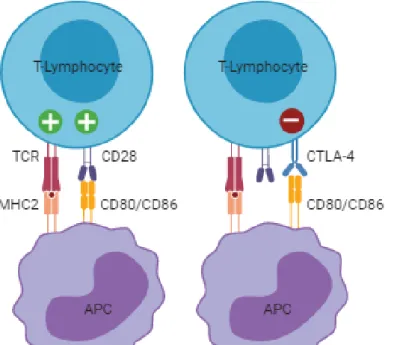
The inflammatory microenvironment surrounding breast cancer cells consists of immune cell infiltrates, cytokines and immune checkpoint molecules that can block anti-tumor immunity (Emens 2012; Yu et al. 2015). Cytotoxic T-lymphocyte associated protein 4 (CTLA-4, CD152), an adhesion molecule from the immunoglobulin superfamily localized on band q33 of human chromosome 2 and on feline chromosome C1, is one of these immune checkpoint molecules. It’s expressed exclusively on lymphocytes and shares a pair of ligands expressed on the surface of antigen-presenting cells (APCs), such as dendritic cells (DCs) and B cells, with its homologue, the cluster of differentiation 28 (CD28) receptor. While CD28 interaction with ligands B7-1 (CD80) and B7-2 (CD86), mediates T-lymphocyte co-stimulation in conjunction with T-cell receptor (TCR) signals, CTLA-4 ligand binding, reduces T-lymphocyte activation, forming a negative feedback loop that is essential to the maintenance of immune self-tolerance and homeostasis (Figure 2).
Figure 2. T-lymphocyte activation and inhibition by the immunoglobulin superfamily receptors cluster of differentiation 28 (CD28) and cytotoxic T-lymphocyte associated protein 4 (CTLA-4). After recognition of the MHC:peptide complex by TCR, T-lymphocytes require a second signal for activation which is provided by binding of CD28 to its ligands CD80/CD86 on APCs. This interaction leads to translocation of CTLA-4 to the cell surface. Because CTLA-4 has higher affinity for CD80/CD86 it can interrupt the activation signal delivered by CD28 and deliver its own signal which downregulates T-lymphocyte function. TCR – T cell receptor; MHC – Major histocompatibility complex; APC – Antigen presenting cell. Figure redrawn from the original in Bell et al. 2018. Created with BioRender.
7
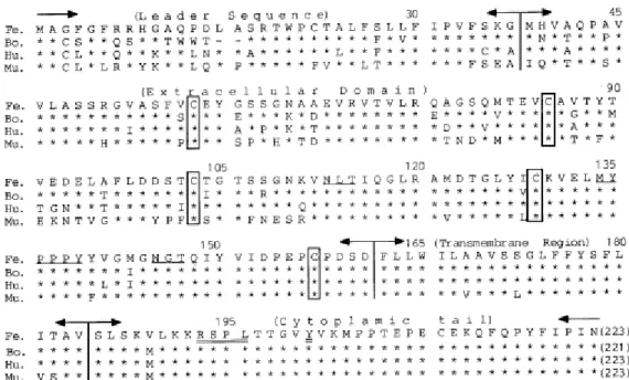
Three isoforms of CTLA-4 have been identified, which result from differential gene splicing: a full-length membrane-bound receptor isoform (mCTLA-4) with an extracellular ligand-binding domain, a transmembrane signal-transducing domain and a cytoplasmic tail; a soluble isoform (sCTLA-4), which does not have the transmembrane domain; and a ligand-independent third isoform, only identified in mice, which lacks the extracellular domain (Wing et al. 2011; Denesha et al. 2014; Yu et al. 2015; Zhang et al. 2016). Feline CTLA-4 shows a high level of sequence homology with the human and murine molecules (86.6% and 76.2% respectively) and the hexapeptide motif (MYPPPY) within the extracellular domain, believed to be responsible for interaction with the B7 ligands, is completely conserved among the studied mammalian species (Ohno et al. 1999) (Figure 3). Upon T-lymphocyte activation, CTLA-4 is transiently translocated to the cell surface where it binds the B7 ligands and initiates inhibitory signals via its intracellular domain (Sharpe and Freeman 2002; Yu et al. 2015). Because CTLA-4 interacts with both B7 ligands, with higher affinity, it can outcompete CD28, attenuating the effector T-lymphocyte response through the inhibition of IL-2 production, blockade of cell cycle progression and therefore, T-lymphocyte proliferation (Walker and Sansom 2015). CTLA-4-expressing cells can also capture B7 ligands from opposing cells by trans-endocytosis and degrade them, resulting in impaired co-stimulation via CD28 (Qureshi et al. 2011).
Figure 3. Alignment of feline (Fe) CTLA-4 amino acid sequence with homologues from bovine (Bo), human (Hu) and mouse (Mu) species. The hexapeptide MYPPPY ligand binding motif on the extracellular domain is underlined. Original in Ohno et al. 1999.
8
sCTLA-4 is generated by alternative splicing of the CTLA-4 mRNA in which the exon that encodes the transmembrane region (exon 2) is spliced out (Figure 4). The deletion causes a shift in the reading frame, producing a unique cytoplasmic tail that is unique to the sCTLA-4 molecule. The alternative splicing also results in the loss of the membrane proximal cysteine residue required for covalent homodimerization, making sCTLA-4 a monomer (Oaks et al. 2000). Both the full-length and the sCTLA-4 transcripts are expressed in CD4+ T-lymphocytes but mCTLA-4 is the predominant among CD8+ subsets of T-lymphocytes, as well as B-lymphocytes. mCTLA-4 is also the predominant transcript on activated T-lymphocytes, however on resting cells or at the post-activated state sCTLA-4 predominates (Oaks et al. 2000).
sCTLA-4 is secreted in a similar manner to mCTLA-4. Upon TCR stimulation, secretory granules are translocated to the central supramolecular activation cluster (cSMAC) within the immunological synapse releasing sCTLA-4 which can interact with the B7 ligands, excluding CD28 from the cSMAC thus inhibiting early T-lymphocyte responses to antigens (Wing et al. 2011; Yu et al. 2015). Translocation is fully dependent on ligand binding but does not require high amounts of ligand in the cSMAC which indicates that sCTLA-4 can control T-lymphocyte
Figure 4. Generation of full-length, soluble and ligand-independent cytotoxic T-lymphocyte associated protein 4 (CTLA-4) mRNA. The CTLA-4 gene encodes a transcript with four exons. Splicing generates the full-length transcript (mCTLA-4). Alternative splicing generates two shorter transcripts: the sCTLA-4 transcript that skips exon 2 (binding domain) and a ligand-independent transcript, only identified in mice, that skips exon 3 (transmembrane domain). Figure redrawn from the original in (Simone and Saverino 2009). Created with BioRender.
9
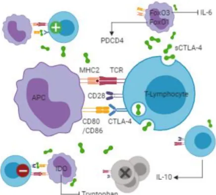
activation even when access to co-stimulatory molecules is limited (Wing et al. 2011). sCTLA-4 signaling may also affect the adhesion and motility of T-lymphocytes to APCs, inhibiting the TCR-mediated signal through dephosphorylation of the TCR signaling proteins via it’s cytoplasmic tail, and may induce production of indoleamine 2,3-dioxygenase (IDO) and immunosuppressive kynurenine in the latter (Ward et al. 2013; Pico de Coaña et al. 2014). IDO is an enzyme system that depletes the amino acid L-tryptophan, establishing a microenvironment which impairs the growth and survival of T-lymphocytes. Finally, sCTLA-4 may induce nuclear localization of the transcription factor Forkhead box (Fox)O3, which inhibits production of inflammatory cytokines, including IL-6 (Dejean et al. 2009; Ward et al. 2013), and can induce increased production of IL-10, an immunosuppressive cytokine (Dahal et al. 2016), thereby constraining T-lymphocyte survival. Recently sCTLA-4 was also implicated in the induction of the translational inhibitor programmed cell death-4 (PDCD4) as a result of FoxO1 nuclear re-localization, which attenuates effector T-lymphocyte responses (Lingel et al. 2017).
Figure 5. Possible mechanisms of immune regulation by soluble cytotoxic T-lymphocyte associated protein 4 (sCTLA-4). Interference with CTLA-4-lingand interactions enhances T-lymphocyte reactivity. By contrast interference with CD28-ligand interactions may result in T-T-lymphocyte inhibition. sCTLA-4 may also induce nuclear localization of FoxO3 and FoxO1 with consequent inhibition of inflammatory cytokine production and induction of PDCD4. Finally, sCTLA-4 may induce expression of IDO and immunosuppressive cytokine IL-10 establishing a microenvironment which impairs the growth and survival of T-lymphocytes. APC – Antigen presenting cell; MHC – Major histocompatibility complex; TCR – T cell receptor; CD – Cluster of differentiation; IL – Interleukin; IDO – Indoleamine 2,3-dioxygenase; PDCD – Programmed cell death; Fox – Forkhead box. Figure redrawn from the original in Dahal et al. 2018. Created with BioRender.
10
Elevated serum CTLA-4 levels have been reported in several cancers, including esophageal (Zhang et al. 2016), lung (Liu, Xie, et al. 2017), and breast carcinoma (Erfani et al. 2010). Furthermore, a molecular study of CTLA-4 genotypes and haplotypes by Erfani and colleagues, among others, clearly demonstrated association of CTLA-4 gene variants with cancer. Indeed, CTLA-4 expression appears to be an important mechanism of tumor immune evasion. Deletion of the CTLA-4 gene in Treg cells in mice was shown to produce potent tumor immunity (Wing et al. 2008). Up-regulated expression of CTLA-4 in tumor cells was also
recently identified as one of the three most prevalent mechanisms of immune evasion in human
breast cancer, the other two being the presence of immunosuppressive factors (i.e. IL-10,
transforming growth factor beta – TGF-β, C-C motif chemokine 22 – CCL22), and tumor expression of a soluble decoy receptor (DcR3) which binds to FasL and inhibits FasL-induced apoptosis (Bou-Dargham et al. 2018). Taken together, these findings show that CTLA-4 plays a crucial role in suppression of tumor immunity.
However, the clinical implications of CTLA-4 in the tumor microenvironment are still controversial. Various studies indicate increased levels of sCTLA-4 in several autoimmune diseases (Simone et al. 2014). A recent study on breast cancer patients found an association between elevated sCTLA-4 levels and improved survival (Liu, Hu, et al. 2017) and several other studies have showed significant correlations between CTLA-4 and OS in non-small cell lung carcinoma, nasopharyngeal carcinoma, esophageal carcinoma, malignant hematologic diseases, glioblastoma and malignant pleural mesothelioma (Liu, Xie, et al. 2017). These findings seem counterintuitive because an increase in sCTLA4 should inhibit T-lymphocyte activity. Some researchers have suggested as an explanation that in the resting T-lymphocytes in which only sCTLA-4 is expressed, CD28-ligand interactions are inhibited. But in a later phase, where mCTLA4 is overexpressed, sCTLA4 interferes with mCTLA-4 ligand interactions, enhancing T-lymphocyte reactivity by preventing the transduction of inhibitory signals (Saverino et al. 2007; Pérez-García et al. 2013; Simone et al. 2014). It has also been suggested that CTLA-4 can mediate negative signal into tumor cells, comparable to those observed in T-lymphocytes (Salvi et al. 2012). Salvi and colleagues observed that established non-small cell lung carcinoma cell lines undergo apoptotic death upon CTLA-4 engagement with soluble B7 (CD80/CD86) ligands. They hypothesize that CTLA-4 expressed by tumor cells may interact with B7 ligands expressed by cells of the tumour micro-environment, thus leading to inhibition of lung cancer cell proliferation and/or induction of apoptotic cell death. These findings may support a role for CTLA-4 as a negative regulator of tumor proliferation, important for cancer biology.
11
1.4. Cellular components of the Tumor microenvironment: the role of Tregs,
TAMs and MDSCs
Most solid tumors contain several subtypes of immune cell infiltrates, including both myeloid- and lymphoid-lineage cells. Human breast cancer, in particular, shows significant levels of tumor infiltrating lymphocytes (TILs). Similarly, lymphocytic infiltration is a common finding in feline mammary carcinoma, though its functional role is not yet fully established (Wiese, Thaiwong, Yuzbasiyan-Gurkan, & Kiupel, 2013). TIL populations dominated by T-lymphocytes (CD3+) are the most commonly reported (Ruffell et al., 2011), being usually associated in some molecular subtypes, namely TNBC and HER-2 positive, with improved survival (Adams et al., 2014; Desmedt et al., 2014; Dieci et al., 2015; Loi et al., 2013; Stanton & Disis, 2016). However, the phenotype of the T-lymphocyte response can influence clinical outcome. While type 1 CD4+ T-helper (Th1) lymphocytes and CD8+ cytotoxic T-lymphocytes are generally associated with a favorable prognosis, type 2 CD4+ T-helper (Th2) lymphocytes inhibit effector T-lymphocyte responses and support proliferation of B-lymphocytes, promoting an anti-inflammatory immune response that may enhance tumor growth (Stanton & Disis, 2016; Ward et al., 2013; Zitvogel, Galluzzi, Kepp, Smyth, & Kroemer, 2015).
Th2 regulatory T-lymphocytes (Tregs) are a subset of CD4+ T-lymphocytes that highly express the IL-2 receptor α chain (CD25) and FoxP3. Tregs also express CTLA-4 whose expression is controlled by FoxP3 and which in Tregs, unlike other T-lymphocyte subsets, is expressed constitutively (reviewed in Wing et al. 2011; Ward et al. 2013). Although their main function is to prevent autoimmune disorders by suppressing effector T-lymphocyte activation, Tregs are known to highly infiltrate various tumor types in both humans and felines (Sparger et al. 2018). Several studies show that Tregs can suppress tumor specific T-lymphocyte immunity, contributing to tumor growth, invasion and metastasis and reduced survival (Curiel et al. 2004; Tan et al. 2011; Emens 2012).
Tregs suppress effector T-lymphocytes via several mechanisms, including Fas/Fas ligand (FasL)-mediated apoptosis, granzyme B/perforin-mediated cytotoxicity and IL-2 deprivation through expression of high levels of CD25 (Pandiyan et al. 2007; Wang et al. 2017). Another highly relevant mechanism whereby Tregs are thought to control effector T-lymphocytes is the CTLA-4-dependent downregulation of B7 ligands on DCs upon antigenic stimulation which is significantly impaired in mice with Treg-specific deficiency of CTLA-4 (reviewed in Wing et al. 2011). CTLA-4 interaction with DCs can also induce expression of IDO (Adams et al. 2014), providing further evidence that CTLA-4 is vital for Treg-lymphocyte mediated suppression. Tregs also produce cytokine IL-35, VEGF and TGF-β which act together to promote angiogenesis and prevent the activation of adaptive and innate immune
12
cellsand may induce polarization of M2 macrophages (Jarnicki et al. 2006; Collison et al. 2007; reviewed in Wang et al. 2017).
It should be noted, however, that Treg infiltration can correlate with a positive prognosis in certain types of cancer. A study in a mouse model of colorectal cancer, showed that under the influence of IL-10, Tregs prevented the development of tumors and rapidly induced tumor regression, at least in part through the inhibition of COX-2 (Erdman et al. 2005). Studies conducted on head and neck, esophageal and hematologic cancers came to similar conclusions (Shang et al. 2015). Tregs have also been shown to suppress inflammation triggered by innate immune cells, such as macrophages and monocytes, in mice and in human cancers (Shang et al. 2015) and seem to be the primary source of sCTLA-4 (Ward et al. 2013) which correlates with improved prognosis in breast cancer patients. These findings raise the possibly of a protective role for Tregs in cancer and warrant further investigation.
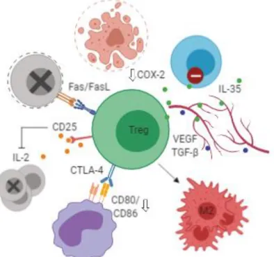
Figure 6. Functions of regulatory T-lymphocytes (Tregs) in the tumor microenvironment. Tregs are capable of suppressing effector T-lymphocyte responses through several mechanisms, including Fas/FasL-mediated apoptosis, granzyme B/perforin-mediated cytotoxicity, IL-2 deprivation through expression of high levels of CD25 and expression of immunosuppressive cytokines like IL-35. Tregs also reduce T-lymphocyte co-stimulatory signals through depletion of CD80/CD86 on dendritic cells via CTLA-4 mediated trans-endocytosis. Tregs may induce polarization of M2 macrophages and promote angiogenesis by secretion of VEGF and TGF-β. Tregs may also have tumor-suppressive effects, for example via inhibition of COX-2. IL – Interleukin; CD – Cluster of differentiation; FasL – Fas ligand; COX – Cyclooxygenase; VEGF – Vascular endothelial growth factor; TGF – Transforming growth
factor; CTLA – cytotoxic T-lymphocyte associated protein. Figure redrawn from the original in Wang et
13
Similar to Tregs, the role of IL-17 producing CD4+ cells (Th17) in the pathogenesis of cancer needs further elucidation. A study on melanoma in a mouse model found that IL-17 signaling was critical for tumor development, with direct effects on tumor and stromal cell-induced production of IL-6 which led to activation of STAT3 (Wang et al. 2009). Dysregulation of the IL-6-mediated STAT3 signaling pathway is also closely related to the development of breast cancer in humans. Other studies, however, have revealed a potential tumor-suppressive role for this cell type. Th17 cells promoted tumor-specific cytotoxic T-lymphocyte activation in a model of lung melanoma (Martin-Orozco et al. 2009) and were positively associated with a more favorable prognosis in human breast carcinoma (Yang et al. 2012).
Myeloid-lineage cells like tumor associated macrophages (TAMs) and myeloid-derived suppressor cells (MDSCs) also play a role in tumor development. In contrast to T-lymphocytes, TAM infiltration is often associated with poor prognosis in human breast cancer (DeNardo et al. 2009; reviewed in Mantovani et al. 2017) and canine mammary tumors (Raposo et al. 2014). These macrophages usually exhibit an M2 phenotype induced by Th2 lymphocytes (Jackute et al. 2018) and may block T-lymphocyte responses through the production of immunosuppressive molecules IL-10, TGF-β and the arginine-degrading enzyme arginase-1 (Arg-1) as well as induce differentiation and recruitment of Tregs via C-C motif chemokine 22 (Curiel et al. 2004; Brown et al. 2017). Moreover, they may support tissue repair and angiogenesis through the production of VEGF or epidermal growth factor (EGF) (Brown et al. 2017). MDSCs, a heterogeneous group of immature cells also seem to be significantly increased in the peripheral blood of breast cancer patients, associated with more aggressive molecular subtypes such as TNBC, advanced stage and positive lymph node status (Safarzadeh et al. 2019). They can influence the tumor microenvironment through multiple mechanisms, including production of reactive oxygen species (ROS) which induces the loss of the TCR ζ-chain leading to T-lymphocyte anergy, and production of Arg1, and IDO, all of which lead to cell cycle arrest of T-lymphocytes (Kumar, Patel, Tcyganov, & Gabrilovich, 2016; Markowitz, Wesolowski, Papenfuss, Brooks, & Carson, 2013; Pico de Coaña, Masucci, Hansson, & Kiessling, 2014). MDSCs can also induce differentiation of CD4+ T-lymphocytes into Tregs and Treg expansion through the secretion of inhibitory cytokines IL-10 and TGF-β. (Markowitz et al. 2013; Pico de Coaña et al. 2014; Kumar et al. 2016).
MDSCs and TAMs, however, do not suppress all aspects of antitumor immune responses. Like TAMs, MDSCs exhibit two distinct phenotypes, the tumor-suppressing M1-like and the tumor-promoting M2-M1-like (Ma et al. 2011). Whereas M2 MDSCs inactivate effector T-lymphocytes and recruit Tregs, M1 MDSCs have the opposing effect. They express higher quantities of pro-inflammatory cytokines and can activate NK cells to produce high amounts of interferon (IFN)-γ (Nausch et al. 2008). IFN-γ induces iNOS expression which generates high amounts of nitric oxide (NO). Because NOs cellular activities are concentration-dependent,
14
with higher levels producing cytotoxicity and antitumorigenic effects (Hussain and Harris 2007), this suggests that M1 MDSCs may have direct tumor-killing activities. Unlike M2 TAMs, the presence of M1 TAMs in the tumor microenvirnoment is associated with increased survival (Jackute et al. 2018). They also direct T-lymphocytes towards Th1 tumor-suppressive responses and interact with NK cells promoting apoptosis in tumor cells through expression of iNOS or TNF-α (Cui et al. 1994; O’Sullivan et al. 2012). Finally, a study on the effects of myeloid-derived VEGF in a mouse model of mammary tumorigenesis, determined that although this factor does increase vascular density in tumors, this change acts to retard not promote tumor progression, as was previously thought (Stockmann et al. 2008).
It is evident that many immune cell types are capable of displaying both tumor-promoting and tumor-suppressive capabilities. However, the cellular and molecular mechanisms that positively or negatively regulate their phenotype and biological functions are not yet fully understood and further study is warranted.
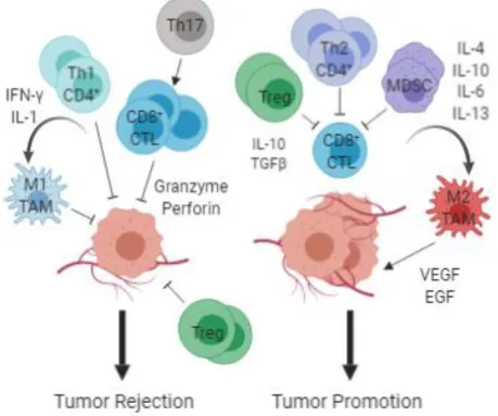
Figure 7. Contrasting functions of immune cells in the tumor microenvironment. Th1 CD4+ and CD8+ T-lymphocytes may directly regulate tumor cell cytotoxicity, while indirectly polarizing immune cells, such as M1 tumor associated macrophages (TAM) and Th17 T-lymphocytes, towards tumor suppression. Th2 CD4+ T-lymphocytes, regulatory T-lymphocytes (Treg) and myeloid derived suppressor cells (MDSC) in contrast, may suppress CD8+ cytotoxicity and induce polarization of immune cells, such as M2 TAMs, which provide a rich proangiogenic and pro-tumoral microenvironment. CTL – Cytotoxic T-Lymphocyte; IFN – interferon; IL – interleukin; TGF – Transforming Growth Factor; VEGF – Vascular Endothelial Growth Factor; EGF – Epidermal Growth Factor. Figure redrawn from the original in (DeNardo and Coussens 2007). Created with BioRender.
15
1.5. Soluble mediators of cancer related inflammation: TNF-α and IL-6
Cytokines are a non-cellular component of the tumor inflammatory microenvironment that can exert rather paradoxical effects during cancer development. The action of inhibitory cytokines has long been implicated in the lack of an effective immune response observed in breast cancers. They also have been reported to promote tumor growth and aggressiveness by influencing aromatase activity and estrogen synthesis directly in the tumor vicinity (reviewed in Knüpfer and Preiß 2007). Certain cytokines, however, have been demonstrated to promote the generation and/or efficacy of anti-tumor effector cells, including DCs and NK cells, causing inhibition of tumor growth and even tumor regression (Knüpfer and Preiß 2007). Whether they exert tumor-promoting or tumor suppressive effects is highly dependent on a number of factors, including the array of cytokines present, their relative concentration, and presence of other modulating factors, such as cytokine receptor expression patterns and the activation state of the cells that express them (Knüpfer and Preiß 2007).
Briefly, cytokines are small pleiotropic proteins that act by altering the function of their target cells in a paracrine or autocrine manner. They are mainly secreted by lymphocytes and macrophages and can be broadly classified as pro-inflammatory or anti-inflammatory. Of the pro-inflammatory cytokines, TNF-α and IL-6 have emerged as central players linking inflammation and cancer.
TNF-α is a member of the TNF cytokine superfamily and is a key molecule regulating inflammation and host defense. It is predominantly produced by immune cells, namely macrophages, T-lymphocytes and NK cells, but also, in low amounts, by fibroblasts, smooth muscle cells and tumor cells (Tse et al. 2012). There are two TNF receptors, TNF receptor type 1 (TNFR1), which is ubiquitously expressed and TNF receptor type 1 (TNFR2), which is mainly expressed on innate immune cells. Activation of TNF receptors can trigger NF-κB and downstream immunosuppressive survival pathways or can activate caspase 8 and the associated apoptotic signal (Wang and Lin 2008) (Figure 8). These activities vary under different physiological conditions and in a cell-type-dependent manner. For example, in rapidly regenerating tissues, TNF-induced NF-κB activity is anti-tumorigenic, whereas in slowly regenerating tissues it seems to be pro-tumorigenic (Wang and Lin 2008). There is also evidence that while chronic synthesis of low amounts of TNF-α promotes tumor growth and angiogenesis, higher doses may stimulate antitumor immunity, induce necrosis of tumor cells, and trigger vascular collapse (Tse et al. 2012). It is likely that differential expression of the TNF receptors is also involved. TNFR1 is a death domain-containing receptor and transduces both proapoptotic and prosurvival signals. TNFR2 does not possess a death domain and is mainly responsible for the promotion of proliferation, although it can mediate a cell death signal which may be indirect through TNFR1 (Wang and Lin 2008). In breast cancer, recent investigations
16
strongly suggest that chronic TNF-α expression supports tumor growth. Expression of TNF-α in inflammatory breast carcinoma correlated with increased tumor grade and lymph node involvement, and patients with more progressed tumor phenotypes were shown to have significantly higher serum concentrations of TNF-α (Ben-Baruch 2003). However, it cannot be ascertained from these studies whether the elevated TNF-α contributes to disease progression, or is a reflection of advanced disease, and more research into its clinical diagnostic and prognostic utility is required.
IL-6 is produced mainly by myeloid cells, including monocytes and macrophages, but also T-lymphocytes, fibroblasts and tumor cells (Fisher et al. 2015). As a secreted protein, IL-6 can be detected in serum, and increased levels have been found in breast cancer patients, associated with worse prognosis (Knüpfer and Preiß 2007). IL-6 signaling is initiated through binding to the IL-6 receptor, a heterodimer consisting of the IL-6 receptor α subunit (IL-6Rα; CD126) and a glycoprotein 130 (gp130) β subunit, whose activation triggers phosphorylation of the Janus kinases (JAK) and its downstream effectors, including the STAT proteins STAT1 and STAT3 (Figure 8). STAT phosphorylation allows dimerization, nuclear translocation, and activation of specific target genes which are mainly involved in cell cycle progression and suppression of apoptosis (Lin and Karin 2007). STAT3 has a predominant role in IL-6 signal transduction and its roles in tumor cell proliferation and survival are well documented (Kim et al. 2014; Chang et al. 2015). Chang and colleagues, for example, report that STAT3 activation initiated by IL-6 promotes recruitment of myeloid cells and induces their ability to express growth factors in a feed-forward loop that positively regulates angiogenesis and induces metastasis. While the predominant view of IL-6 in breast cancer is as a driver of malignancy, recent studies have highlighted its beneficial role in promoting anti-tumor immunity. IL-6 plays a vital role in the development of T-lymphocyte responses, being required for T-lymphocyte priming, the induction of a productive IFN-γ response, protection of T-lymphocytes from the suppressive activities of Tregs, and the acquisition of the ability to provide help to B cells (Fisher et al. 2015). IL-6 signaling also influences lymphocyte trafficking to lymph nodes and to tumor tissues stimulating anti-tumor activities within the tumor microenvironment (Fisher et al. 2015). Taken together, these findings underscore the pleiotropic characteristic of this cytokine and further studies are necessary to elucidate its role in tumor development and progression.
17
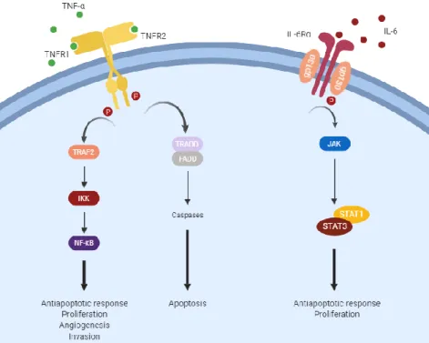
Figure 8. Signal transduction pathways and major biological responses of tumor necrosis factor alpha (TNF-α) and interleukin 6 (IL-6). TNFR – Tumor Necrosis Factor Receptor 1; gp – glycoprotein; TRAF – TNF receptor associated factor; TRADD – TNF receptor type 1-associated death domain; JAK – Janus Kinase; STAT – Signal Transducer and Activator of Transcription. Redrawn from the original in Lin and Karin 2007. Created with BioRender.
2. Retrospective Study
2.1. Objective
Several reports have indicated that CTLA-4 is elevated in the sera from patients with several inflammatory human disorders. The association between CTLA-4 and cancer has also been extensively investigated in recent years. These investigations often point to the link between cancer and inflammation, and the role of the tumor microenvironment in cancer initiation, promotion and progression. Immune cells and inflammatory mediators, such as pro-inflammatory cytokines, are an important part of this microenvironment. In the present study, our main objective was to investigate the profiles of serum CTLA-4 and pro-inflammatory cytokines IL-6 and TNF-α in cats with mammary carcinoma. In addition, we sought to determine whether an association between CTLA-4 and pro-inflammatory cytokine serum levels exists. We hope our findings will contribute to the clarification of the complex interactions that occur within the mammary tumor microenvironment and further validate the cat as a model for the study of human breast cancer.
18
2.2. Materials and Methods
2.2.1. Study Population
Sera from 57 female cats were used in this study. All animals had a fully documented history of feline mammary carcinoma and were followed up at the Faculty of Veterinary Medicine Teaching Hospital (HEV) between June 2011, and September 2013. Available historical data included age, clinical stage (TMN), malignancy grade, tumor burden and size, regional lymph node involvement, presence of tumor necrosis, lymphatic vessel invasion, lymphocyte infiltration or cutaneous ulceration and histopathological classification (ER status,
PR status, HER-2 status, basal status, Ki67 index) (Table 1). Serum samples were collected
at time of admission, aliquoted and stored at -80ºC. Serum samples from twelve healthy cats
were used as controls for the cytokine and sCTLA-4 analysis.
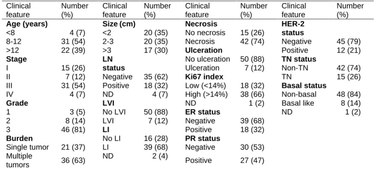
Table 1. Clinicopathological characteristics of female cats with mammary carcinoma
(n=57). LN – Lymph Node; LVI – Lymphatic Vessel Invasion; LI – Lymphocyte Infiltration; ER – Estrogen
Receptor; PR – Progesterone Receptor; HER – Epidermal Growth Factor Receptor; TN – Triple Negative; ND – Not Determined.
Clinical feature Number (%) Clinical feature Number (%) Clinical feature Number (%) Clinical feature Number (%)
Age (years) Size (cm) Necrosis HER-2
status <8 4 (7) <2 20 (35) No necrosis 15 (26) 8-12 31 (54) 2-3 20 (35) Necrosis 42 (74) Negative 45 (79) >12 22 (39) >3 17 (30) Ulceration Positive 12 (21) Stage LN status No ulceration 50 (88) TN status I 15 (26) Ulceration 7 (12) Non-TN 42 (74)
II 7 (12) Negative 35 (62) Ki67 index TN 15 (26)
III 31 (54) Positive 18 (32) Low (<14%) 18 (32) Basal status
IV 4 (7) ND 4 (7) High (>14%) 38 (66) Non-basal 48 (84)
Grade LVI ND 1 (2) Basal like 8 (14)
1 3 (5) No LVI 50 (88) ER status ND 1 (2)
2 8 (14) LVI 7 (12) Negative 39 (68)
3 46 (81) LI Positive 18 (32)
Burden No LI 16 (28) PR status
Single tumor 21 (37) LI 39 (68) Negative 30 (53) Multiple
tumors 36 (63)
ND 2 (4)
Positive 27 (47)
2.2.2. Measurement of serum CTLA-4 and cytokine levels
Serum samples were kept frozen at -80ºC and thawed shortly before determination of CTLA-4, TNF-α and IL-6. Commercially available immunoassay kits from R&D Systems (R&D Systems, Minneapolis, MN, USA) were used according to the manufacturers' instructions. CTLA-4 levels were determined with the Mouse CTLA-4 DuoSet® ELISA immunoassay kit (code DY476); TNF-α levels were determined with the Feline TNF-α DuoSet® ELISA
19
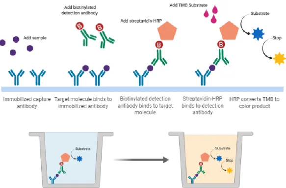
immunoassay kit (code DY2586); and IL-6 levels were determined with the Feline IL-6 DuoSet® ELISA immunoassay kit (code DY2305). Given the high level of sequence homology between the feline and murine CTLA-4 molecules (76.2%) we estimated that the murine kit would be adequately sensitive. All kit components were stored at 4ºC. A seven-point standard curve was prepared for each assay by making serial dilutions from a stock of recombinant mouse CTLA-4, feline TNF-α and feline IL-6 provided in the kits. The immunoassays use a solid-phase sandwich enzyme-linked immunosorbent assay (ELISA) technique (Figure 9). Briefly:
• A 96-well microplate is prepared by adding Capture Antibody to each well, after which the plate is sealed and incubated at room temperature overnight. The antibody is removed, and unbound molecules washed away by washing the plate three times with wash buffer;
• To prevent nonspecific binding the plate is blocked by adding 1% BSA in PBS to each well, and the plate is sealed and incubated at room temperature for one hour. After incubation, blocking agent is removed and the plate washed three times as before with wash buffer.
• Diluted serum samples are added to each well, and the plate is sealed and incubated at room temperature for two hours. After incubation, samples are removed, and unbound molecules washed away by washing three times;
• Biotinylated Detection Antibody is added to each well, and the plate is sealed and incubated at room temperature for another two hours. After incubation, antibody is removed, and unbound molecules washed away by washing three times;
• A working solution of Streptavidin Conjugated to Horseradish Peroxidase (HRP) is added to each well, and the plate is sealed and incubated for 20 minutes at room temperature. After incubation, Streptavidin-HRP is removed, and the plate washed three times as before with wash buffer. During the last wash, the substrate solution is prepared by mixing equal volumes of 3,3’,5,5’-tetramethylbenzidine and H2O2 solutions.
• Substrate solution is added to each well and incubated for 20 minutes at room temperature.
20
Figure 9. Sandwich enzyme-linked immunosorbent assay (ELISA) technique. HRP –
Horse radish peroxidase; TMB – 3,3’,5,5’-Tetramethylbenzidine. Created with BioRender.
The optical density was determined using a FLUOstar OPTIMA microplate reader from BMG Labtech, set to 450 nm. To correct for optical imperfections in the plate a second reading was performed at 570 nm and readings were subtracted from the readings at 450 nm. The data were linearized by plotting the log of the mean absorbance against the log of the concentration using Microsoft® Excel® version 1904 for Windows (Microsoft Corporation, Redmond, WA, USA). CTLA-4, TNF-α and IL-6concentrations were determined using the
curve fit equation (y = mx + c) generated. The correlation coefficient between the fitted data and the actual data was greater than 0.99 for all assays.
2.2.3. Statistical Analysis
Statistical analysis was carried out using GraphPad Prism version 8.11 for Windows (GraphPad Software, La Jolla, CA, USA). The values p < 0.05 (*), p < 0.01 (**) and p < 0.001
(***) were considered statistically significant. A Kruskal-Wallis test was used to assess
significance between serum cytokine and serum CTLA-4 levels and clinicopathological data. Pearson correlation was used to assess the correlation between CTLA-4 and IL-6/TNF-α serum levels. Survival curves were plotted using the Kaplan–Meier (KM) method and the statistical significance between groups determined by the Log-rank test. Receiver operating characteristic (ROC) analysis was used to determine optimal cut-point values for the cytokines.
21
2.3. Results
2.3.1. Serum CTLA-4 levels
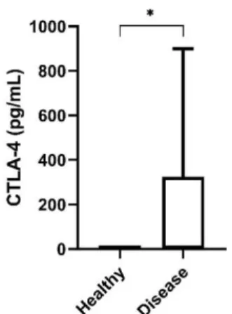
CTLA-4 levels were detectable in 23 (43%) of the 54 cats assessed, showing a median of 459.4 pg/mLwhen detectable (range 77–999.3 pg/mL). In the following analysis, CTLA-4 serum level is considered to be 0 pg/mL for the 23 patients whose serum level was below the detection limit (31.3 pg/mL). The data were tested for associations with clinicopathological criteria (Table 1). Serum CTLA-4 levels in the cats with mammary carcinoma were significantly higher than those in the healthy group (P=0.022; Figure 10)
Figure 10. Box plot analysis of serum cytotoxic T-lymphocyte associated protein 4 (CTLA-4) levels in healthy cats and cats with mammary carcinoma. * P < 0.05; ** P < 0.001; *** P < 0.0001.
Serum CTLA-4 levels were significantly increased in older cats (P=0.009; Figure 11a), cats with stage I (P=0.002; Figure 11b) and stage II (P=0.049; Figure 11b) tumors, smaller tumors (P<0.001; Figure 11c), no tumor necrosis (P<0.001; Figure 11d), no lymphatic vessel invasion (P=0.006; Figure 11e) and no lymph node involvement (P=0.007; Figure 11f).
No significant correlation was found between CTLA-4 serum levels and either tumor grade (P=0.061), tumor burden (P=0.523), lymphocyte infiltration (P=0.141) or cutaneous ulceration (P=0.056) (data not shown).