146 Abdelhamid Elaissari and Hatem Fessi
Advances in the Biomedical Applications of Reactive Colloids
Abdelhamid Elaissari
Universit´e de Lyon, Universit´e Lyon 1, CNRS, UMR 5007, Laboratoire d’Automatique et de Gnie des Proc´ed´es, 43,
Bd. 11 Nov. 1918, 69622 Villeurbanne Cedex, France
Hatem Fessi
Universit´e de Lyon, Universit´e Lyon 1, CNRS, UMR 5007, Laboratoire d’Automatique et de G´enie des Proc´ed´es, 43,
Bd. 11 Nov. 1918, 69622 Villeurbanne Cedex, France (Received on 1 July, 2008)
This short review aimed to give to reader’s brief applications of polymer colloids in biomedical area such as therapy and medical diagnosis as also developed in our laboratory. Indeed, the polymer particles and com-posite particles are commonly used in immunoassays as solid phase supports, for the encapsulation of active agents and for the immobilization of biomolecules such as oligonucleotides, proteins or antibodies. In the area of composite particles, magnetic particles bearing immobilized biomolecules are used in biomedical diagno-sis such as immunoassay, specific nucleic acids concentration, cell labelling and separation and in numerous biotechnological applications.
Keywords: colloid, biomedical, in-vivo, in-vitro, biotechnology, polymer, latex, diagnostic, drug-delivery
1. INTRODUCTION
Polymer colloids have received an increasing interest as solid-phase supports in numerous applications, especially in the biomedical [1, 2], due to the versatility of the het-erophase elaboration processes (emulsion, dispersion, precip-itation, physical processes) for making well-defined micro-spheres with appropriate particle sizes and surface reactive groups [3, 4].
The inorganic colloidal nanoparticles are used in dipsticks as label in the detection step [2, 5]. In this field, gold nanopar-ticles [6, 8] are used in order to enhance and to facilitate the read of the results via the apparition of intense coloured line [9].
Polymer based colloids are elaborated using numerous processes [10]. The more examined particles are latexes and principally polystyrene based particles. The synthesis of those classical latexes mainly hydrophobic in nature is performed using radical polymerization in dispersed media such as: emulsion, miniemulsion, dispersion microemulsion etc. . . Recently special attention has been dedicated to the preparation of hydrophilic, smart (sensitive to the pH, salinity and temperature) [11] particles. Those particles are used as models for in vivo applications in drug delivery and also in in-vitro biomedical diagnostic area [12]. The magnetic particles [2] are specially designed in order to replace the heavy pro-cesses used during the separation of particles from the con-tinuous phase (centrifugation and filtration). In fact, using any classical permanent magnet, the magnetic particles can be collected and concentrated in small volume. The elabo-ration of magnetic particles can be performed using different processes and now days, the market offer a variety of mag-netic nanparticles, microspheres and beads bearing reactive groups [1, 13, 14].
Colloidal particles are used in both in-vivo and in-vitro biomedical applications. But before any real application, the particles are first conjugated with appropriate receptor or biomolecules in order to target the specificity of the appli-cation. Then, the obtained particles-biomolecules conjugates
are evaluated in targeted biomedical applications such as local targeting15 in drug delivery system [16], in-vivo diagnostic [17], immunology [18], specific capture of nucleic acid [19] molecules, cell sorting [20] and identification, bacteria isola-tion [21] and detecisola-tion, viruses [22] extracisola-tion, concentraisola-tion and detection [1].
In this short and non-exhaustive revue, the aim is to give to the reads some information related to particles for in vivo applications and for in vitro biomedical diagnosis.
2. PARTICLES FOR IN-VIVO APPLICATIONS
Drug targeting is a novel approach in pharmaceutical tech-nology receiving so much attention in medical research field with a great progress these last decades [16]. For such medical applications, nanoparticles (i.e. nanoliposomes, nanospheres and nanocapsules. . . ) [16, 23, 24] should be biodegradable polymers particles in nature. They can be de-veloped as a matrix incorporating a drug in the whole sys-tem (like nanospheres) or capsule-like syssys-tem with a poly-meric shell surrounding a core where the drug is encapsu-lated. These nanoparticles for drug delivery system offer many application possibilities such as in the field of medicine, biotechnology, cosmetic and also in both agriculture and the industry fields. For illustration, the schematic presentation of colloidal particles commonly used or studied in this field is below presented.
Brazilian Journal of Physics, vol. 39, no. 1A, April, 2009 147
FIG. 1: Schematic presentation of: (A) liposomes, (B) polymer par-ticles (spheres and capsules), (C) micelles, (D) dendrimer, (E) hy-drogel.
3. PARTICLES FOR IN VITRO APPLICATIONS
3.1. Classical polymer particles
Non magnetic polystyrene latexes have been largely used (as carrier for antigen and antibody reaction) in immuno-agglutination assay as described in 1956 by Singer [8] and first used for rheumatoid factor detection. A given anti-body is chemically [27] or physically [28] immobilized onto polymer-based particles such as polystyrene latex particles. The immobilization performed using well-defined condition such as pH, salinity, temperature and antibody/particles ratio etc. The presence of any specific antigen in the biological sample reacts immediately with the antibody, which induces rapid flocculation of the latex particles via bridging floccu-lation mechanism [29, 30]. The formed clusters can be evi-denced by naked eyes as illustrated in figure 2. The immuno-agglutination [30] assay is acceptably specific and sensitive but not quantitative.
FIG. 2: Immuno-agglutination assay of antibody containing parti-cles. The agglutination is induced by the specific capture of the tar-geted antigen molecules [31].
3.2. Magnetic carriers
Magnetic particles and magnetic latexes are widely used in bionanotechnology based applications and principally in biomedical diagnosis such as in immunoassays, molecular bi-ology, cell sorting, and bacteria and viruses isolation [2]. In addition, the magnetic property is also used to enhance the concentration of the targeted biomolecules and consequently the sensitivity of the biomedical diagnostic.
Increasing interest has been dedicated to the preparation of magnetic particles and magnetic latex particles for diagnostic applications purpose. The pioneer works in this domain were reported by Ugelstad [32] by reporting not only on the prepa-ration of magnetic latexes, but also their use in biomedical di-agnosis. Other approaches were developed in order to prepare well-defined reactive magnetic particles such as; (i) thermally sensitive magnetic particles by Kondo et al. [33], (ii) batch emulsion polymerization of styrene in the presence of mag-netic iron oxides by Charmot et al [34] and (iii) miniemulsion polymerization of styrene containing organic ferrofluid and more recently, (iv) transformation of oil in water magnetic droplet by Elaissari et al. [35] via seed emulsion polymer-ization process. It is interesting to notice, that only this last process leads to highly magnetic submicron latex particles.
FIG. 3: Transmission Electron Microscopy analysis of structured core-shell morphology magnetic latexes as obtained from o/w mag-netic emulsion transformation [26].
3.3. Nucleic acids extraction, concentration and detection
In in-vitro biomedical diagnosis, magnetic particles or beads are generally used in sample preparation and in some cases in order to separate easily the particles from the aque-ous phase. In this area, two processes where used for nucleic acids extractions:
3.3.1. Non-specific capture of nucleic acids
148 Abdelhamid Elaissari and Hatem Fessi
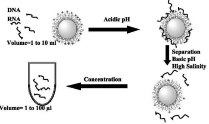
them in small volume which leads to purification and con-centration processes by using cationic magnetic particles [2]. The extracted nucleic acid molecules can then be amplified on the magnetic beads or after desorption step and removal of the magnetic particles using PCR (polymerase chain reaction) [36] in the case of DNA molecule or RT-PCR (Reverse Tran-scriptase PCR) [37] in the case of RNA as below illustrated [38].
FIG. 4: Illustration of non-specific capture, purification and concen-tration of nucleic acid molecules. The adsorption of nucleic acid molecules is performed at acidic pH using cationic magnetic par-ticles. After magnetic separation of magnetic particles and removal of the supernatant, the adsorbed nucleic acid molecules are desorbed using basic pH and high salinity medium. The released nucleic acids are then collected in small volume.
3.3.2. Specific capture of nucleic acids
The specific capture of nucleic acids [39, 40] using mag-netic particles is generally performed as follows. The capture probe of well-defined sequence is chemically immobilized on the magnetic latex particles. A given biological sample (or the above purified nucleic acids) is mixed with the magnetic particles-ODN (ODN for oligonucleotide) conjugates. The target is then specifically captured via hybridization process (specific hydrogen binding). The detection is performed by adding the labelled detection probes (i.e. oligonucleotide la-belled with enzyme) [41]. The addition of substrate is oxi-dized by the enzyme, which lead to coloured supernatant as in immunoassay. This specific capture of nucleic acid molecules combined with well-optimized detection process lead to the enhancement of this molecular biology based diagnosis [42].
4. CONCLUSION
The preparation of colloidal particles should solve specific questions related to the targeted applications. In fact,
col-loidal particles bearing reactive groups such as (−COOH, −NH2, −SH etc.. . . ) are suitable for the covalent binding of biomolecules in order to be used as a solid support for spe-cific capture of targets and also suitable for the encapsulation of active molecules (i.e. drug) or biomolecules (peptides, nu-cleic acids. . . ).
FIG. 5: Illustration of specific capture and detection of targeted nu-cleic acids. Particles bearing chemically grafted ODN (Oligonu-cleotide) are mixed with a given biological sample containing the target. After the specific capture of the target, the particles are ex-tracted and washed before adding detection probe (ODN bearing en-zyme). The evidence of the specific capture is performed by adding substrate which reacts with enzyme in order to lead to coloured medium [4].
To target any biomedical application (in-vivo or in-vitro use), well-appropriate colloidal particles need to be used. The elaboration of polymer-based particles can be performed us-ing well established formulation recipes and polymerization processes. Before any application, the physical chemistry and the colloidal properties of the particles are of great interest. In fact, the examination of those properties is suitable in order to control the interaction between the particles and the used active agents, biomolecules etc. . .
To prepare suitable solids support, many criteria should be considered, the particles, the size distribution, the surface po-larity of the particles, the surface charge density, the chemical composition of the particles, the compatibility (the degrad-ability or the possible bio-elimination), the internal and exter-nal morphologies, the colloidal stability, the swelling ability and finally the intrinsic properties of the particles.
The polymerization kinetics and the colloidal character-ization are conducted as systematic studies. The char-acterization of the final particles is of great importance, since well-characterized particles helps the investigation of biomolecules interactions with the colloidal support.
[1] Arshady Citus Books2001, 3.
[2] U. H¨afeli, W. Sch¨utt, J. Teller, M. Zborowski, Plenum Press: New York, 1997.
Brazilian Journal of Physics, vol. 39, no. 1A, April, 2009 149
[4] A. Elaissari, R. Veyret, B. Mandrand, J. Chatterjee in Col-loidal Biomolecules, Biomaterials, and Biomedical Applica-tions, Edited by Elaissari, Marcel Dekker Edition 2003, Sur-factant Science Series, Volume 116, 1-26.
[5] J.W.M. Bulte, R.A. Brooks, In Scientific and clinical applica-tions of magnetic carriers; Al., H. e., Ed.; Plenum Press: New York, 1997; pp 527-543.
[6] A. Perrin, A. Theretz, B. Mandrand, Anal.Biochem. 1997, to be published.
[7] W.L. Shaiu, D.D. Larson, J. Vesenka, E. Henderson, Nucleic Acids Res. 1993, 21, 99-103.
[8] J.M. Singer and C.M. Plotz. Am. J. Med. 21 (1956), p. 888. [9] A. Elaissari, Wely 2008.
[10] R; Arshady, Colloid Polym.Sci. 1992, 270, 717-732.
[11] A. Elaissari, W. Yang, W. Smart Nano and Microparticles, The MML serie, Volume 7, Kenji Kono, Reza Arshady, (Editors), Kentus Books, United Kingdom 2006, 7.
[12] S. Stainmesse, A.M. Orecchioni, E. Nakache, E.; Puisieux, F.; Fessi, H. Colloid Polym.Sci. 1995, vol. 273, 505-511. [13] Grttner, C.; Teller, J.; Schtt, W.; Westphal, F.; Schmichen, C.;
Paulke, B.-R. In Scientific and clinical applications of magnetic carriers; Al., H. e., Ed.; Plenum Press: New York, 1997; pp 53-67.
[14] Elaissari, A.; Sauzedde, F.; Montagne, F.; Pichot, C. in Col-loidal Polymers, Synthesis and Characterization, Edited by Elaissari, Marcel Dekkert Edition 2003, Surfactant Science Se-ries Volume 115, 285-318.
[15] Kumar, M. N. V.; Sameti, M.; Mohapatra, S. S.; Kong, X.; Lockey, R. F.; Bakowsky, U.; Lindenblatt, G.; Schmidt, H.; Lehr, C.-M. Journal of nanoscience and nanotechnology 2004, 4.
[16] Couvreur, P.; Dubernet, C.; Puisieux, F. european journal of pharmaceutics and biopharmaceutics 1995, vol. 41, 2-13. [17] Bridot, J.; Faure, A.; Laurent, S.; Rivire, C.; Billotey, C.; Hiba,
B.; Janier, M.; Josserand, V.; Coll, J.; VL., E.; Muller, R.; Roux, S.; Perriat, P.; Tillement, O. Journal of American Chem-ical Society 2007, Apr 25; 129 (16), 5076-5084.
[18] Kriz, K.; Gehrke, J.; Kriz, D. Biosens.Bioelectron. 1998, 13, 817-823.
[19] Inomata, Y.; Wada, T.; Handa, H.; Fujimoto, K.; Kawaguchi, H. J.Biomater.Sci.Polym.Edn. 1994, 5 (4), 293-302.
[20] Furdui, V. I.; Harrison, D. J. Lab Chip 2004, 4, 614-618. [21] Mitchell, B. A.; Milbury, J. A.; Brookins, A. M.; Jackson, B. J.
Journal of Foods Protection 1994, 57,n◦8, 743-745.
[22] Veyret, R.; Elaissari, A.; marianneau, P.; Sall, A. A.; Delair, T.
Annalyical Biochemistry 2005 59-68.
[23] Bouchemal, K.; Brianon, S.; Fessi, H.; Chevalier, Y.; Bonnet, I.; Perrier, E. materials Science and Engineering 2006, 26, 472-480.
[24] Bouchemal, K.; Couenne, F.; Brianon, S.; Fessi, H.; Tayakout, M. AIChE Journa 2006, 52.
[25] Hamoudeh, M.; Fessi, H. Journal of colloid and interface sci-ence 2006, 300, 584-590.
[26] Arshady, R. Biomaterials 1993, 14(1), 5-15.
[27] Kandzia, J.; Scholz, W.; Anderson, M. J. D.; Mller-Ruchholtz, W. J.Immunol.Methods 1984, 75, 31-41.
[28] Suzawa, T.; Shirahama, H. Advances in Colloid and Interface Science 1991, 35, 139-172.
[29] Ouali, L.; Pefferkorn, E.; Elaissari, A.; Pichot, C.; Mandrand, B. J.Colloid Interface Sci. 1995, 171, 276-282.
[30] Stoll, S.; Lanet, V.; Pefferkorn, E. J.Colloid Interface Sci. 1993, 157, 302-311.
[31] Polpanich, D.; Tangboriboonrat, P.; Elaissari, A.; Udomsang-petch, R. Analytical Chemistry 2007, 4690-4695.
[32] Nustad, K.; Funderud, S.; Ellingsen, T.; Berge, A.; Ugelstad, J. In Scientific Methods for the Study of Polymers Colloids and their Applications; Candau, F., Ottewill, R. H., Eds.; Kluver Academic Publishers: the Netherlands, 1990; pp 517-527. [33] Kondo, A.; Fukuda, H. J.Ferment.Bioeng. 1997, 84, 337-341. [34] Charmot, D. Progr.Colloid Polym.Sci. 1989, 76, 94-100. [35] Montagne, F.; Mondain-Monval, O.; Pichot, C.; Elaissari, A.
Journal of Polymer Science: Part A: Polymer Chemistry 2006, 44 2642-2656.
[36] Yamada, O.; Matsumoto, T.; Nakashima, M.; Hagari, S.; Kamahora, T.; Ueyama, H.; Kishi, Y.; Uemura, H.; Kurimura, T. Journal of Virological methods 1990, 27, 203-210.
[37] Mallet, F.; Oriol, G.; Mary, C.; Verrier, B.; Mandrand, B. BioTechniques 1995, 18, 678-687.
[38] Elaissari, A. e-Polymer 2005, 28.
[39] Mallet, F.; Hebrard, C.; Livrozet, J. M.; Lees, O.; Tron, F.; Touraine, J. L.; Mandrand, B. J.Clin.Microbiol. 1995, 33(12), 3201-3208.
[40] Mallet, F.; Hebrard, C.; Brand, D.; Chapuis, E.; Cros, P.; Besnier, J. M.; Barin, F.; Mandrand, B. J.Clin.Microbiol. 1993, 31(6), 1444-1449.
[41] Elaissari, A.; Rodrigue, M.; Meunier, F.; Herve, C. Journal of Magnetism and Magnetic Materials 2001, 127-133.
![FIG. 3: Transmission Electron Microscopy analysis of structured core-shell morphology magnetic latexes as obtained from o/w mag-netic emulsion transformation [26].](https://thumb-eu.123doks.com/thumbv2/123dok_br/18983288.457875/2.977.97.448.842.1120/transmission-electron-microscopy-analysis-structured-morphology-magnetic-transformation.webp)