Spray-drying technique to prepare innovative nanoparticulated formulations for drug
administration: a brief overview
S.S. Guterres∗
Programa de P´os-Graduac¸˜ao em Ciˆencias Farmacˆeuticas, Faculdade de Farm´acia, Universidade Federal do Rio Grande do Sul,
Av Ipiranga, 2752, Porto Alegre, 90610-000, RS, Brazil
R. C. R. Beck
Departamento de Farm´acia Industrial, Centro de Ciˆencias da Sa´ude,
Universidade Federal de Santa Maria, Av. Roraima, 1000, Santa Maria, 97105-900, RS, Brazil
A. R. Pohlmann
Departamento de Qu´ımica Orgˆanica, Universidade Federal do Rio Grande do Sul, PBOX 15003, Porto Alegre, 91501-970, RS, Brazil (Received on 15 July, 2008)
Polymeric nanoparticle systems (nanocapsules and nanospheres) present potential applications as drug deliv-ery systems. Nevertheless, their full applications have not been exploited due to their limited shelf life when stored in aqueous medium. Drying polymeric nanoparticles using spray-drying represents a promising plat-form to improve the physicochemical stability of plat-formulations and/or to control the release of hydrophilic and lipophilic drugs. This article presents a brief overview of the most recent and ongoing research in the use of spray-drying process to prepare and/or to dry polymeric nanoparticles formulations intended for drug adminis-tration.
Keywords: microparticles; nanocapsules; nanospheres; spray-drying
I. INTRODUCTION
Spray-drying is a common technique used in pharmaceu-ticals to produce a dry powder from a liquid phase [1]. This technique has also been employed as a microencapsulation method because it can be adapted to the development of dif-ferent systems, microspheres or microcapsules, depending on the initial aqueous formulation, a solution, a suspension or an emulsion [2]. Another application is its use as a preserva-tion method, increasing the storage stability due to the water elimination.
Nanoparticle, submicronic colloidal carriers, is a general name to describe nanocapsules and nanospheres. Nanocap-sules correspond to a polymeric wall enveloping an oil core, while the nanospheres consist of a polymeric matrix [3]. Nanoparticles have been extensively studied over the last years in order to control the drug release, to increase the drug selectivity and effectiveness, to improve drug bioavailability and decrease the drug toxicity [4,5]. Nevertheless, the full ap-plications of polymeric nanoparticles have not been exploited due to the lack of stability of formulations when conserved in aqueous medium for a long period [6]. During the stor-age, microbiological growth, polymer hydrolysis and physic-ochemical instability as a consequence of particle aggregation can take place [7]. According to Tewa-Tagne and co-workers, efforts to develop more stable nanoparticle formulations have been unsuccessfully carry out for many years [8]. The type and the concentration of surfactants in the formulations have been varied, but the results shown good stability only in the short-term [9,10]. On the other hand, the incorporation of nanoparticles in solid dosage forms resulted in phase separa-tion after the addisepara-tion of different polymeric binders to
aque-∗Electronic address:
ous suspensions of nanoparticles [11]. So, the development of an effective technique to improve the shelf life of nanopar-ticles is a requirement.
Microparticles are generally composed by polymeric ma-terials and present advantages such as a rapid and one step production, ready distribution on a large surface of the body, more constant plasma levels, higher accuracy in reproducibil-ity dose-by dose, less decrease in bioavailabilreproducibil-ity and minor risk of toxicity due to the dose dumping [12,13]. A modern microparticle formulation should be able to provide a con-trolled and well-defined drug release pattern.
In the last years our research group and others have been involved in the study of the potential of the spray drying technique to stabilize polymeric nanoparticle aqueous sus-pensions converting them into powders, as well as to prepare innovative nanocoated-microparticles to control the drug re-lease. So, this article presents a brief overview of most recent and ongoing research in the use of spray-drying process to prepare and/or to dry polymeric nanoparticles formulations intended for drug administration.
II. DRYING POLYMERIC NANOPARTICLE
SUSPENSIONS
particle sizes (200 nm) than those determined by PCS in the suspensions before dehydration process [14].
Drug Aerosil® 200 Polymeric nanoparticles
(matricial or vesicular)
A
B
C
Xerogel
D
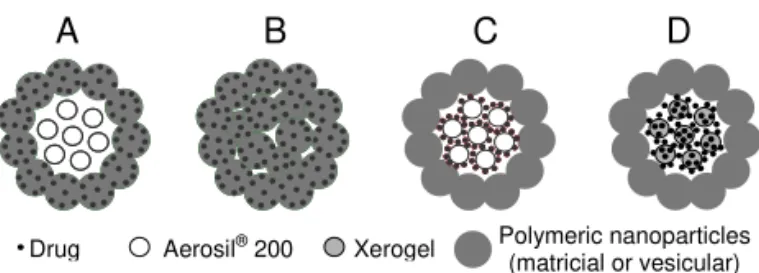
FIG. 1: Illustrative representation of the architecture of different nanostructured microparticles obtained either by sequential assem-bly of silica and polymeric nanoparticles or by drying polymeric nanoparticles: type A is prepared by drying the drug-loaded poly-meric nanoparticles with colloidal silicon dioxide; type B is obtained by drying the polymeric nanoparticles using water soluble materials; and type C and D are prepared by coating the drug-loaded AerosilR 200 or xerogel, respectively, with polymeric nanoparticles
On the other hand, the micro-powders prepared from the nanosphere suspensions presented a reduction in the particle size which decreased from 200 nm to 60-90 nm after dry-ing [15,16]. The polymeric nanoparticles used to coat the inorganic microparticles can be prepared by nanoprecipita-tion or interfacial deposinanoprecipita-tion of pre-formed polymers [17], as well as by emulsification-diffusion technique [18]. Regard-ing the biological effects, after oral administration in rats, the diclofenac-loaded nanocapsule spray-dried powders dis-persed in water were valuable for reducing the gastrointesti-nal irritant effect of the non-steroidal anti-inflammatory drug [19]. In parallel, a pharmacokinetic study in rats was also conducted, showing a complete oral absorption of the drug from spray-dried powders dispersed in water [19].
In addition, those diclofenac-loaded nanoparticle spray-dried powders have been investigated after 14 months of stor-age at ambient temperature. The powders showed higher chemical stability than the drug-loaded nanocapsule suspen-sions. Our study showed that besides the nature of the formu-lation, suspension or powder, the decrease in the drug con-tent is dependent on the type and concentration of the surfac-tant system. An accelerate stability study carried out under UVC light exposition followed by mass spectrometry analysis demonstrated that 2-(2’,6’-dichlorophenyl)aminobenzyl al-cohol and N-(2’,6’-dichlorophenyl)anthranilylaldehyde were the diclofenac degradation products [20].
Nanocapsules and nanospheres containing an anti-inflammatory drug were freeze-dried after addition of col-loidal silicon dioxide to obtain intact dried nanoparticles [21]. The micro-powder surfaces presented a homogeneous nanocoating and satisfactory biological effect (gastrointesti-nal tolerance). However, in spite these promising results, the freeze-drying technique is highly expensive and reserved for products with high added value [22]. So, we continued our study in the use of spray-drying to dry the polymeric nanopar-ticle suspensions. In the case of the nanocapsule spray-dried powders, the oil concentration in the suspension presented an important influence on the homogeneous recovering of the particles. The powders prepared using low oil concentration showed two patterns of nanoparticles on the microparticle surface [23].
Indeed, based on those results we acquired the knowledge needed for the design of homogeneous nanocoating surfaces. The morphologic control of the micro-powder coating is de-termined by the use of either polymeric spheres or vesicular nanoparticles. These studies showed the different behavior of polymeric nanocapsules and nanospheres in the preparation of these organic-inorganic microparticles [15,16,19,23]. The nanosphere suspension (polymeric matrix) or the nanocap-sule suspension (vesicular nanostructure) led to microparti-cles presenting different and homogeneous nanocoating after the drying process [16]. It is important to note that consider-ing this dryconsider-ing strategy the drug is encapsulated in the poly-meric nanoparticles. In spite the evident advantages of the spray-drying in the presence of silicon dioxide as a strategy to dry the aqueous suspensions, the interactions between the polymeric nanoparticles and the silicon dioxide had not been investigated.
Viewing to understand what kind of interactions takes place, Tewa-Tagne and co-workers demonstrated that the in-teractions occurring in the feed are directed by hydrogen bounds and they were more sensitive to the silica concen-tration than that of nanocapsules [8]. SEM analyses of the powders showed spherical separated microparticles formed by the association of nanocapsules and silica when they are mixed at adequate concentrations in the feed before spray-drying. On the other hand, fused agglomerated particles pre-senting nanocapsules at their surface, characterized by irregu-lar shapes and a strong adhesiveness were prepared when the silica concentration was not sufficient.
More recently, the effects of formulations and spray-drying process variables on the powders properties has been studied in order to optimize the process [22]. Due to the high numbers of parameters involved in the spray-drying process, particular attention is required in the process optimization. Responses such as temperature, moisture content, operation yield, par-ticle size and parpar-ticle densities are useful to determine the optimal operational conditions. In the particular case of dry-ing nanocapsules usdry-ing silicon dioxide as an auxiliary agent, the concentrations of both determined the powder characteris-tics. Besides silicon dioxide, soluble supports such as lactose, PVP-K30 and mannitol can also be employed to dry nanocap-sules [24]. Using nanocapnanocap-sules at a concentration of 1% (w/v) and lactose at 10 % (w/v), the powder had adequate morphol-ogy and ease reconstitution in water (Figure 1B). Focusing on the size distribution after reconstitution, satisfactory result has been observed using mannitol and PVP-K30 both at 10 % (w/v).
III. PREPARING INNOVATIVE
NANOPARTICLE-COATED MICROPARTICLES
formulation and controlled the drug release profile in compar-ison with the pure drug [25]. Moreover, a monoexponential model described the release profile and the drug release was governed by swelling, relaxation and dissolution of the poly-mer.
Indeed, coating techniques are largely employed in order to control the drug release from dosage forms for oral ad-ministration, as well as to protect the drug from inactivation or the gastrointestinal mucosa against drug damage. In gen-eral coating processes are carried out on single unit dosage forms, as tablets or capsules, after their production [26]. Sev-eral reports in the literature show that the coating of parti-cles presents more advantages compared to the coating of unit dosage forms [27,28]. These advantages are given by the more reproducible gastrointestinal transport, higher bioavail-ability, more uniformly spread out in the gastrointestinal tract and reduction of the local irritation. The main useful methods in particle coating are air suspension (Wurster process, fluid-bed dryer), centrifugation, spray drying, coacervation (aque-ous phase separation) and interfacial polymerization [26].
Taking into account that coating systems using organic sol-vents need to be avoided due to safety and economic disad-vantages and that the polymeric nanoparticles are aqueous dispersions, we considered that the latter could be also useful to coat the microparticles to control the drug release. In this way, new nanoparticle-coated drug-loaded organic-inorganic microparticles using the spray-drying technique were pro-posed (Figure 1C). In 2004, we reported for the first time the preparation of spray-dried nanoparticle-coated microparticles using diclofenac and unloaded nanoparticles (nanospheres or nanocapsules) prepared with Eudragit S100R as a coating material [29]. The work showed the preparation of these microparticles using a one-step and a two-step method. In the one-step method the microparticles were prepared by the addition of the drug and silicon dioxide directly into the nanoparticle suspension before the spray-drying. On the other hand, using the two-step method, an organic-inorganic core was previously prepared by mixing the drug and silicon diox-ide in acetone followed by solvent evaporation under reduced pressure. This core was redispersed in the nanoparticle sus-pension before the drying process. All formulations showed the presence of nanostructures on their surface forming a covering layer which particle diameters were in accordance with the original suspension. When the diclofenac (sodium salt) was employed as hydrophilic model, the powders pre-pared in two steps (core previously prepre-pared) showed satis-factory gastroresistance. The use of diclofenac (acid form) as hydrophobic model also conducted to powders present-ing good gastroresistance uspresent-ing triacetin as a plasticizer in the nanocapsule formulations [30]. The type of the coat-ing layer (nanospheres or nanocapsules) had also an impor-tant influence on the protective efficiency against gastroin-testinal injury after oral administration of diclofenac, which gastrointestinal side-effects, such as irritation, ulceration and mucosal damage are well known. Following oral administra-tion in rats, nanocapsule-coated diclofenac-loaded micropar-ticles demonstrated a significant protective effect of the gas-trointestinal mucosa against ulceration [30].
The morphological studies showed that these nanoparticle-coated drug-loaded microparticles presented a decrease in the powder surface area compared to the powders before
coating independently on the preparation method (one-step or two-steps) or the drug model (diclofenac salt or acid) [31]. The decrease in the surface area was also dependent on the type of polymeric nanoparticle used as coating mate-rial. Nanocapsule-coated microparticles showed a higher de-crease in the surface area, as well as in the pore size distribu-tion in the mesoporous region, compared to the nanosphere-coated microparticles [30,31]. The effects of spray-drying process variables (inlet temperature and feeding spray rate) on the physicochemical properties of the powders were also studied in order to optimize the coating process. They were dependent on the type of the coating material. The control of these processing variables allowed obtaining nanosphere or nanocapsule-coated microparticles with satisfactory yields, particle sizes, encapsulation efficiencies and low water con-tents [30].
The use of another nanostructured suspension obtained by the dispersion of Eudragit S100R in phosphate buffer pH 7.4 (50 – 400 nm) instead of nanocapsule or nanosphere suspen-sions to prepare nanoparticle-coated microparticles were also reported by our research group [32]. These spray-dried mi-croparticles present similar advantages to nanocapsule and nanosphere-coated microparticles considering the gastroin-testinal protection against diclofenac mucosal damage and control of drug release. The major advantage of this mi-croparticulated system is the absence of organic solvent either in the preparation of the polymeric nanostructure suspension or in the coating step.
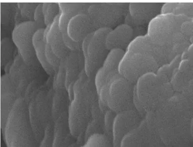
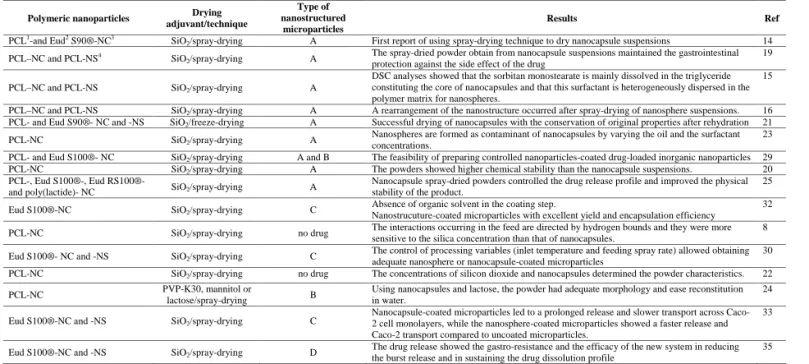
The efficient control of drug release from nanoparticle-coated microparticles was also showed by an in vitro drug transport study across Caco-2 cell monolayers [33]. Nanoparticle-coated dexamethasone-loaded microparticles were prepared and showed efficient coating by the polymeric nanostructures as observed by SEM and atomic force mi-croscopy (Figures 2 and 3). In accordance with the in vitro drug release studies at pH 7.4, nanocapsule-coated micropar-ticles presented lower permeability coefficient values across this human intestinal cell line compared to the free dexam-ethasone solution, mainly in the presence of a plasticizer in the microparticle formulation. This lower permeability across the cell layers is explained by the more internal localization of the drug in this formulation as confirmed by the mathematical modeling of the drug release data [33]. Furthermore, cytotox-icity studies showed that the nanoparticle-coated microparti-cles are non-toxic to the membranes of Caco-2 cells. This recent study reinforced that nanocapsule-coated microparti-cles represent a promising platform for the development of controlled oral drug delivery systems.
Regarding the mechanism of drug release from these nanoparticle-coated microparticles, Domingues and co-workers showed a controlled release of indomethacin, a non-steroidal anti-inflammatory drug, from nanocapsule-coated microparticles prepared in one step compared to the pure drug [34]. The drug release profiles fit to the monoexponential equation and the Korsmeyer-Peppas model, which allowed the proposition of a non-Fickian release mechanism depen-dent on the nanocapsule-coated microparticle desagglomera-tion [34].
FIG. 2: SEM micrographs of nanocapsule-coated dexamethasone-loaded microparticles obtained by spray-drying (photo width = 2.93 µm).
FIG. 3: AFM image of nanocapsule-coated dexamethasone-loaded microparticles (1µm x 1µm image).
the drug release because the drug is also encapsulated in the mesoporous besides its encapsulation in the xerogel macro-pores [35]. Moreover, to diminish the burst release of the drug from the xerogel the particles have been coated with polymeric nanoparticles (Figure 1D). The silica xerogel has been prepared in situ in the presence of the drug, sodium di-clofenac, used as hydrophilic model. Then, the powder has been coated with polymeric nanocapsules by spray-drying. The drug release experiments showed the gastroresistance and the efficacy of the new system in reducing the burst re-lease and in sustaining the sodium diclofenac dissolution pro-file. The nanocapsule-coated drug-loaded xerogel micropar-ticles showed potential use for controlling the release of
hy-drophilic drugs.
Table 1 summarizes the nanostructured microparticles pre-pared by drying polymeric nanoparticles in order to obtain stable products and/or controlled release formulations. The different systems (Figure 1A-D) are able to encapsulate and control the release of drugs which characters are hydrophilic or lipophilic depending on its localization in the phases of microparticles.
IV. CONCLUSION
The nanostructured microparticles that were reviewed in this article are useful to encapsulate either lipophilic or hy-drophilic drugs. In addition, drying polymeric nanoparticles using spray-drying technique is a promising platform to im-prove the physicochemical stability of formulations and/or to control the release of drugs. Finally, the use of different ma-terials leads to well defined complex architectures and deter-mines the routes of drug administration.
V. ACKNOWLEDGMENTS
Table 1. Nanostructured microparticles: composition, preparation technique, model of structure and main results.
Polymeric nanoparticles Drying adjuvant/technique
Type of nanostructured
microparticles
Results Ref
PCL1
-and Eud2
S90®-NC3
SiO2/spray-drying A First report of using spray-drying technique to dry nanocapsule suspensions 14
PCL–NC and PCL-NS4
SiO2/spray-drying A
The spray-dried powder obtain from nanocapsule suspensions maintained the gastrointestinal protection against the side effect of the drug
19
PCL–NC and PCL-NS SiO2/spray-drying A
DSC analyses showed that the sorbitan monostearate is mainly dissolved in the triglyceride constituting the core of nanocapsules and that this surfactant is heterogeneously dispersed in the polymer matrix for nanospheres.
15
PCL–NC and PCL-NS SiO2/spray-drying A A rearrangement of the nanostructure occurred after spray-drying of nanosphere suspensions. 16
PCL- and Eud S90®- NC and -NS SiO2/freeze-drying A Successful drying of nanocapsules with the conservation of original properties after rehydration 21
PCL-NC SiO2/spray-drying A
Nanospheres are formed as contaminant of nanocapsules by varying the oil and the surfactant concentrations.
23
PCL- and Eud S100®- NC SiO2/spray-drying A and B The feasibility of preparing controlled nanoparticles-coated drug-loaded inorganic nanoparticles 29
PCL-NC SiO2/spray-drying A The powders showed higher chemical stability than the nanocapsule suspensions. 20
PCL-, Eud S100®-, Eud RS100®-
and poly(lactide)- NC SiO2/spray-drying A
Nanocapsule spray-dried powders controlled the drug release profile and improved the physical stability of the product.
25
Eud S100®-NC SiO2/spray-drying C
Absence of organic solvent in the coating step.
Nanostrucuture-coated microparticles with excellent yield and encapsulation efficiency
32
PCL-NC SiO2/spray-drying no drug
The interactions occurring in the feed are directed by hydrogen bounds and they were more sensitive to the silica concentration than that of nanocapsules.
8
Eud S100®- NC and -NS SiO2/spray-drying C
The control of processing variables (inlet temperature and feeding spray rate) allowed obtaining adequate nanosphere or nanocapsule-coated microparticles
30
PCL-NC SiO2/spray-drying no drug The concentrations of silicon dioxide and nanocapsules determined the powder characteristics. 22
PCL-NC PVP-K30, mannitol or
lactose/spray-drying B
Using nanocapsules and lactose, the powder had adequate morphology and ease reconstitution in water.
24
Eud S100®-NC and -NS SiO2/spray-drying C
Nanocapsule-coated microparticles led to a prolonged release and slower transport across Caco-2 cell monolayers, while the nanosphere-coated microparticles showed a faster release and Caco-2 transport compared to uncoated microparticles.
33
Eud S100®-NC and -NS SiO2/spray-drying D
The drug release showed the gastro-resistance and the efficacy of the new system in reducing the burst release and in sustaining the drug dissolution profile
35
1
PCL: Poly(ε-caprolactone), 2
Eud: Eudragit®, 3
NC: nanocpasules, 4
NS: nanospheres.
[1] J. Broadhead, S. K. Edmond Rouan, and C. T. Rhodes, Drug Dev Ind. Pharm.18, 1169 (1992).
[2] M. I. R´e, Dry. Technol.24, 433 (2006).
[3] P. Couvreur, C. Dubernet, and F. Puisieux, Eur. J. Pharm. Bio-pharm.41, 2 (1995).
[4] P. Couvreur, G. Barratt, E. Fattal, P. Legrand, and C. Vauthier, Crit. Rev. Ther. Drug1999 (2002).
[5] S. R. Schaffazick; L. L. Freitas, A. R. Pohlmann, and S. S. Guterres, Quim. Nova 26 (2003).
[6] M. D. Coffin and J. W. Mcginity, Pharm. Res.9200 (1992). [7] B. Magenheim and S. Benita, STP Pharma Sci.1, 221 (1991). [8] P. Tewa-Tagne, S. Briancon, and H. Fessi, Int. J. Pharm.325,
63 (2006).
[9] V. C. F Mosqueira, P. Legrand, H. Pinto-Alphandary, F. Puisieux, and G. Barratt, J. Pharm. Sci.89, 614, 2000. [10] R. B. Friedrich, M. C. Fontana, R. C. R. Beck, A. R. Pohlmann,
and S. S. Guterres, Quim. Nova (2008), in press.
[11] C. Schmidt and R. Bodmeier, J. Control. Release 57, 115 (1999).
[12] S. Lin and Y. Kao, Pharm. Res.8, 919 (1991).
[13] Y. Kawashima, T. Iwamoto, T. Niwa, H. Takeuchi, and T. Hino, Int. J. Pharm.89, 9 (1993).
[14] C. R. M¨uller, V. L. Bassani, A. R. Pohlmann, C. B. Michalowski, P. R. Petrovick, and S. S. Guterres, Drug Dev. Ind. Pharm.26, 343, (2000).
[15] C. R. M¨uller, S. R. Schaffazick, A. R. Pohlmann, L. De Lucca Freitas, N. Pesce Da Silveira, T. Dalla Costa, and S. S. Guter-res, Pharmazie56, 864 (2001).
[16] A. R. Pohlmann, V. Weiss, O. Mertins, N. Pesce Da Silveira, and S. S. Guterres, Eur. J. Pharm. Sci.16, 305 (2002). [17] H. Fessi, F. Puisieux, and J. P. Devissaguet, European Patent
0274961, A1 (1988).
[18] D. Quintanar-Guerrero, E. All´emann, H. Fessi, and E. Doelker, Drug Dev. Ind. Pharm.24, 1113 (1998).
[19] S. S. Guterres, C. R. M¨uller, A. R. Pohlmann, and T. Dalla Costa, S.T.P. Pharma. Sci.11, 229 (2001).
[20] C. R. M¨uller, S. E. Haas, V. L. Bassani, S. S. Guterres, H. Fessi,
M. C. R. Peralba, and A. R. Pohlmann, Quim. Nova27, 555 (2004).
[21] S. R. Schaffazick, A. R. Pohlmann, T. Dalla Costa, and S. S. Guterres, Eur. J. Pharm. Biopharm.56, 501 (2003).
[22] P. Tewa-Tagne, G. Degobert, S. Briancon, C. Bordes, J. Y. Gau-vrit, P. Lanteri, and H. Fessi, Pharm. Res.24, 650 (2007). [23] R. P Raffin, E. S. Obach, G. Mezzalira, A. R. Pohlmann, and
S. S. Guterres, Acta Farm Bonaerense22, 163 (2003). [24] P. Tewa-Tagne, S. Briancon, and H. Fessi, Eur. J. Pharm. Sci.
30, 124 (2007)
[25] S. R. Schaffazick, A. R. Pohlmann, G. Mezzalira, and S. S. Gutterres, J. Braz. Chem. Soc.17, 562 (2006).
[26] H. A. Lieberman and L. Lachman, Pharmaceutical Dosage Forms: Tablets (Marcel Decker, New York, 1982).
[27] E. A. Hosny, G. M. El-Mahrouk, and M. W. Gorda, Drug Dev. Ind. Pharm.24, 661 (1998).
[28] K. Walter, K. and A. von Nieciecki, Arzneimittel-Forsch.51, 643 (2001).
[29] R. C. R. Beck, A. R. Pohlmann, and S. S. Guterres, J. Microen-capsulation21, 499 (2004).
[30] R. C. R. Beck, S. E. Hass, S. S Guterres, M. I. R´e, E. V. Ben-venutti, and A. R. Pohlmann, Quim. Nova29, 990 (2006). [31] R. C. R. Beck, M. I. Z. Lionzo, T. M. H. Costa, E. V.
Ben-venutti, M. I. R´e, M. R. Gallas, A. R. Pohlmann, and S. S Guterres, Braz. J. Chem. Eng.25, 389 (2008).
[32] R. C. R. Beck, A. R. Pohlmann, E. V. Benvenutti, T. Dalla Costa, and S. S. Guterres, J. Braz. Chem. Soc.16, 1233 (2005). [33] R. C. R. Beck, A. R. Pohlmann, C. Hoffmeister, M. R. Gallas, E. Collnot, U. F. Schaefer, S. S. Guterres, and C. M. Lehr, Eur. J. Pharm. Biopharm.67, 18 (2007).
[34] G. S. Domingues, S. S. Guterres, R. C. R. Beck, and A. R. Pohlmann, Quim. Nova (2008), in press.
[35] L. S. Fonseca, R. P. Silveira, A. M. Deboni, E. V. Benvenutti, T. M. H. Costa, S. S. Guterres, and A. R. Pohlmann, Int. J. Pharm.