Article
Printed in Brazil - ©2014 Sociedade Brasileira de Química0103 - 5053 $6.00+0.00
A
*e-mail: [email protected]
Novel Luminescent Eu
3+-Indandionate Complexes Containing Heterobiaryl Ligands
João B. M. Resende Filho,a Jannine C. Silva,a Juliana A. Vale,a Hermi F. Brito,b
Wagner M. Faustino,a José G. P. Espínola,a Maria C. F. C. Felintoc and
Ercules E. S. Teotonio*,a
aDepartamento de Química, Universidade Federal da Paraíba, 58051-970 João Pessoa-PB, Brazil
bDepartamento de Química Fundamental, Instituto de Química da Universidade de São Paulo,
05508-900 São Paulo-SP, Brazil
cInstituto de Pesquisas Energéticas e Nucleares, Av. Prof. Lineu Prestes, 2242,
Cidade Universitária, 05508-000 São Paulo-SP, Brazil
Novos complexos indandionatos de fórmulas [Ln(aind)3L(H2O)] e [Ln(bind)3L]·H2O, (L: 1,10-fenantrolina (phen) ou 4,7-dimetil-1,10-fenantrolina (dmphen), Ln3+: Eu3+ ou Gd3+, aind: 2-acetil-1,3-indandionato e bind: 2-benzoil-1,3-indandionato) foram sintetizados e caracterizados por análise elementar, espectroscopia de absorção na região do infravermelho e análise termogravimétrica. Os dados de caracterização são consistentes com a presença de uma molécula de água de cristalização nas estruturas dos compostos [Ln(bind)3L]·H2O, enquanto que nos complexos [Ln(aind)3L(H2O)] a molécula de água atua como ligante. As geometrias dos complexos do íon Eu3+, otimizadas pelo modelo SPARKLE/AM1, foram consistentes com os resultados experimentais de luminescência. As propriedades luminescentes dos compostos de Eu3+ são discutidas em termos de parâmetros de intensidade experimentais (Ω
2 e Ω4), taxas radiativas (Arad) e não-radiativas (Anrad) e eficiência quântica de emissão (η). Os maiores valores de η foram obtidos para os compostos [Eu(bind)3L]·H2O, refletindo a ausência de moléculas de água na primeira esfera de coordenação do íon Eu3+.
Novel Ln3+-indandionate complexes of formulas [Ln(aind)
3L(H2O)] and [Ln(bind)3L]·H2O (L: 1,10-phenanthroline (phen) or 4,7-dimethyl-1,10-phenanthroline (dmphen), Ln3+: Eu3+ or Gd3+, aind: 2-acetyl-1,3-indandionate and bind: 2-benzoyl-1,3-indandionate) were synthesized and characterized by elemental analysis, infrared spectroscopy, and thermogravimetric analyses. The characterization data are consistent with the presence of a water lattice molecule in the [Ln(bind)3L]·H2O compounds. However, the data also suggest that the water acts as a coordinated molecule in the [Ln(aind)3L(H2O)] ones. Theoretical geometries of the Eu3+-complexes have been optimized via the SPARKLE/AM1 Model for lanthanide complexes that are consistent with their luminescence experimental data. The photoluminescence properties of the Eu3+-compounds have been discussed in terms of experimental intensity parameters (Ω2 and Ω4), radiative (Arad), and non-radiative (Anrad) spontaneous emission coefficients. The higher values of emission quantum efficiency (η) of the [Eu(bind)3L]·H2O compounds reflect the absence of luminescence-quenching water molecule in their first coordination spheres.
Keywords: lanthanides, 2-acyl-1,3-indandionates, 4,7-dimethyl-1,10-phenanthroline
Introduction
2-acyl-1,3-indandiones (acind) compounds have been frequently used as precursor’s molecules for the synthesis of novel compounds exhibiting biological activities and spectroscopic properties.1-3 They are well known for their
microbial, anti-tumor, anti-inflammatory, anticoagulants, herbicides, and insecticide activities.4,5 Therefore,
2-acyl-1,3-indandiones have been inspired intensive theoretical and experimental investigations.6-11
The significant interest in the investigation of the indandionate complexes has been increased in the last decade.6-12 This kind of cyclic ligands presents a good
groups, which has contributed to obtain different d-metal complexes with high thermodynamic and photophysical stabilities. Most of the studies on d-metal complexes with the acind ligand have been concerned with structural and magnetic properties.6-10 However, there are only a few
works reporting on Ln3+-complexes with these ligands,
for example, the first structural characterization and photoluminescent as well as electroluminescent properties of the Ln3+-complexes containing 2-acyl-1,3-indandionates
ligands have been reported by our group.11,12 Moreover,
Li et al.13 have reported similar study on the Eu3+-complexes
containing the 2-(2,2,2-trifluoroacethyl)-1-indone (TFI) ligand, which presents correlated structure with the 2-acyl-1,3-indandionate ligands.
Lanthanide diketonate complexes containing heterobiaryl ligands have been the subject of extensive studies in the literature ranging from synthesis, characterization, and their photoluminescent properties to sensor applications.14-17 Although the great coordinating
ability of bidentate 1,10-phenanthroline and 4,7-dimethyl-1,10-phenanthroline ligands through the two nitrogen atoms to the Ln3+ ions, up to now, to our knowledge, no work
has been reported on luminescent properties of complexes containing indandionate and heterobiaryl as ancillary ligands. Furthermore, the relatively rigid structure of the heteroaryl ancillary ligands may contribute to minimize the luminescence quenching of the europium ion via thermal vibrations.
Based on the above considerations, in this work, we have reported the synthesis and spectroscopic studies of the novel [Ln(aind)3(L)(H2O)] and [Ln(bind)3(L)]·H2O
complexes, where aind = 2-acetyl-1,3-indandione, bind = 2-benzoyl-1,3-indandione, Ln = Eu3+ or Gd3+,
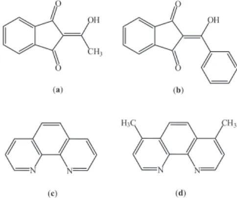
and L = 1,10-phenanthroline or 4,7-dimethyl-1,10-phenanthroline (Figure 1). In addition, theoretical structures of the Eu3+ complexes were optimized by
semi-empirical quantum-mechanical calculations and their
optical properties have been investigated on the basis of experimental intensity parameters.
Experimental
Materials and methods
The solvents (acetone and ethanol), 1,10-phenanthroline, 4,7-dimethyl-1,10-phenanthroline and lanthanide oxides (Ln2O3) were purchased from Aldrich Co. and used without
previous treatment. Hexahydrated lanthanide chlorides, LnCl3⋅6H2O, were prepared by reaction between Ln2O3
and hydrochloric acid purchased from Aldrich Co.18
2-acetyl-1,3-indandione (aind), 2-benzoyl-1,3-indandione (bind) ligands, and [Ln(aind)3(H2O)2], [Ln(bind)3(H2O)2]
precursor complexes were synthesized according to the procedures as reported in the literature.4,11
Elemental analyses (C, H, and N) were carried out using a Perkin Elmer 2400 elemental microanalyzer. Infrared (IR) spectra were recorded from 400 to 4000 cm−1 on a Shimadzu FT-IR spectrophotometer
model IRPRESTIGE-21 using KBr pellets. Excitation and emission spectra of the Ln3+-complexes in solid
state were recorded at liquid nitrogen temperature on a Fluorolog-3 spectrofluorometer (Horiba) with excitation and emission double grating 0.22 m monochromators, and a 450 W Xenon lamp as excitation source and R928P PMT as detector. All spectra were recorded using a detector mode correction. The second-order diffraction of the source radiation was eliminated using a cut-off filter. Luminescence decay curves of the Eu3+-complexes in the
solid state were recorded at low temperature (77 K) using the same equipment but operating on the phosphorescence mode with pulsed Xenon lamp as excitation source. The luminescence instruments were fully controlled by the FluorEssence program. All the luminescence data of the samples were measured in quartz tube of 2 mm in diameter. TG curves were recorded from 30 to 900 °C using a Shimadzu DTG-60H thermobalance with a heating rate 10 °C min−1 under a nitrogen atmosphere.
Synthesis of the Ln3+-complexes with heteroaromatic ligands
Lanthanide indandionate complexes containing ancillary ligands were synthesized by mixing acetonic solutions of the corresponding hydrated complexes, [Ln(aind)3(H2O)2] or [Ln(bind)3(H2O)2], with phen or
dmphen ligands in the molar ratio of complex:ligand (1:1.3). The resulting solutions were stirred for 1 h at room temperature (ca. 27 °C). Later on, these solutions were stand up until the total evaporation of solvent. The prepared
Figure 1. Structural formulas of the ligands: (a) 2-acetyl-1,3-indandione
yellow solids were washed thoroughly with cold ethanol and dried under reduced pressure.
[Eu(aind)3(phen)(H2O)] (1): 0.2482 g, yield 68%;
FT-IR (KBr, cm−1): 3485(w), 3443(w), 3057(w), 2951(w),
2920(w), 2856(w), 1681(m), 1643(m), 1622(s), 1585(s), 1467(s), 1363(m), 1340(m), and 732(s). Anal. Calc. for C45H31EuN2O10: C, 59.28, H, 3.43, and N, 3.07. Found: C,
59.71; H, 3.35, and N, 2.74.
[Eu(bind)3(phen)]·H2O (2): 0.4230 g, yield 72%;
FT-IR (KBr, cm−1): 3429(w), 3055(w), 2952(w),
2920(w), 1694(m), 1618(s), 1587(s), 1568(s), 1564(s), 1448(s), 1420(s), 1340(m), and 741(m). Anal. Calc. for C60H37EuN2O10: C, 65.64; H, 3.40, and N, 2.55. Found: C,
65.35, H, 3.44, and N, 2.84.
[Gd(aind)3(phen)(H2O)] (3): 0.2088 g, yield 56%;
FT-IR (KBr, cm−1): 3524(w), 3433(w), 3057(w), 2951(w),
2922(w), 2866(w), 2856(w), 1682(m), 1643(m), 1622(s), 1587(s), 1467(s), 1364(m), 1340(m), and 735(s). Anal. Calc. for C45H31GdN2O10: C, 58.94, H, 3.41, and N, 3.05.
Found: C, 59.63, H, 4.15, and N, 2.66.
[Gd(bind)3(phen)]·H2O (4): 0.2042 g, yield 58%; FT-IR
(KBr, cm−1): 3445(w), 3053(w), 3032(w), 2926(w),
1694(m), 1636(m), 1616(s), 1587(s), 1568(s), 1564(s), 1448(s), 1423(s), 1340(m), and 741(m). Anal. Calc. for C60H37GdN2O10: C, 65.32, H, 3.38, and N, 2.54. Found: C,
65.55, H, 3.34, and N, 3.24.
[Eu(aind)3(dmphen)(H2O)] (5): 0.1050 g, yield 57%;
FT-IR (KBr, cm−1): 3564(w), 3502(w), 3068(w), 1691(m),
(s), 1643(s), 1620 (s), 1583(s), 1467(s), 1359(m), 1340(m), and 725(s). Anal. Calc. for C47H35EuN2O10: C, 60.06, H,
3.73, and N, 2.98. Found: C, 60.26, H, 3.71, and N, 3.01.
[Eu(bind)3(dmphen)]·H2O (6): 0.1030 g, yield 58%;
FT-IR (KBr, cm−1): 3435(w), 3055(w), 1693(m), 1629(m),
1612(m), 1585(s), 1564(s), 1448(s), 1421(s), 1361(m), and 736(m). Anal. Calc. for C62H41EuN2O10: C, 66.13, H, 3.64,
and N, 2.49. Found: C, 66.28, H, 3.48, and N, 2.52.
[Gd(aind)3(dmphen)(H2O)] (7): 0.1630 g, yield 60%;
FT-IR (KBr, cm−1): 3442(w), 3068(w), 1685(m), 1645(m),
1620(s), 1585(s), 1467(m), 1357(m), 1340(m), and 729(m). Anal. Calc. for C47H35GdN2O10: C, 59.74, H, 3.71, and N,
2.97. Found: C, 60.32, H, 3.61, and N, 3.12.
[Gd(bind)3(dmphen)]·H2O (8): 0.0980 g, yield 55%;
FT-IR (KBr, cm−1): 3441(w), 3053(w), 1691(m), 1629(s),
1612(s), 1585(s), 1564(s), 1450(s), 1421(s), 1342(m), and 736(m). Anal. Calc. for C62H41GdN2O10: C, 65.84, H, 3.63,
and N, 2.47. Found: C, 65.92, H, 3.57, and N, 2.67.
Results and Discussion
Characterization
The results of the elemental analysis (C, H, and N) are consistent with the monohydrated formulas of Ln(aind)3L⋅H2O and Ln(bind)3L⋅H2O complexes, where
Ln = Eu3+ or Gd3+.
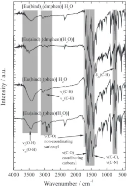
The infrared spectra of the Ln3+-complexes (Figure 2)
exhibit two bands at around 1560 and 1690 cm−1 assigned to
the ν(C=O) stretching modes, due to the coordinated and non-coordinated carbonyl groups, respectively. These data are in agreement with the FT-IR spectral data of precursor-hydrated complexes,11 suggesting that the 2-acyl-1,3-indandione
ligands are coordinated to Ln3+ ions through oxygen atoms
of the carbonyl groups in bidentate mode. Moreover, the absorption bands at around 1468 and 1423 cm−1 indicate
that the phen and dmphen ligands act as chelating agents, coordinating to the Ln3+ ion through the nitrogen atoms.19,20
The complexes also exhibit absorption bands in the spectral range 4000-3200 cm−1 assigned to ν(O−H) stretching of the
water molecule (Figure 2). Interestingly, FT-IR spectra of the [Ln(aind)3L⋅H2O] compounds exhibit narrow peaks at
around 3560 cm−1, while the [Ln(bind)
3L]⋅H2O compounds
show only one broad absorption band. These data give
Figure 2. FT-IR absorption spectra of the Eu3+-complexes recorded in the
evidences that the water molecule is coordinated to the Ln3+ ion only in the [Ln(aind)
3L⋅H2O] complexes. Similar
spectral profiles have also been observed for La3+ and Tb3+
complexes using EDTA as ligand that present coordinated and non-coordinated water molecules, respectively.21,22
Thermogravimetric curves of the [Eu(aind)3(dmphen)
(H2O)] and [Eu(bind)3(dmphen)]·H2O compounds
(Figure 3) show that these systems undergo thermal decomposition in consecutive steps. The curves show the first weight loss in the temperature range from 60 to 110 oC
that correspond to 1.6% for [Eu(bind)3(dmphen)]·H2O and
2.1% for [Eu(aind)3(dmphen)(H2O)], which are in agreement
with the release of water molecules. However, it can be observed in Figure 3 that the [Eu(aind)3(dmphen)(H2O)]
complex exhibits a shorter dehydration interval (60-130 oC)
than the [Eu(bind)3(dmphen)](H2O) complex, in which the
dehydration process occurs from 40 to 150 oC (see insert
Figure 3). These data corroborate with the FT-IR results, indicating that the water molecules are acting as ligand in the former complex. Similar behavior has been observed in the thermal and FT-IR studies of the Ln3+-EDTA complexes,
as reported by Gigante et al..22
Figure 3 also shows that the anhydrous compounds undergo consecutive weight loss starting from 250 oC,
yielding the lanthanide oxides, for example, Eu2O3, that is
in agreement with theoretical values: 15.3% (calc. 18.7%) and 16.4% (calc. 15.6%) for [Eu(aind)3(dmphen)(H2O)]
and [Eu(bind)3(dmphen)]·H2O compounds, respectively.
Theoretical structural studies
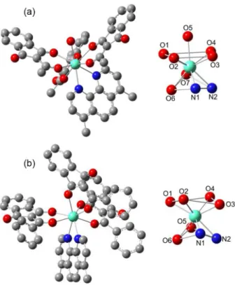
T h e o r e t i c a l m o l e c u l a r s t r u c t u r e s o f t h e [Eu(aind)3L(H2O)] and [Eu(bind)3L]·H2O complexes,
where L = phen or dmphen, were optimized using the reparameterized latest version of the SPARKLE/AM1
model for Eu(III) and other lanthanide ions implemented in the software package MOPAC2012.23,24
Water molecule was not considered in the last complexes, since the FT-IR and thermogravimetric data indicate that the water molecule is out of the first coordination sphere. The calculated structures of the [Eu(aind)3(dmphen)(H2O)] and
[Eu(bind)3(dmphen)]·H2O complexes are shown in Figure 4,
while those ones for the similar complexes with phen ligand are presented in Supporting Information (Figure S1).
The optimized structures reveal that the indandionate ligands are characterized by the planarity of 1,3-indandione moieties. In addition, the substituent phenyl groups in the bind ligands are twisted from the 1,3-indandione moieties at around angles of 29 and 40°. The less pronounced twist is observed in the phenyl group that belongs to the bonded molecule more distant from heteroaromatic ligand. This behavior reflects the steric hindrance on the ligands due to the heteroaromatic one in the first coordination sphere. Thus, the phenyl groups are orientated to minimize steric repulsion among ligands. Therefore, the steric hindrance due to the substituent phenyl groups of the ligand is enough to avoid water coordination to the lanthanide ion compared to the [Eu(aind)3L(H2O)] complexes.
The coordination polyhedron, EuN2O6, for the
[Eu(bind)3L]⋅H2O complexes can be described as distorted Figure 3. Thermogravimetric curves of the [Eu(aind)3(dmphen)(H2O)]
(solid line) and [Eu(bind)3(dmphen)]·H2O (dashed line) complexes.
Figure 4. Theoretical geometries of the Eu3+-complexes optimized
square antiprismatic geometries (Figure 4b, Figure S1). A square face contains four oxygen atoms from indandionate ligands, another one presenting two oxygen atoms from indandionate and two nitrogen atoms belong to phen or dmphen ligand.
The coordination polyhedron of the [Eu(aind)3L(H2O)]
complex, EuN2O7, is better described as a distorted
monocapped square antiprismatic geometry, with oxygen atom from water molecule occupying the capping position (Figure 4a, Figure S1). The bond lengths of Eu3+-O(indandionate), and Eu3+-N(heteroaromatic) in
the complexes are around 2.38 and 2.51 Å, respectively. These structural data are close to those obtained for [Eu(isovind)3(EtOH)(H2O)],11 as well as other
Eu3+-compounds with bipy and phen ligands.25-28
Photoluminescent properties of the Gd3+-compounds
The phosphorescence spectra of the analogous [Gd(aind)3L(H2O)] and [Gd(bind)3L]·H2O compounds
were recorded at 77 K with excitation monitored at 370 nm for S0→S1 transition, in order to estimate the
triplet state energies (T1) of the indandionate ligands. The
Gd3+-compounds were used for this propose because the
lowest excited energy level of Gd3+ ion (6P
7/2) is too high
to receive energy from the ligands. In addition, the Gd3+
radius is similar to the Eu3+ ion, therefore, Gd3+-complexes
mimetize both geometry and intraligand energy level structures of Eu3+-complexes. Figure S2 shows the
phosphorescence spectra of the Gd3+-compounds, which
exhibit broad emission bands in the spectral range from 440 to 650 nm assigned to the T→S0 transition centered
on the indandionate ligands.
The first T1 excited state energies of the ligands were
estimated as the energies correspond to the 0-0 phonon transitions from the phosphorescence spectra of the [Gd(aind)3L(H2O)], and [Gd(bind)3L]·H2O compounds
with values of 21906 cm−1 (456.5 nm), and 22371 cm−1
(447.0 nm), respectively. These results suggest that both the aind and bind ligands have appropriated T1 state energies,
which play most important role in intramolecular ligand-to-metal energy transfer process. Although, the S1 and T1 energy
levels of the 1,10-phenanthroline ligand and its derivative are usually located at around 30000 and 22000 cm−1,
respectively,29 the main role of these ligands in lanthanide
diketonate complexes is only to act as ancillary ones.30
Photoluminescent properties of the Eu3+-compounds
Figure 5 shows the excitation spectra of the [Eu(aind)3L(H2O)] and [Eu(bind)3L]·H2O compounds
recorded at 77 K in the spectral range 250-590 nm with emission monitored on the 5D
0→7F2 hypersensitive
transition at 611 nm. These spectra are dominated by two intense broad absorption bands in the spectral range from 270-470 nm, which are assigned to the S0→S1 and S0→S2
transitions centered on the acind ligands. These bands are overlapped with the absorption bands assigned to the S0→S1
transition of the phen and dmphen ligands at 300 nm. The spectral profiles of the Eu3+-compounds (Figure 5) indicate
that the S0→S1 and S0→S2 transitions are approximately
located in the 1,3-indandionate moieties. These results are in agreement with those ones observed for similar complexes, as reported in the literature.11
Figure 5 also presents some characteristic narrow absorption bands assigned to the 7F
0→2S+1LJ
intraconfigurational transitions of the Eu3+ ion. These bands
exhibit lower intensity than those ones, which correspond to the intraligand transitions, corroborating the efficient sensitization process via antenna effect.
The emission spectra of the [Eu(aind)3L(H2O)] and
[Eu(bind)3L]·H2O compounds were recorded at 77 K in Figure 5. Excitation spectra of the Eu3+-compounds recorded at
the spectral range from 420 to 730 nm, under excitation centered on indandionate ligand, S0→S1 transitions (at
370 nm), as shown in Figure 6. The luminescent spectra display only the characteristic emission narrow bands that are assigned to the 5D
0→7FJ transitions of the Eu3+ ion,
where J = 0, 1, 2, 3, and 4.31,32
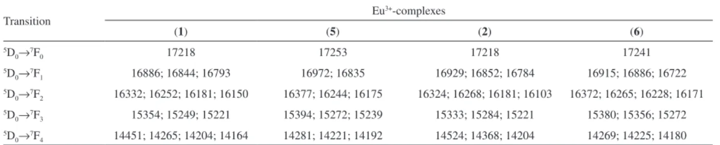
The local ligand field effect of the Eu3+-complexes has
been qualitatively investigated based on the splitting and relative emission intensities of the 5D
0→7FJ(0-4) transitions.
Table 1 presents the intraconfigurational-4f6 transitions
and their Stark (2J + 1)-components. The presence of the only one emission band assigned to 5D
0→7F0, as well
as three stark components for 5D
0→7F1 indicate that the
Eu3+ is located at low symmetry environment (C
n, Cnv or
Cs).33 These optical data corroborate with the fact that the
intensity of the band assigned to 5D
0→7F2 transition is
higher than that of the assigned to 5D
0→7F1 one,27 which
are in close agreement with the distorted coordination
polyhedra obtained from theoretical modeling for both Eu3+-complexes (Figure 4, Figure S1).
The absence of broad phosphorescence bands arising from the indandionate ligands (Figure 6) emphasizes the presence of an efficient luminescence sensitization occurring via antenna effect. The efficient intramolecular energy transfer mechanism from aind and bind ligands to the Eu3+
ion in the complexes is characterized by: (i) strong absorption of S0→S1 transition of the 2-acyl-1,3-indandione ligands;
(ii) intersystem cross by the non-radiative processes S1→T; (iii) energy transfer of the T1→(5D
1, 5D0), and (iv) efficient
radiative process from the 5D
0→7FJ(0-4) transitions.34-35
Judd-Ofelt intensity parameters (Ωλ = 2.4), radiative (Arad), non-radiative (Anrad) rates, and emission quantum
efficiency (η) of the 5D
0 emitting level of the Eu3+ complexes
have been quantitatively obtained from the luminescence decay curves and emission spectral data (Table 2). The luminescence decay curves (Figure S3) were recorded by monitoring the emission intensity of the hypersensitive
5D
0→7F2 transition under excitation of the S0→S1 transition
centered on the 2-acyl-1,3-indandionate ligands at 370 nm. The curves were well-adjusted to a single exponential function, yielding the 5D
0 lifetimes values (τ) equal to 0.459,
0.465, 0.229, and 0.594 ms for [Eu(aind)3(phen)(H2O)],
[Eu(bind)3(phen)]·H2O, [Eu(aind)3(dmphen)(H2O)] and
[Eu(bind)3(dmphen)]·H2O (Table 2), respectively. While
using these data, the total decay rates were determined according to the expression: Atotal = 1/τ. The radiative rates
(Arad) were determined using the 5D0→7F2,4 transitions
(forced electric dipole) and the 5D
0→7F1 transition
(magnetic-dipole) as internal reference.36,37 The values of
both radiative and noradiative rates for [Eu(aind)3L(H2O)],
and [Eu(bind)3L]·H2O complexes are presented in Table 2.
When τ values of these complexes were compared with the corresponding ones of precursor hydrated complexes, it was observed a considerable decrease in both Arad and
Anrad rates. The decrease in Anrad values might be attributed
to the water molecules substitution by the auxiliary phen and dmphen ligands in the first coordination sphere of the Eu3+ ion (Table 2), that reduce the multiphonon decays,
Table 1. Energy levels of the Eu3+-complexes assigned to the transitions (5D
0→7FJ , J = 0-4) (in cm−1)
Transition Eu
3+-complexes
(1) (5) (2) (6)
5D
0→7F0 17218 17253 17218 17241
5D
0→7F1 16886; 16844; 16793 16972; 16835 16929; 16852; 16784 16915; 16886; 16722
5D
0→7F2 16332; 16252; 16181; 16150 16377; 16244; 16175 16324; 16268; 16181; 16103 16372; 16265; 16228; 16171
5D
0→7F3 15354; 15249; 15221 15394; 15272; 15239 15333; 15284; 15221 15380; 15356; 15272
5D
0→7F4 14451; 14265; 14204; 14164 14281; 14221; 14192 14524; 14368; 14204 14269; 14225; 14180
(1) [Eu(aind)3(phen)(H2O)]; (2) [Eu(bind)3(phen)]·H2O; (5) [Eu(aind)3(dmphen)(H2O)]; (6) [Eu(bind)3(dmphen)]·H2O. Figure 6. Emission spectra of the Eu3+-complexes recorded at 77 K in the
also observed for the other Eu3+-indandionate complexes,
as reported in the literature.11,12 The lower values of A nrad
for [Eu(bind)3L]·H2O compounds compared to those
observed for [Eu(aind)3L(H2O)] are a direct consequence
of the multiphonon decay, because the water molecule is not coordinated to the Eu3+ ion.
Moreover, the luminescence quantum efficiency was determined using the expression η = Arad/(Arad + Anrad).
Table 2 shows that the higher values were observed for the [Eu(bind)3L]⋅H2O compounds than the [Eu(aind)3L(H2O)]
ones, indicating the absence of water molecules in the first coordination of Eu3+ ion, which decrease significantly the
luminescence quenching effect.
The experimental intensity parameters Ω2 and Ω4 for the
complexes were directly determined from the Arad values of
the 5D
0→7F2 and 5D0→7F4 transitions, respectively.35,36 These
parameters depend on the local geometry and the ligating atoms polarizabilities in the first coordinating sphere around the metal central ion.37 As observed in Table 2, the values of
Ω2 for the complexes containing heteroaromatic phen and
dmphen ligands are lower than those ones calculated for the hydrated precursor compounds.11 This result suggests that
the chemical environment polarizabilities have decreased when phen and dmphen ligands substitute water molecules in the first coordination sphere of the Eu3+ ion, which have
contributed to a significant decrease in the emission rate assigned to the 5D
0→7F2 transition. These values are similar
to those found for europium 2-acyl-1,3-indandionate complexes containing triphenylphosphine oxide ligand.11
The values of Ω4 exhibit similar behavior, suggesting that
the sensibility of the 5D
0→7F4 transition has also been
changed with the polarizability and chemical environment.
Conclusions
In the current work, we have reported the synthesis, characterization, and photoluminecent properties of
[Ln(aind)3L(H2O)] and [Ln(bind)3L]·H2O coordination
compounds (Ln : Eu3+ or Gd3+). The FT-IR data suggest
that the water molecule is coordinated to the Ln3+ ion in the
case of 1, 3, 5, and 7 complexes. The photoluminescence
data are consistent with coordination polyhedra calculated
via SPARKLE/AM1 model for the [Ln(aind)3L(H2O)] and
[Ln(bind)3L]·H2O complexes. The structural differences
and the absence of water molecules in the first coordination sphere have great impact on the luminescence quantum efficiency (η) of the Eu3+-complexes, decreasing the
luminescence quenching in the intramolecular energy transfer process.
Supplementary Information
The luminescence decay curves of the Eu3+-complexes are
available free of charge at http://jbcs.sbq.org.br as PDF file.
Acknowledgements
This research was supported by grants from the Conselho Nacional de Desenvolvimento Científico e Tecnológico (CNPq), Instituto Nacional de Ciência e Tecnologia-Nanotecnologia para Marcadores Integrados (INCT-INAMI), FACEPE-PRONEX, Fundação de Amparo à Pesquisa do Estado de São Paulo (FAPESP No. 2003/07178-8) and, Financiadora de Estudos e Projetos (FINEP).
References
1. Yue, E. W.; Higley, C. A.; DiMeo, S. V.; Carini, D. J.; Nugiel, D. A.; Benware, C.; Benfield, P. A.; Burton, C. R.; Cox, S.; Grafstrom, R. H.; Sharp, D. M.; Sisk, L. M.; Boylan, J. F.; Muckelbauer, J. K.; Smallwood, A. M.; Chen, H.; Chang, C.; Seitz, S. P.; Trainor, G. L.; J. Med. Chem.2002, 45, 5233. 2. Sloop, J. C.; Bumgardner, C. L.; Loehle, W. D.; J. Fluorine
Chem.2002, 118, 135.
Table 2. Experimental intensity parameters (Ωλ = 2.4), radiative (Arad), non-radiative (Anrad) rates, and emission quantum efficiency (η) of the 5D0 emitting
level determined for the 2-acyl-1,3-indandionate Eu3+-compounds
Eu3+-complexe Ω
2 / (10−20cm2) Ω4 / (10−20cm2) τ / ms Arad / s−1 Anrad / s−1 Atot / s−1 η / %
[Eu(aind)3(H2O)2] 42.1 14.8 0.108 1532 7739 9261 16.5
[Eu(aind)3(tppo)2] 35.5 8.9 0.531 1255 628 1883 66.7
[Eu(aind)3(phen)(H2O)] 4.4 6.6 0.459 280 1897 2177 12.8
[Eu(aind)3(dmphen)(H2O)] 16.6 3.9 0.229 611 3755 4367 14.0
[Eu(bind)3(H2O)2] 40.5 14.2 0.054 1482 17080 18560 8.0
[Eu(bind)3(tppo)2] 29.4 15.9 0.318 1165 1978 3143 37.1
[Eu(bind)3(phen)]·H2O 29.5 8.8 0.465 1064 1086 2150 49.5
3. Enchev, V.; Ivanova, G.; Pavlovic, G.; Rogojerov, M.; Ahmedova, A.; Mitewa, M.; J. Mol. Struct.2003, 654, 11. 4. Kilgore, L. B.; Ford, J. H.; Wolfe, W. C.; Ind. Eng. Chem. Res.
1942, 34, 494.
5. Liu, Y.; Saldivar, A.; Bess, J.; Solomon, L.; Chen, C.; Tripathi, R.; Barrett, L.; Richardson, P. L.; Molla, A.; Kati, W.; Kohlbrenner, W.; Biochemistry2003, 42, 8862.
6. Ahmedova, A.; Rusanov, V.; Hazell, A.; Wolny, J. A.; Gochev, G.; Trautwein, A. X.; Mitewa, M.; Inorg. Chim. Acta
2006, 359, 3123.
7. Ahmedova, A.; Cador, O.; Sorace, L.; Ciattini, S.; Gatteschi, D.; Mitewa, M.; J. Coord. Chem.2008, 61, 3879.
8. Ahmedova, A.; Marinova, P.; Stoyanov, N.; Ciattini, S.; Springborg, M.; Mitewa, M.; Struct. Chem.2009, 20, 101. 9. Ahmedova, A.; Marinova, P.; Stoyanov, N.; Ciattini, S.;
Springborg, M.; Mitewa, M.; Polyhedron2010, 29, 1687. 10. Ahmedova, A.; Atanasov, V.; Marinova, P.; Stoyanov, N.;
Mitewa, M.; Cent. Eur. J. Chem.2009, 7, 429.
11. Teotonio, E. E. S.; Brito, H. F.; Viertler, H.; Faustino, W. M.; Malta, O. L.; Sá, G. F.; Felinto, M. C. F. C.; Santos, R. H. A.; Cremona, M.; Polyhedron2006, 25, 3488.
12. Teotonio, E. E. S.; Brito, H. F.; Cremona, M.; Quirino, W. G.; Legnani, C.; Felinto, M. C. F. C.; Opt. Mater.2009, 32, 345. 13. Li, J.; Li, H.; Yan, P.; Chen, P.; Hou, G.; Li, G.; Inorg. Chem.
2012, 51, 5050.
14. Romanova, K. A.; Datskevich, N. P.; Taidakov, I. V.; Vitukhnovskii, A. G.; Galyametdinov, Yu. G.; Russ. J. Phys. Chem. A2013, 87, 2108.
15. Greco, C.; Moro, G.; Bertini, L.; Biczysko, M.; Barone, V.; Cosentino, U.; J. Chem. Theory Comput.2014, 10, 767. 16. Yang, C. L.; Xu, J.; Li, J. Y.; Lu, M. G.; Li, Y. B.; Wang, X. L.;
Sens. Actuators, B2014, 196, 133.
17. Yang, C. L.; Xu, J.; Ma, J. Y.; Zhu, D. Y.; Zhang, Y. F.; Liang, L. Y.; Lu, M. G.; Polym. Chem.2012, 3, 2640.
18. Bemquerer, M. P.; Bloch, C.; Brito, H. F.; Teotonio, E. E. S.; Miranda, M. T. M.; J. Inorg. Biochem.2002, 91, 363. 19. Nakamoto, K.; Infrared and Raman Spectra of Inorganic and
Coordination Compounds, Applications in Coordination,
Organometallic, and Bioinorganic, 6th ed.; Willey: New Jersey,
2009.
20. Gerasimova, T. P.; Katsyuba, S. A.; Dalton Trans.2013, 42, 1787.
21. Wagner, C. C.; Baran, E. J.; Spectrochim. Acta, Part A 2010, 75, 807.
22. Gigante, A. C.; Caires, F. J.; Gomes, D. J. C.; Lima, L. S.; Treu-Filho, O.; Pivatto, M.; Ionashiro, M.; J. Therm. Anal. Calorim.2014, 115, 127.
23. Stewart J. J. P. “MOPAC2012”, Stewart Computational Chemistry, Colorado Springs, CO, USA. Available: http:// OpenMOPAC.net, accessed in April 2014.
24. Freire, R. O.; Rocha, G. B.; Simas, A. M.; Inorg. Chem.2005, 44, 3299.
25. Sá, G. F.; Batista, H. J.; Andrade, A. V. M.; Longo, R. L.; Simas, A. M.; Ito, N. K.; Thompson, L. C.; Inorg. Chem.1998, 37, 3542.
26. Li, X.; Bian, Z.; Jin, L.; Lu, S.; Huang, S.; J. Mol. Struct.2000,
522, 117.
27. Chen, X.-F.; Zhu, X.-H.; Xu, Y.-H.; Raj, S. S. S.; Öztürk, S.; Fun, H.-K.; Ma, J.; You, X. Z.; J. Mater. Chem.1999, 9, 2919. 28. Lü, Y.-G.; Li, G.; Shi, C.-H.; Yu, L. S.; Luan, F.; Zhang, F. J.;
Trans. Nonferrous Met. Soc. China2010, 20, 2336. 29. Yu, X.; Su, Q.; J. Photoch. Photobio. A2003, 155, 73. 30. Brito, H. F.; Malta, O. L.; Felinto, M. C. F. C.; Teotonio, E. E. S.
In The Chemistry of Metal Enolates, v. 1; Zabicky, J., ed.; John Wiley & Sons, Ltd.: Chichester, 2009, ch. 3.
31. Binnemans, K.; Chem. Rev.2009, 109, 4283.
32. Gruber, J. B.; Valiev, U. V.; Burdick, G. W.; Rakhimov, S. A.; Pokhrel, M.; Sardar, D. K.; J. Lumin.2011, 131, 1945. 33. Forsberg, J. H.; Coord. Chem. Rev.1973, 10, 195.
34. Sá, G. F.; Malta, O. L.; Donegá, C. M.; Simas, A. M.; Longo, R. L.; Santa-Cruz, P. A.; Silva Júnior, E. F.; Coord. Chem. Rev.
2000, 196, 165.
35. Faustino, W. M.; Nunes, L. A.; Terra, I. A. A.; Felinto, M. C. F. C.; Brito, H. F.; Malta, O. L.; J. Lumin.2013, 137, 269. 36. Werts, M. H. V.; Jukes, R. T. F.; Verhoeven, J. W.; Phys. Chem.
Chem. Phys.2002, 4, 1542.
37. Bünzli, J.-C. G.; Eliseeva, S. V. In Lanthanide Luminescence: Photophysical, Analytical and Biological Aspects VII; Hänninen, P.; Härmä, H., eds.; Springer: Berlin, 2010, ch. 1.
Submitted on: March 13, 2014
Published online: August 26, 2014
![Figure 5 shows the excitation spectra of the [Eu(aind) 3 L(H 2 O)] and [Eu(bind) 3 L]·H 2 O compounds](https://thumb-eu.123doks.com/thumbv2/123dok_br/18997972.462799/5.892.455.793.503.980/figure-shows-excitation-spectra-eu-aind-bind-compounds.webp)
![Table 2 shows that the higher values were observed for the [Eu(bind) 3 L]⋅H 2 O compounds than the [Eu(aind) 3 L(H 2 O)]](https://thumb-eu.123doks.com/thumbv2/123dok_br/18997972.462799/7.892.69.803.163.357/table-shows-higher-values-observed-bind-compounds-aind.webp)
