Influence of treatment including second molars on final
and postretention molar angulation
Luiz Filiphe Gonçalves Canuto1, Karina Maria Salvatores de Freitas2, Marcos Roberto de Freitas3, Rodrigo Hermont Cançado4
How to cite this article: Canuto LFG, Freitas KMS, Freitas MR, Cançado RH. Influence of treatment including second molars on final and postretention molar angulation. Dental Press J Orthod. 2013 Sept-Oct;18(5):121-7.
Submitted: September 20, 2011 - Revised and accepted: December 27, 2011
» The authors report no commercial, proprietary or financial interest in the prod-ucts or companies described in this article.
Contact address: Luiz Filiphe Gonçalves Canuto
Rua José Bonifácio, 205 – Sl 109 – Madalena – Recife/PE, Brazil CEP: 50.710-000 – E-mail: [email protected]
1 Professor of graduate program in Orthodontics at the Brazilian Dental
Association / Pernambuco (ABO-PE).
2 Professor of the graduate program in Orthodontics at UNINGÁ.
3 Full professor of Pediatric Dentistry, Orthodontics and Public Health at USP,
Bauru Dental School.
4 Associate professor at UNINGÁ.
Objective:Evaluate axial mesiodistal inclinations of the mandibular molars in orthodontically treated cases, analyzing
whether inclusion of second mandibular molars in treatment mechanics has any influence on final and postretention molars angulations. Methods: The sample comprised 150 panoramic radiographs of 50 patients. Patients were treated with extraction of four first premolars and divided into 2 groups: Group 1 comprised 25 subjects without inclusion of mandibular second molars during orthodontic treatment, whereas Group 2 comprised 25 subjects with inclusion of mandibular second molars. Panoramic radiographs at three observation times were evaluated: pretreatment, post-treatment and postretention. The statistical analysis included one-way analysis of variance (ANOVA) for intragroup evaluation and independent t-tests for intergroup comparisons. Results: Intragroup analysis demonstrated significant uprighting of mandibular first and second molars during treatment in Group 2, which remained stable during the postretention stage. Intergroup comparison demonstrated that Group 2 presented first and second molars significantly more uprighted in relation to Group 1 in both post-treatment and postretention stages. Conclusions: It was con-cluded that inclusion of mandibular second molars in the orthodontic mechanics is relevant not only to correct the angulation of these teeth, but also to aid mandibular first molars uprighting.
Keywords:Panoramic X-ray. Tooth angulation. Tooth movement.
Objetivo:analisar a influência da inclusão dos segundos molares inferiores durante a mecânica ortodôntica nas
an-gulações dos molares ao final do tratamento e na fase de pós-contenção. Métodos: a amostra consistiu em 150 radio-grafias panorâmicas de 50 pacientes avaliados antes, após o tratamento e no período de pós-contenção. Os pacientes foram tratados com extrações dos quatro primeiros pré-molares, e divididos em dois grupos: grupo 1, composto por 25 pacientes com segundo molares incluídos na mecânica ortodôntica; grupo 2, 25 pacientes cujos segundos molares não foram incluídos na mecânica ortodôntica. As angulações dos primeiros e segundos molares inferiores foram com-paradas nas fases estudadas utilizando-se a Análise de Variância (análise intragrupo) e o teste t independente (análise
intergrupos). Resultados: a análise intragrupo, realizada no grupo 2, demonstrou que ocorreu uma verticalização significativa dos primeiros e segundos molares inferiores durante o tratamento, que se manteve estável na fase de pós--contenção. Os resultados da análise intergrupos demonstraram diferenças significativas na angulação dos primeiros e segundos molares após o tratamento e na fase de pós-contenção. Conclusão: a inclusão dos segundos molares inferiores à mecânica ortodôntica apresenta-se relevante, não apenas para corrigir a angulação desses dentes, mas, também, para auxiliar a correção da angulação dos primeiros molares permanentes.
INTRODUCTION
The importance of appropriate mesiodistal teeth angulation in orthodontic patients has been empha-sized by many clinicians. In 1972, Andrews reported that tooth angulation is one of the 6 keys to be
evalu-ated in ideal static occlusions.1
It has been reported that the final spatial orienta-tion of each tooth should be such that it can best with-stand the forces during function. Corrected angula-tion is universally accepted, and other several related parameters have been studied. These include
peri-odontal health,1,2,3 even distribution of occlusal forces
through contact points, tight posterior occlusion, no spaces as well as retention and stability of
orthodonti-cally closed extraction sites.1,4-7 The American Board
of Orthodontics has included assessment of mesio-distal angulation in panoramic radiographs as a pa-rameter for evaluating finished cases for orthodontists aspiring to be board diplomate.
Adequate mesiodistal teeth angulations with roots parallel to each other are frequently mentioned in the
literature1,3,8,9-13 as a fact that not only improves teeth
alignment stability in their apical bases, but also allows
normal maxilomandibular occlusion.6 Moreover, an
adequate mesiodistal positioning allows a uniform dis-tribution of occlusal forces through contact points and
contributes to overall treatment stability.3,6,10
Given the importance of appropriate mesiodis-tal teeth angulation in orthodontic patients regarding quality and stability of treatment, the authors aimed to investigate the inluence of mandibular second molars inclusion in orthodontic mechanics on inal and postre-tention molar angulations.
MATERIAL AND METHODS
Material
The sample comprised 150 panoramic radiographs of 50 young patients of both genders. The radio-graphs were taken from the files of Pediatric Dentist-ry, Orthodontics and Public Health Department at University of São Paulo (USP), Bauru Dental School. Each case was evaluated at three stages:
pretreat-ment (T1), posttreatment (T2) and after a minimum
of 3 years of follow-up (T3).
When selecting the sample, the following inclusion criteria were applied: cases initially presenting Class I or Class II malocclusion, treated with ixed Edgewise
ap-pliances and extraction of four irst premolars. All sub-jects had all permanent teeth erupted except the third molars at the pretreatment stage. Other inclusion crite-ria were patients with no history of previous interceptive orthodontic treatment, absence of root dilaceration or mandibular skeletal asymmetries.
Ater active treatment, all patients wore a modiied Hawley retainer in the maxillary arch, full time for the irst 12 months and during sleep for the next 6 months. A lingual canine-to-canine mandibular bonded retainer was placed and let for a mean period of 3 years.
Methods
The sample was divided into two groups:
» Group 1: 25 patients (14 female; 11 male) in whom the mandibular second molars were not included in treatment mechanics.
» Group 2: 25 patients (15 female; 10 male) with man-dibular second molars included in treatment mechanics.
The mean pretreatment age was 13.29 ± 1.44
years for Group 1 and 12.95 ± 1.26 years for Group 2.
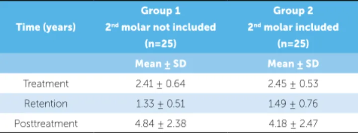
The mean treatment, retention and postretention evalu-ation times of each group are shown in Table 1.
First and second molar angulations were evaluated with panoramic radiographs (orthopantomography) traced manually by a single investigator, in acetate paper
(Ultraphan Paper®, Berlin, Germany).
Time (years)
Group 1
2nd molar not included
(n=25)
Group 2
2nd molar included
(n=25)
Mean ± SD Mean ± SD
Treatment 2.41 ± 0.64 2.45 ± 0.53
Retention 1.33 ± 0.51 1.49 ± 0.76
Posttreatment 4.84 ± 2.38 4.18 ± 2.47
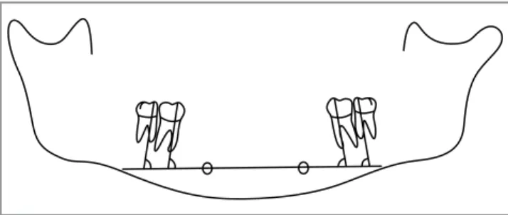
Tracing method
The tracing procedure of the initial, posttreat-ment and postretention radiographs was conducted in four phases: a) Delineation of dentoskeletal structures; b) Deinition of reference points; c) Deinition of hori-zontal and vertical reference lines; d) Measurement of tooth angulation (Fig 1).
a) Delineation of dentoskeletal structures:
The external outline of the mandible, mental fora-men, and outlines of mandibular irst and second molars roots and crowns were traced.
b) Definition of landmarks:
The deinition of the landmarks was performed as
proposed by Tavano et al:14
1) Right mental foramen (RMF) – The central point of the right mental foramen.
2) Let mental foramen (LMF) – The central point of the let mental foramen.
c) Tracing of horizontal and vertical reference lines:
1) Intermental line (IL): Line passing through the centers of the right and let mental foramens. 2) First and second molars long axes: The long
axes of the mentioned teeth were determined as the mean of the images of mesial and distal
root canals13.
d) Angles measurement:
The angles formed by the Intermental line (IL) and the long axes of the irst and second mandibular molars were then measured (Fig 1).
Statistical analyses
Statistical analysis was performed with Statistica sot-ware (Statistica for Windows, version 7.0, StatSot Inc). To avoid type I error (probability of accepting the alternative hypothesis H1 and be wrong) sample was
calculated considering α = 5% (type I error), β = 20%
(type II error), an estimated variability (s) of 5 degrees and a minimum detectable diference (d) of 5 degrees.
In each group, means and standard deviations for the mesiodistal inclination of the four evaluated teeth (let mandibular irst molar-36; let mandibular second mo-lar-37; right mandibular irst molar-46; and right man-dibular second molar-47) were determined. The intra-group comparison of these variables at the three ob-servation stages was performed by one-way dependent ANOVA and Tukey tests as a second step. For inter-group comparison, t-tests were used. Prior to the use of ANOVA and t-tests, analyses of data normality and homoscedasticity of the groups was performed with Kolmogorov-Smirnov and Levene tests, respectively.
Method error
Within a week interval from the irst measure-ment, 30 randomly selected radiographs were retraced and remeasured by the same examiner. The random error was calculated according to Dahlberg’s formula
(Se2= Σd2/2n) and the systematic error was calculated
with dependent t-tests, for p<0.05.
RESULTS
Results for power analysis showed that a sample with 23 patients in each group would give a 80% ability to detect diferences, whereas a sample comprising 26 pa-tients in each group would give 85%.
Results for the data distribution evaluation performed by the Kolmogorov-Smirnov test showed p > 0.05 for both groups, for all variables evaluated, indicating that the data had normal distribution. Levene test was used to verify homoscedasticity. All results exhibited p> 0.05 for both groups, during the three stages evaluated. Thus, it was concluded that there was homogeneity of variables and that the ANOVA test could be applied for intragroup analysis.
The results for intragroup comparison in Group 1 (#37 and #47 teeth not included) demonstrated no statistically signiicant diferences between the mean values for the mesiodistal inclinations of the teeth (36, 37, 46 and 47) at
random and systematic errors were within acceptable parameters, thus, not inluencing the results and con-clusions of the present study.
The methodology of this study was based on
pre-vious researches9,13,14 that also used panoramic
radio-graphs to obtain tooth angulation measurements. Pan-oramic radiographs are ordinarily used in orthodontic practice to provide signiicant information about teeth, axial inclinations, maturation periods, and
surround-ing tissues.8,12,13,19 Some authors suggest that dental
ax-ial inclinations be radiographically checked at the
be-ginning and end of orthodontic treatment.3,5,7-10,13,16,17,19
Panoramic radiographs may be the technique of choice since it provides signiicant amount of diagnostic in-formation obtained by viewing all teeth as well as the basal bone at once. In addition, it is the best option to evaluate teeth axial inclinations and root parallelism
ater orthodontic treatment.2,5-7,11,12,13,15,17,24
As occurring in other radiographic methods, the dimensions of structures in panoramic radiographs can
be magniied5,7,8,11,12,13,15-19 and due to distortions,
hori-the three evaluated stages. On hori-the ohori-ther hand, results for the intragroup comparison in Group 2 (#37 and #47 teeth included) demonstrated signiicant uprighting of mandib-ular irst and second molars throughout treatment, which remained stable during the postretention stage (Table 2).
Intergroup comparison demonstrated that Group 2 presented the irst and second molars signiicantly up-righted in relation to Group 1 at both posttreatment and postretention stages (Table 3).
No signiicant systematic errors were detected and the major random error was of 1.27 degrees for the let mandibular irst molar mesiodistal inclination.
DISCUSSION
Throughout this research, signiicant eforts were expended in order to minimize, or at least control the errors deriving from the procedures involved in pan-oramic radiograph tracings, demarcation of landmarks and measurement of the variables investigated. Knowl-edge of the methodology precision provided more reli-able results. It was observed that the results obtained for
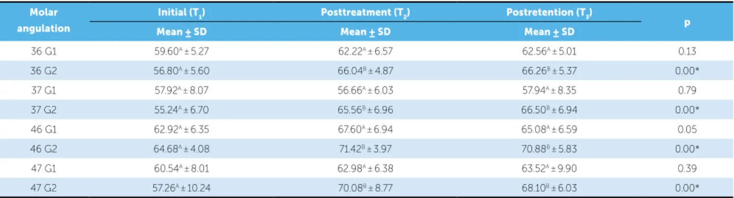
Table 2 - Means and standard deviations of mesiodistal inclinations of the teeth (36, 37, 46 and 47) at the three evaluation stages (T1, T2 and T3) for Groups 1 (second molars not included in treatment, n = 25) and 2 (second molars included, n = 25) and results of dependent ANOVA and Tukey tests.
*Statistically significant for p < 0.05. Different letters mean a statistically significant difference between the phases. Molar
angulation
Initial (T1) Posttreatment (T2) Postretention (T3)
p
Mean ± SD Mean ± SD Mean ± SD
36 G1 59.60A ±5.27 62.22A ±6.57 62.56A ±5.01 0.13
36 G2 56.80A ±5.60 66.04B ±4.87 66.26B ±5.37 0.00*
37 G1 57.92A ±8.07 56.66A ±6.03 57.94A ±8.35 0.79
37 G2 55.24A ±6.70 65.56B ±6.96 66.50B ±6.94 0.00*
46 G1 62.92A ±6.35 67.60A ±6.94 65.08A ±6.59 0.05
46 G2 64.68A ±4.08 71.42B ±3.97 70.88B ±5.83 0.00*
47 G1 60.54A ±8.01 62.98A ±6.38 63.52A ±9.90 0.39
47 G2 57.26A ±10.24 70.08B ±8.77 68.10B ±6.03 0.00*
Table 3 - Means and standard deviations of mesiodistal inclinations of the teeth (36, 37, 46 and 47) at the three evaluation stages (T1, T2 and T3) for Group 2 (second molars included) and results of dependent ANOVA and Tukey tests.
*Statistically significant for p < 0.05. Different letters mean a statistically significant difference between the phases. Molar
angulation
Initial (T1) Posttreatment (T2) Postretention (T3)
p
Mean ± SD Mean ± SD Mean ± SD
36 56.80A ±5.60 66.04B ±4.87 66.26B ±5.37 0.00*
37 55.24A ±6.70 65.56B ±6.96 66.50B ±6.94 0.00*
46 64.68A ±4.08 71.42B ±3.97 70.88B ±5.83 0.00*
comparison demonstrated signiicant uprighting of man-dibular irst and second molars during treatment, which remained stable at the postretention stage (Table 2). Re-garding the assessment of changes in mesiodistal dental inclination as a result of orthodontic treatment, there are few studies that could be used for comparisons, and most of them are related to patterns of normal occlusion.
In an attempt to establish a basis for quantitative evaluation of mesiodistal axial inclinations of permanent
teeth ater orthodontic treatment, Ursi et al13 conducted
a study that determined the normal mean values for den-tal angulations through panoramic radiographs. For the authors, the mesiodistal root angulations of high quality orthodontic treatment exhibited in the inal panoramic radiographs should be similar to normal occlusion values. In the present study, it was noted that the mean values obtained for the mesiodistal inclinations of the teeth (36, 37, 46 and 47) at posttreatment and postretention phases in Group 2 (with inclusion of the second molars) were
closer to the normal values proposed by Ursi et al.13
In 2002, Brandão9 evaluated if alterations in the
mesiodistal axial inclination of the mandibular anterior teeth would present any inluence in the relapse of their crowding. The panoramic radiographic and dental casts
of each patient were evaluated at the beginning (T1), at
the end (T2) and ive-year posttreatment (T3) phases.
Results showed that the mesiodistal axial inclinations of the teeth at the beginning of treatment were diferent from those observed in normal occlusion cases in 85% of the evaluated teeth. However, 45% of the teeth at the end, and 55% at the ive-year posttreatment phase showed mean values similar to those of normal occlu-sion. Evaluation of mesiodistal axial inclination stability at the ive-year posttreatment phase demonstrates that 75% of the teeth proved to maintain the angulation ob-tained at the end of the treatment, regardless of being similar or not to the normal values. The changes in the
mesiodistal axial inclination between T2 and T3 did not
inluence the relapse of mandibular anterior crowding.
In 2006, Almeida-Pedrin et al8 evaluated, through
panoramic radiographs, the mesiodistal axial incli-nations of the maxillary anterior teeth at the begin-ning and end of nonextraction orthodontic treatment. The experimental sample comprised 40 Caucasian pa-tients who were treated orthodontically with a standard Edgewise technique, without extractions. The mesio-distal axial inclinations of the maxillary anterior teeth of
zontal measurements are unreliable.17,19 In this study,
panoramic radiograph magniication did not inlu-ence the results, as the same radiographic equipment and similar techniques were used for both groups. Thus, when Groups 1 and 2 were compared, the pos-sible inluence of this variable was eliminated.
Accuracy of tooth length and angulation measure-ments on panoramic radiographs is thought to be highly
dependent on head positioning technique.18,20
Stramo-tas et al18 noted a signiicant error (p < 0.05) in such
measurements when the occlusal plane was tilted up anteriorly by 8 degrees. A lateral cant of the occlusal plane less than 10 degrees without an upward anterior rotation showed no signiicant efect on the measure-ments. Regarding angular measurements, the literature reports that the analysis of dental angulations through panoramic radiographs can be performed with good
reliability8,11,12,13,15,17,18,19 and that there is some tolerance
of variation in head position.18 During the radiographic
examination, all patients who comprised the sample were positioned with both the occlusal plane parallel and the sagittal plane perpendicular to the ground.
Recent studies have compared the accuracy of as-sessing mesiodistal root angulations with posttreatment panoramic radiographs and with cone-beam computed tomography (CT). The results show that CT is the most
accurate method for assessing dental angulation.21,22
Thus, assessment of mesiodistal tooth angulations with panoramic radiograph should be approached with cau-tion and reinforced by a thorough clinical examinacau-tion
of the dentition.23 However, due to economic as well
as biological reasons, CT should not be considered for clinical routine, but rather only for mesiodistal root an-gulations evaluation, before, during or ater orthodon-tic treatment. The use of panoramic radiograph as data source may be considered a limitation of this study. The use of CT could result not only in a more accurate assessment of the mesiodistal root angulations, but it could also enable tridimensional evaluation of the teeth. Another limitation of this research is the fact that it eval-uated the mandibular molars changes, only.
Results of Group 1 (#37 and #47 teeth not included) intragroup comparison demonstrated no statistically sig-niicant diferences between the mean values for the me-siodistal inclinations of the teeth (36, 37, 46 and 47) at the
three evaluation stages (T1, T2 and T3) (Table 2).
the experimental group at T1 were diferent from those of the control group for 50% of the evaluated teeth.
In contrast, the inclinations at T2 were consistent with
the normal anatomical coniguration of the controls. The authors concluded that panoramic radiograph is an efective tool for evaluating the mesiodistal axial inclina-tions of maxillary anterior teeth.
In 2009, Sella et al.25 compared the normal mean values
of mesiodistal axial angulations, proposed by Ursi et al,13
with mesiodistal axial angulations of canine teeth, premo-lars and inferior mopremo-lars in individuals aged between 18 and 25 years old, with and without the presence of the mandib-ular third molars. The authors concluded that the groups presented similar angular values for the canine teeth, pre-molars and inferior pre-molars in such a way that the presence of the third molars did not inluence dental angulations.
The intergroup comparison (Table 4) demonstrated statistically signiicant diferences with regards to mesio-distal inclinations of mandibular irst and second molars
at T2. Patients with mandibular second molars included
in treatment mechanics presented mandibular irst and
second molars more uprighted. At T3, mesiodistal
incli-nations of the molars remained signiicantly diferent
be-tween groups, except for the mean values for angulation
of mandibular right second molars (#47 – T3) that, despite
not statistically signiicant (p = 0.05), were, on average, ap-proximately 5 degrees more uprighted in comparison to Group 1. There are some limitations hindering compari-son between these intergroup results with other studies, namely: the nonexistence of previous studies with similar objectives in the literature and their methodological dif-ferences. However, based on the results of this research, it may be inferred that the inclusion of second mandibu-lar momandibu-lars in orthodontic mechanics beneits not only the mandibular second molars, but also irst molars upright-ing, as the mandibular irst molars in Group 2 were more uprighted at posttreatment and postretention stages (Tables 2 and 3). Additionally, the results suggest that the inclusion of second molars in orthodontic mechanics probably con-sists in a distal support that improves irst molar uprighting. There are some doubts and controversies about the necessity of second molars inclusion during orthodon-tic treatment. Two of the major goals of treatment con-sist in leveling the curve of Spee and correcting overbite. Thus, nothing is more rational than using the second molars to provide an anchorage that allows anterior teeth
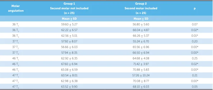
Table 4 - Intergroup comparison of mesiodistal inclinations of the teeth (36, 37, 46 and 47) at the three evaluation stages (T1, T2 and T3) with t-test.
*Statistically significant for p < 0.05. Molar
angulation
Group 1
Second molar not included (n = 25)
Group 2
Second molar included (n = 25)
p
Mean ± SD Mean ± SD
36 T1 59.60 ± 5.27 56.80 ± 5.60 0.07
36 T2 62.22 ± 6.57 66.04 ± 4.87 0.02*
36 T3 62.56 ± 5.01 66.26 ± 5.37 0.01*
37 T1 57.92 ± 8.07 55.24 ± 6.70 0.20
37 T2 56.66 ± 6.03 65.56 ± 6.96 0.00*
37 T3 57.94 ± 8.35 66.50 ± 6.94 0.00*
46 T1 62.92 ± 6.35 64.68 ± 4.08 0.25
46 T2 67.60 ± 6.94 71.42 ± 3.97 0.02*
46 T3 65.08 ± 6.59 70.88 ± 5.83 0.00*
47 T1 60.54 ± 8.01 57.26 ± 10.24 0.21
47 T2 62.98 ± 6.38 70.08 ± 8.77 0.00*
1. Andrews LF. The six keys to normal occlusion. Am J Orthod. 1972;62(3):296-309. 2. Dempster WT, Adams WJ, Duddles RA. Arrangement in the jaws of the roots
of the teeth. J Am Dent Assoc. 1963;67:779-97.
3. Hatasaka HH. A radiographic study of roots in extraction sites. Angle Orthod. 1976;46(1):64-8.
4. Beyron H. Optimal occlusion. Dent Clin North Am. 1969;13(3):537-54. 5. Ingervall B. Functionally optimal occlusion: the goal of orthodontic
treatment. Am J Orthod. 1976;70(1):81-90.
6. Mayoral G. Treatment results with light wires studied by panoramic radiography. Am J Orthod. 1982;81(6):489-97.
7. Williams R. Eliminating lower retention. J Clin Orthod. 1985;19(5):342-9. 8. Almeida-Pedrin RR, Pinzan A, Almeida RR, Ursi W, Almeida MR. Panoramic
evaluation of mesiodistal axial inclinations of maxillary anterior teeth in orthodontically treated subjects. Am J Orthod Dentofacial Orthop. 2006;130(1):56-60.
9. Brandão AG. Estudo ortopantomográico longitudinal das inclinações axiais mesiodistais em pacientes tratados ortodonticamente com extrações dos quatro primeiros pré-molares [dissertação]. Bauru (SP): Faculdade de Odontologia de Bauru; 2002.
10. Edwards JG. The prevention of relapse in extraction cases. Am J Orthod. 1971;60(2):128-44.
11. Jesuino FA, Costa LR, Valladares-Neto J. Mesiodistal root angulation of permanent teeth in children with mixed dentition and normal occlusion. J Appl Oral Sci. 2010;18(6):625-9.
12. Lucchesi MV, Wood RE, Nortje CJ. Suitability of the panoramic radiograph for assessment of mesiodistal angulation of teeth in the buccal segments of the mandible. Am J Orthod Dentofacial Orthop. 1988;94(4):303-10. 13. Ursi WJ, Almeida RR, Tavano O, Henriques JF. Assessment of mesiodistal axial
inclination through panoramic radiography. J Clin Orthod. 1990;24(3):166-73. 14. Tavano O, Ursi WJS, Almeida RR, Henriques JFC. Determinação de linhas de
referência para medições angulares em radiograias ortopantomográicas. Odontol Mod. 1989;16(9):22-5.
15. Akcam MO, Altiok T, Ozdiler E. Panoramic radiographs: a tool for investigating skeletal pattern. Am J Orthod Dentofacial Orthop. 2003;123(2):175-81.
REFERENCES
16. McKee IW, Glover KE, Williamson PC, Lam EW, Heo G, Major PW. The efect of vertical and horizontal head positioning in panoramic radiography on mesiodistal tooth angulations. Angle Orthod. 2001;71(6):442-51.
17. McKee IW, Williamson PC, Lam EW, Heo G, Glover KE, Major PW. The accuracy of 4 panoramic units in the projection of mesiodistal tooth angulations. Am J Orthod Dentofacial Orthop. 2002;121(2):166-75. 18. Stramotas S, Geenty JP, Petocz P, Darendeliler MA. Accuracy of linear and
angular measurements on panoramic radiographs taken at various positions in vitro. Eur J Orthod. 2002;24(1):43-52.
19. Larheim TA, Svanaes DB. Reproducibility of rotational panoramic radiography: mandibular linear dimensions and angles. Am J Orthod Dentofacial Orthop. 1986;90(1):45-51.
20. McDavid WD, Welander U, Brent Dove S, Tronjje G. Digital imaging in rotational panoramic radiography. Dentomaxillofac Radiol. 1995;24(2):68-75. 21. Bouwens DG, Cevidanes L, Ludlow JB, Phillips C. Comparison of
mesiodistal root angulation with posttreatment panoramic radiographs and cone-beam computed tomography. Am J Orthod Dentofacial Orthop. 2011;139(1):126-32.
22. Van Elslande D, Heo G, Flores-Mir C, Carey J, Major PW. Accuracy of mesiodistal root angulation projected by cone-beam computed tomographic panoramic-like images. Am J Orthod Dentofacial Orthop. 2010;137(4 Suppl):S94-9.
23. Owens AM, Johal A. Near-end of treatment panoramic radiograph in the assessment of mesiodistal root angulation. Angle Orthod. 2008;78(3):475-81.
24. Strang RHW. Factors associated with successful orthodontic treatment. Am J Orthod. 1952:38(10):790-800.
25. Sella RC, Mendonça MR, Cuoghi OA. Avaliação ortopantomográica das angulações mesiodistais de caninos, pré-molares e molares inferiores com e sem a presença dos terceiros molares. Rev Dental Press Ortod Ortop Facial. 2009;14(6):97-108.
26. Janson M. Ortodontia em adultos e tratamento interdisciplinar. 2a ed. Maringá: Dental Press; 2010.
intrusion and correction of the curve of Spee. When us-ing Class II elastics, the second molar inclusion increases arch length. Therefore, there is not only an increase in the horizontal component of force, but also a decrease of the vertical component. This fact is generally favorable because it facilitates sagittal interarch adjustment and prevents irst molars extrusion. In extraction cases, mandibular second molars inclusion provides posterior anchorage improve-ment and avoids inclination and rotation of the irst molars. It is also indicated for crossbites, proclined or rotated
sec-ond molars cases as well as surgical cases. However, there are clinical situations in which inclusion of second molars may be contraindicated, such as in patients with initial an-terior open bite and vertical facial growth tendency.
CONCLUSION