Corrosion behavior of self-ligating and conventional metal brackets
Lúcio Henrique Esmeraldo Gurgel Maia1, Hibernon Lopes Filho1, Antônio Carlos de Oliveira Ruellas2, Mônica Tirre de Souza Araújo2, Delmo Santiago Vaitsman3
How to cite this article: Maia LHEG, Lopes Filho H, Ruellas ACO, Araújo MTS, Vaitsman DS. Corrosion behavior of self-ligating and conventional metal brack-ets. Dental Press J Orthod. 2014 Mar-Apr;19(2):108-14. doi: http://dx.doi. org/10.1590/2176-9451.19.2.108-114.oar
Submitted: September 19, 2012 - Revised and accepted: December 05, 2012
Contact address: Mônica Tirre de Souza Araújo
Avenida Professor Rodolpho Paulo Rocco, 325 – Ilha do Fundão – Departamento de Ortodontia / UFRJ. CEP: 21941-590 – Rio de Janeiro/RJ – Brazil
E-mail: [email protected]
1 PhD resident of Orthodontics, Federal University of Rio de Janeiro (UFRJ). 2 Associate professor, Department of Pediatric Dentistry and Orthodontics,
UFRJ.
3Adjunct professor, Department of Analytical Chemistry, LaDA - IQ/UFRJ.
» The authors report no commercial, proprietary or financial interest in the prod-ucts or companies described in this article.
Objective: To test the null hypothesis that the aging process in self-ligating brackets is not higher than in con-ventional brackets. Methods: Twenty-five conventional (GN-3M/Unitek; GE-GAC; VE-Aditek) and 25 self-ligating (SCs-3M/Unitek; INs-GAC; ECs-Aditek) metal brackets from three manufacturers (n = 150) were submitted to aging process in 0.9% NaCl solution at a constant temperature of 37 ± 1°C for 21 days. The content of nickel, chromium and iron ions in the solution collected at intervals of 7, 14 and 21 days was quantified by atomic absorption spectrophotometry. After the aging process, the brackets were analyzed by scanning electron microscopy (SEM) under 22X and 1,000X mag-nifications. Results: Comparison of metal release in self-ligating and conventional brackets from the same manufacturer proved that the SCs group released more nickel (p < 0.05) than the GN group after 7 and 14 days, but less chromium (p < 0.05) after 14 days and less iron (p < 0.05) at the three experimental time intervals. The INs group released less iron (p < 0.05) than the GE group after 7 days and less nickel, chromium and iron (p < 0.05) after 14 and 21 days. The ECs group released more nickel, chromium and iron (p < 0.05) than the VE group after 14 days, but released less nickel and chromium (p < 0.05) after 7 days and less chromium and iron (p < 0.05) after 21 days. The SEM analysis revealed altera-tions on surface topography of conventional and self-ligating brackets. Conclusions: The aging process in self-ligating brackets was not greater than in conventional brackets from the same manufacturer. The null hypothesis was accepted.
Keywords:Corrosion. Orthodontic brackets. Metal.
Objetivo: testar a hipótese nula de que o processo de envelhecimento em braquetes autoligáveis não é superior ao de braquetes convencionais. Métodos: Vinte e cinco braquetes metálicos convencionais (GN, 3M/Unitek; GE, GAC; VE, Aditek) e 25 autoligáveis (SCs, 3M/Unitek; INs, GAC; ECs, Aditek) de três fabricantes (n = 150) foram submetidos ao envelhecimento em solução de NaCl à temperatura constante de 37 ± 1°C, durante 21 dias. O conteúdo de íons níquel, cromo e ferro na solução coletada com 7, 14 e 21 dias foi quantificado por meio de espectrofotometria de absorção atô-mica. Depois de completado o processo de envelhecimento, os braquetes foram analisados com microscópio eletrônico de varredura (MEV), em magnificações de 22x e de 1.000x. Resultados: comparando-se a liberação de metais por braquetes autoligáveis e convencionais do mesmo fabricante, observou-se que o grupo SCs liberou mais níquel (p < 0,05) que o grupo GN após 7 e 14 dias, mas menos cromo (p < 0,05) após 14 dias e menos ferro (p < 0,05) nos três períodos experimentais. O grupo INs liberou menos ferro (p < 0,05) que o grupo GE após 7 dias, e menos níquel, cromo e ferro (p < 0,05) após 14 e 21 dias. O grupo ECs liberou mais níquel, cromo e ferro (p < 0,05) que o grupo VE após 14 dias; entretanto, liberou menos níquel e cromo (p < 0,05) após 7 dias e menos cromo e ferro (p < 0,05) após 21 dias. A análise no MEV demonstrou alterações na topografia da superfície de braquetes convencionais e autoligáveis. Conclusões: o processo de envelhecimento em braquetes autoligáveis não foi superior ao de braquetes convencionais do mesmo fabri-cante. A hipótese nula está aceita.
INTRODUCTION
Metal alloys are frequently used in Orthodontics to fabricate brackets, bands, wires and tubes. These alloys
are made of austenitic stainless steel,1,2 such as AISI
303, 304 and 316L,3 and have nickel, chromium and
iron in their composition.
When exposed to the oral environment, metal orthodontic accessories are subjected to degradation, such as corrosion by pits, fracture due to fatigue, in-crease in the coefficient of friction or microbiological
degradation.4,5 When the corrosive process occurs,
metal ions are released into the oral medium or
trans-formed into oxides.2
Self-ligating brackets have been widely used during
the last decade by supposing that they have advantages6
such as reduced treatment time as a result of reduced friction, and a more irregular morphology due to the connection system and larger volume in comparison with conventional brackets. For this reason, it is possible that they are more susceptible to corrosion.
Orthodontic appliance biodegradation is
undesir-able, and can hinder sliding mechanics,7 cause
reac-tions of hypersensitivity due to the release of nickel and
chromium,8,9 stain the enamel as a result of
incorporat-ing metal ions,10 or even damage the appliance.2
In self-ligating brackets, corrosion is also capable of altering
the connection system and reducing its efectiveness.11
In the active ligation system, it may hinder the capacity
of pressing the wire into the slot.11 Conversely, in the
passive one, it may hamper opening or closing of the
connection system.12
The aim of this study was to assess the aging pro-cess in conventional and self-ligating metal brackets. To this end, the null hypothesis assumed that the ag-ing process in self-ligatag-ing brackets is not greater than in conventional brackets.
MATERIAL AND METHODS
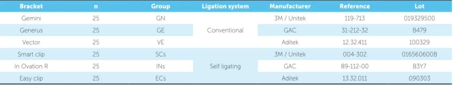
Table 1 shows the distribution of the analyzed samples. The sample consisted of 150 metal brackets, 75 self-ligating and 75 conventional for maxillary right central incisors, from three diferent manufacturers (3M Unitek, Aditek and GAC) (25 brackets each). The conventional brackets were Gemini (3M/Unitek, Monrovia, CA, USA), Generus (GAC, Islandia, NY, USA) and Vector (Aditek, Cravinhos, SP, Brazil); whereas the self-ligating brackets were Smart Clip (3M/ Unitek), In Ovation R (GAC) and Easy Clip (Aditek).
The 25 brackets of each group were divided into ive subgroups, with an equal number of samples each, numbered from one to ive.
The brackets of each subgroup were stored in previ-ously sterilized Petri dishes, without coming into con-tact with one another. They were subjected to corrosion
in 20 ml of sterile 0.9% NaCl solution13,14 for a period
of 21 days. The Petri dishes were kept in an incuba-tor (Quimis - Quimis Aparelhos Cientíicos LTDA., Diadema-SP, Brazil) at an unchanged temperature of
37 ± 1°C.15 Every 7 days ± 1 hour, the brackets were
re-moved and transferred to another container illed with
a new solution.1,13
At the end of each experimental time interval (7 days, 14 days and 21 days), the solution remaining in each container was analyzed by atomic absorption spec-trophotometry in a Varian spectrophotometer, Model AA-1475 (Varian Indústria e Comércio Ltda., São Pau-lo/SP, Brazil) with a view to determining their nickel, chromium and iron contents.
At the end of the experiment, ive brackets from each group (one from each subgroup) were randomly select-ed and their surface topography was analyzselect-ed by scan-ning electron microscopy – SEM (Jeol JSM 6460 LV, Japan) and compared with the surface of new brackets.
Bracket n Group Ligation system Manufacturer Reference Lot
Gemini 25 GN
Conventional
3M / Unitek 119-713 019329500
Generus 25 GE GAC 31-212-32 B479
Vector 25 VE Aditek 12.32.411 100329
Smart clip 25 SCs
Self ligating
3M / Unitek 004-302 016560600B
In Ovation R 25 INs GAC 89-112-00 B3Y7
Easy clip 25 ECs Aditek 13.32.011 090303
SEM was operated at 20 kV, and readouts were taken under 22X and 1,000X magniication. The 22X magniication allowed a complete view of the bracket. The 1,000X magniication was performed at the slot of each bracket with the connection system opened so as to analyze the area for wire insertion. No treatment was performed on the brackets at the end of the experiment in order to prevent any possible oxides, deposited on
the bracket surface as a result of the corrosion process, from being removed. Energy-dispersive X-ray spectros-copy (EDS) was used to identify atypical depositions on bracket surface.
In each group and at each time interval, nickel, chro-mium and iron concentrations as well as the total amount released during the experiment were statistically assessed. The Kolmogorov-Smirnov test was used to verify the
Identical letters reveal no statistical diference (p > 0.05). Intragroup signiicance - Comparison of the diferent time intervals in each group with ANOVA test and Tukey
post hoc-test. Intergroup Signiicance - Comparison among the groups in each time interval with Kruskal-Wallis test and Wilcoxon post hoc-test.
Table 2 - Nickel content (ppM) after the different experimental time intervals.
Nickel release (ppM)
7 days 14 days 21 days Total
Group Mean ± SD Intra.
Sig.
Inter.
Sig. Mean ± SD
Intra. Sig.
Inter.
Sig. Mean ± SD
Intra. Sig.
Inter.
Sig. Mean ± SD
Inter. Sig.
GN 0.00 a A 0.00 a A 0.00 a A 0.0 (0.0) A
GE 0.14 ± 0.28 a ABC 0.92 ± 0.03 b B 0.68 ± 0.02 b B 1.74 ± 0.27 B
VE 0.53 ± 0.03 a B 0.00 ± 0.01 b A 0.00 ± 0.01 b A 0.54 ± 0.03 C
SCs 0.11 ± 0.03 a C 0.07 ± 0.01 b C 0.00 c A 0.18 ± 0.01 D
INs 0.14 ± 0.01 a C 0.39 ± 0.02 b D 0.00 ± 0.01 c A 0.54 ± 0.03 C
ECs 2.79 ± 0.03 a D 1.24 ± 0.03 b E 0.00 ± 0.01 c A 4.03 ± 0.03 E
Table 3 - Chromium content (ppM) after the different experimental time intervals.
Chromium release(ppM)
7 days 14 days 21 days Total
Group Mean ± SD Intra.
Sig.
Inter.
Sig. Mean ± SD
Intra. Sig.
Inter.
Sig. Mean ± SD
Intra. Sig.
Inter.
Sig. Mean ± SD
Inter. Sig.
GN 0.50 ± 0.11 a AB 1.03 ± 0.01 b A 1.68 ± 0.03 c A 3.21 ± 0.12 AB
GE 0.89 ± 0.10 a C 1.29 ± 0.02 b B 1.95 ± 0.03 c B 4.13 ± 0.10 C
VE 1.15 ± 0.02 a D 1.03 ± 0.05 b A 1.95 ± 0.06 c B 4.13 ± 0.11 C
SCs 0.45 ± 0.01 a A 0.30 ± 0.03 a C 2.05 ± 0.35 b AB 2.81 ± 0.33 D
INs 0.76 ± 0.02 a C 1.03 ± 0.08 b A 1.42 ± 0.02 c C 3.21 ± 0.09 A
ECs 0.63 ± 0.02 a B 1.55 ± 0.03 b D 1.16 ± 0.03 c D 3.34 ± 0.05 B
Identical letters indicate no statistical diference (p > 0.05). Intragroup signiicance - Comparison of the diferent time intervals in each group with ANOVA test and
Tukey post hoc-test. Intergroup Signiicance - Comparison among the groups in each time interval with Kruskal-Wallis test and Wilcoxon post hoc-test.
Table 4 - Iron content (ppM) after the different experimental time intervals.
Iron release (ppM)
7 days 14 days 21 days Total
Group Mean ± SD Intra.
Sig.
Inter.
Sig. Mean ± SD
Intra.
Sig.
Inter.
Sig. Mean ± SD
Intra. Sig.
Inter.
Sig. Mean ± SD
Inter. Sig.
GN 0.35 ± 0.03 a A 0.44 ± 0.03 b A 1.02 ± 0.02 c A 1.81 ± 0.04 A
GE 0.93 ± 0.05 a B 1.02 ± 0.02 b B 1.28 ± 0.02 c B 3.23 ± 0.08 B
VE 1.01± 0.05 a B 0.77 ± 0.04 b C 1.02 ± 0.02 a A 2.80 ± 0.09 CD
SCs 0.00 ± 0.01 a C 0.01 ± 0.02 a D 0.94 ± 0.02 b C 0.96 ± 0.04 E
INs 0.77 ± 0.02 a D 0.94 ± 0.02 b E 1.01 ± 0.03 c A 2.72 ± 0.02 C
ECs 1.02 ± 0.03 a B 0.94 ± 0.02 b E 0.94 ± 0.04 b D 2.90 ± 0.08 D
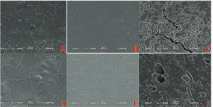
Figure 1 - Surface topography of new brackets visualized by SEM under 22X magnification. A) GN; B) GE; C) VE; D) SCs; E) INs; F) ECs.
Figure 3 - Surface topography of new brackets visualized by SEM under 1000X magnification. A) GN; B) GE; C) VE; D) SCs; E) INs; F) ECs.
Figure 2 - Surface topography of brackets visualized by SEM under 22X magni-fication after 21 days. A) GN; B) GE; C) VE; D) SCs; E) INs; F) ECs.
Figure 4 - Surface topography of brackets visualized by SEM under 1000X magnification after 21 days. A) GN; B) GE; C) VE; D) SCs; E) INs; F) ECs. sample normality of distribution. As normal distribution
was not found, the non-parametric ANOVA test with Tukey post-test were applied for intragroup assessment, whereas the Kruskal-Wallis test with Wilcoxon post-test were applied for intergroup assessment. The data were statistically analyzed using SPSS 17.0 sotware (Statisti-cal Package for Social Sciences, SPSS Inc., Chicago, IL, USA). The signiicance level was set at 5%.
RESULTS
During the experimental period, the release of nickel, chromium and iron metal ions was observed in all groups, except for nickel ion in the GN group, which was not detected at any of the time intervals (Tables 2, 3 and 4).
There was greater release of nickel in the initial period of the experiment, both for conventional and self-ligating brackets, with a trend towards no release of this ion at the end of the experiment (p < 0.05). Only group GN and group GE behaved diferently with
greater release of nickel on the 14th and 21st days. The
experimental groups revealed a trend towards increasing release of ions, such as chromium and iron, from the irst to the third week of the experiment (p < 0.05). The only exception was group VE, in which a great amount of iron release was found in the irst week. Metal release was similar in self-ligating and conventional brackets from the same manufacturer. Group SCs released more nickel (p < 0.05) than group GN ater 7 and 14 days, but group GN released more chromium (p < 0.05) ater 14 days and more iron (p < 0.05) at the three experimental time intervals. Group INs released less iron (p < 0.05) than group GE ater 7 days; and less nickel, chromium and iron (p < 0.05) ater 14 and 21 days. Group ECs released more nickel, chromium and iron (p < 0.05) than group VE ater 14 days, but released less nickel and chromium (p < 0.05) ater 7 days and less chromium and iron (p < 0.05) ater 21 days.
DISCUSSION
Bracket corrosion with consequent release of metal ions during orthodontic treatment may hinder
orth-odontic mechanics,2 trigger hypersensitivity reactions
with hyperplasia and gingival tissue inlammation,4,8,9
and contribute to iatrogenic staining of the enamel with
the incorporation of metals.10,16
A corroded bracket oten presents a more irregular surface and can accumulate products resulting from corrosion (Fig 4). Thus, friction between the bracket and wire during sliding mechanics increases, mak-ing it necessary to apply force of greater magnitude to overcome the friction and produce physiologic tooth
movement.7,17,18 When excessive force is applied to the
bracket, there is loss of mechanical control and increased
probability of root resorption.5,9,19,21
One of the advantages attributed to self-ligating brackets is the lower degree of friction that the ligation system imposes on the orthodontic wire when
com-pared with conventional tying.6,22,24 However, if the
bracket has an increased corrosion potential, friction
also increases,25 afecting this advantage.
In the present study, SEM analysis under 22X mag-niication (Fig 2) revealed that, in general, self-ligating brackets showed more areas with altered surface to-pography due to corrosion, when compared with con-ventional brackets from the same manufacturer, pos-sibly due to their geometry with larger retentive areas. Thus, brackets in the INs group had a more irregular surface than the brackets in the GE group, while the brackets in the ECs group presented greater alterations in comparison to those in the VE group. The self-ligating brackets of the SCs group and the conventional brackets in group GN did not present signiicant al-terations in their surface topography. Energy-dispersive X-ray spectroscopy (EDS) revealed NaCl deposition on the surfaces of the brackets in group GN.
Analysis of the images obtained by SEM under 1000X magniication (Figs 3 and 4) suggests that new brackets with more irregular surfaces were more sus-ceptible to corrosion. Thus, the brackets in group ECs, which presented a surface topography with deicient smoothness and porosity at the beginning of the experi-ment, presented a considerably more irregular surface at the end of it. The brackets in group VE also presented considerably altered surfaces at the end of the experi-ment, due to the formation of oxides. Moreover, the
brackets in group INs, which presented irregularities at the beginning of the experiment, were signiicantly subjected to pitting corrosion. Quantiication of the metal ions released during the experiment corroborates these data. These groups had great release of nickel, chromium and iron ions.
The release of metal ions into the oral
environ-ment may trigger hypersensitivity reactions.4,5,8,9,26
Nickel and chromium are present in the composition of brackets with the goal of increasing resistance to
corrosion.2,4,16,27 These elements are largely responsible
for the aforementioned adverse reactions. Nickel is strongly responsible for triggering more allergic
reac-tions than any other metal.8,26
Under the conditions of this experiment, the release of nickel was considerably lower than the daily
inges-tion of this metal through food (300 to 600 µg/day).13
However, it is worth noting that susceptible patients in contact with small concentrations of this metal are more
likely to sufer hypersensitivity reactions.29
The oral reaction of allergy to nickel is diicult to diagnose, since its clinical signs and symptoms are similar to those of gingivitis caused by poor oral hy-giene. The low number of reports of hypersensitivity to nickel is possibly due to this diiculty in diagnosis. Epidemiological data point towards an incidence of sensitivity to this metal of approximately 20% in the
general population.28,30
Similarly to other in vitro studies,1,15,31 the release of
nickel in this experiment was higher in the irst two weeks, with a trend towards no further release during the third week. The exception was group GE which contin-ued to release nickel ions during the last week (Table 2).
Similarly to previous studies,1,13 chromium and iron
ions had a trend towards increasing release through-out the experiment in all groups, except for group ECs (Tables 3 and 4). In this group, there was a greater release of chromium in the second week, and greater release of iron in the irst week, although the concentration of these ions remained high at the end of the third week.
Within the limitations and conditions of this experi-ment, it is reasonable to conclude that metal release was similar in self-ligating and conventional brackets from the same manufacturer.
7 and 14 days, whereas group GN released more chro-mium (p < 0.05) ater 14 days and more iron (p < 0.05) at the three experimental time intervals. Group INs re-leased less iron (p < 0.05) than group GE ater 7 days and less nickel, chromium and iron (p < 0.05) ater 14 and 21 days. Group ECs released more nickel, chromium and iron (p < 0.05) than group VE ater 14 days, but released less nickel and chromium (p < 0.05) ater 7 days and less chromium and iron (p < 0.05) ater 21 days.
Metal ions released by metal brackets and bands dur-ing orthodontic treatment may be incorporated into tooth
enamel, causing iatrogenic color alteration and stains.10,16
In cases of severe pigmentation, restorative treatment of the vestibular surface of the stained tooth proves
nec-essary.32,33 Special care must be given to patients with
poor oral hygiene, since altered oral environment, with
reduced pH and presence of acidogenic microorganisms potentiates the corrosion of metal accessories. Addition-ally, enamel demineralization and remineralization
pro-cesses may inluence the incorporation of metals.4,10
CONCLUSIONS
» The null hypothesis was accepted.
» The SEM analysis revealed that self-ligating and conventional brackets presented signs of aging. » Metal ions release in self-ligating brackets was
similar to conventional brackets from the same manufacturer.
Acknowledgements
1. Barrett RD, Bishara SE, Quinn JK. Biodegradation of orthodontic appliances. part I. Biodegradation of nickel and chromium in vitro. Am J Orthod Dentofacial Orthop. 1993;103(1):8-14.
2. Toms Ap. The corrosion of orthodontic wire. Eur J Orthod.
1988;10:87-97.
3. Zinelis S, Annousaki O, Eliades T, Makou M. Elemental composition
of brazing alloys in metallic orthodontic brackets. Angle Orthod. 2004;74(3):394-9.
4. Eliades T, Athanasiou AE. In vivo aging of orthodontic alloys: implications for corrosion potential, nickel release, and biocompatibility. Angle Orthod. 2002;72(3):222-37.
5. House K, Sernetz F, Dymock D, Sandy JR, Ireland AJ. Corrosion of
orthodontic appliances--should we care? Am J Orthod Dentofacial Orthop. 2008;133(4):584-92.
6. pizzoni L, Ravnholt G, Melsen B. Frictional forces related to self-ligating brackets. Eur J Orthod. 1998;20(3):283-91.
7. Andreasen GF, Quevedo FR. Evaluation of friction forces in the 0.022 x 0.028 edgewise bracket in vitro. J Biomech. 1970;3(2):151-60.
8. Menezes LM, Campos LC, Quintão CC, Bolognese AM. Hypersensitivity
to metals in orthodontics. Am J Orthod Dentofacial Orthop. 2004;126(1):58-64.
9. Hwang CJ, Shin JS, Cha JY. Metal release from simulated ixed
orthodontic appliances. Am J Orthod Dentofacial Orthop. 2001;120(4):383-91.
10. Maia LHEG, Lima Filho HL, Araujo MVA, Ruellas ACO, Araujo MTS. Incorporation of metal and color alteration of enamel in the presence of orthodontic appliances. Angle Orthod. 2012;82(5):889-93.
11. pandis N, Bourauel C, Eliades T. Changes in the stifness of the ligating mechanism in retrieved active self-ligating brackets. Am J Orthod Dentofacial Orthop. 2007;132(6):834-7.
12. Eliades T. Self-ligating brackets malfunction: is it due to corrosion? Orthod Mat Insider. 2009;21:4-7.
13. Grimsdottir MR, Gjerdet NR, Hensten-pettersen A. Composition and in vitro corrosion of orthodontic appliances. Am J Orthod Dentofacial Orthop. 1992;101(6):525-32.
14. Kerosuo H, Moe G, Kleven E. In vitro release of nickel and chromium from diferent types of simulated orthodontic appliances. Angle Orthod. 1995;65(2):111-6.
15. park HY, Shearer TR. In vitro release of nickel and chromium from simulated orthodontic appliances. Am J Orthod. 1983;84(2):156-9. 16. Maijer R, Smith DC. Corrosion of orthodontic bracket bases. Am J
Orthod. 1982;81(1):43-8.
17. Angolkar pV, Kapila S, Duncanson MG Jr, Nanda RS. Evaluation of friction between ceramic brackets and orthodontic wires of four alloys. Am J Orthod Dentofacial Orthop. 1990;98(6):499-506.
18. Kapila S, Angolkar pV, Duncanson MG Jr, Nanda RS. Evaluation of friction between edgewise stainless steel brackets and orthodontic wires of four alloys. Am J Orthod Dentofacial Orthop. 1990;98(2):117-26.
REFERENCES
19. Drescher D, Bourauel C, Schumacher HA. Frictional forces between bracket and arch wire. Am J Orthod Dentofacial Orthop. 1989;96(5):397-404.
20. Lin MC, Lin SC, Lee TH, Huang HH. Surface analysis and corrosion resistance of diferent stainless steel orthodontic brackets in artiicial saliva. Angle Orthod. 2006;76(2):322-9.
21. Reitan K. Initial tissue behavior during apical root resorption. Angle Orthod. 1974;44(1):68-82.
22. Berger JL. The inluence of the SpEED bracket’s self-ligating design on force levels in tooth movement: a comparative in vitro study. Am J Orthod Dentofacial Orthop. 1990;97(3):219-28.
23. Thorstenson GA, Kusy Rp. Resistance to sliding of self-ligating brackets versus conventional stainless steel twin brackets with second-order angulation in the dry and wet (saliva) states. Am J Orthod Dentofacial Orthop. 2001;120(4):361-70.
24. Thomas S, Sherrif M, Birnie D. A comparative in vitro study of the frictional characteristics of two types of self-ligating brackets and two types of pre-adjusted edgewise brackets tied with elastomeric ligatures. Eur J Orthod. 1998;20(5):589-96.
25. Marques IS, Araujo AM, Gurgel JA, Normando D. Debris, roughness and friction of stainless steel archwires following clinical use. Angle Orthod. 2010;80(3):521-7.
26. Bishara SE, Barrett RD, Selim MI. Biodegradation of orthodontic appliances. part II. Changes in the blood level of nickel. Am J Orthod Dentofacial Orthop. 1993;103(2):115-9.
27. Maijer R, Smith DC. Biodegradation of the orthodontic bracket system. Am J Orthod Dentofacial Orthop. 1986;90(3):195-8.
28. Bass JK, Fine H, Cisneros GJ. Nickel hypersensitivity in the orthodontic patient. Am J Orthod Dentofacial Orthop. 1993;103(3):280-5.
29. prystowsky SD, Allen AM, Smith RW, Nonomura JH, Odom RB, Akers WA. Allergic contact hypersensitivity to nickel, neomycin, ethylenediamine, and benzocaine. Relationships between age, sex, history of exposure, and reactivity to standard patch tests and use tests in a general population. Arch Dermatol. 1979;115(8):959-62.
30. Kerosuo H, Kullaa A, Kerosuo E, Kanerva L, Hensten-pettersen A. Nickel allergy in adolescents in relation to orthodontic treatment and piercing of ears. Am J Orthod Dentofacial Orthop. 1996;109(2):148-54.
31. Menne T, Brandup F, Thestrup-pedersen K, Veien NK, Andersen JR, Yding F, et al. patch test reactivity to nickel alloys. Contact Dermatitis. 1987;16(5):255-9.
32. Romano FS, Correr Sobrinho L, Consani S, Araújo MBB, Nouer DF. Oxidação de bráquetes metálicos. Rev Dental press Ortod Ortop Facial. 2005;10(6):24-5.