B B O CA S E RE P O R T
Class III malocclusion with unilateral
posterior crossbite and facial asymmetry*
Silvio Rosan de Oliveira**
This article reports on the orthodontic treatment performed on a 36-year-old female patient with skeletal and dental Class III pattern, presenting with a left unilateral poste-rior crossbite and mandibular asymmetry, and a relatively significant difference between maximum intercuspation (MIC) and centric relation (CR). The treatment was performed with maxillary dental expansion, mandibular dental contraction and anterior crossbite correction, eliminating the difference between MIC and CR. Results were based on care-ful diagnosis and planning of orthodontic compensation without surgical intervention in the maxilla, at the request of the patient. This case was presented to the Brazilian Board of Orthodontics and Facial Orthopedics (BBO) as representative of Category 5, i.e., mal-occlusion with a transverse problem, presenting with a crossbite in at least one of the quadrants, as part of the requirements for obtaining the BBO Certificate.
Abstract
Keywords: Angle Class III. Crossbite. Facial asymmetry. Adult patient. Corrective Orthodontics.
** Specialist in Orthodontics, School of Dentistry, Rio de Janeiro State University - UERJ. MSc in Orthodontics, School of Dentistry, Rio de Janeiro State University - UERJ. Diplomate of the Brazilian Board of Orthodontics and Dentofacial Orthopedics (BBO).
* Case report, Category 5 - approved by the Brazilian Board of Orthodontics and Facial Orthopedics (BBO).
HISTORY AND ETIOLOGY
The patient sought orthodontic treatment at 36 years of age, in good general health and without significant medical history. Her chief complaint concerned anterior and posterior crossbites and chronic pain in the left temporo-mandibular joint. She showed good oral hy-giene, overall healthy-looking gingiva and some poorly fitting amalgam restorations.2 She had no history of orthodontic intervention. When orthognathic surgery was suggested the patient expressed her unwillingness to undergo surgery to correct the malocclusion.
DIAGNOSIS
As regards dental pattern (Figs 1 and 2), she presented with an Angle Class III, subdivision left malocclusion, no mandibular dentoalveolar discrep-ancy, 3 mm overbite, 2 mm overjet, crowding in the upper anterior region, U-shaped maxillary arch, contracted on the right side, lower arch slightly ex-panded on the right side, posterior crossbite on the left5, less than 3 mm lower midline shift to the left and inclined lower occlusal plane.
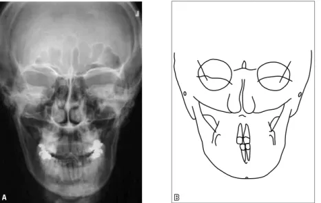
FIGURE 1 - Initial facial and intraoral photographs.
Regarding functional occlusion, at MIC she pre-sented with a 5 mm mandibular deviation to the left side (Fig 5) and a 2 mm difference between MIC and CR. At CR, contact existed only between tooth 23 (left upper canine) and tooth 33 (left lower canine) with reduced mandibular deviation.
On clinical examination, bilateral clicks were observed in the TMJ with mandibular deviations on opening and closing movements and no crepita-tion or mandibular defleccrepita-tion at maximum open-ing. Palpation examination showed more intense pain in the left than in the right TMJ, regardless
of whether the mouth was open or closed.3,6 A maximum opening of 52 mm was recorded.
The analysis of panoramic and periapical ra-diographs (Fig 3) showed that the patient did not present with any condition that might compro-mise her orthodontic treatment.
A
B C
Class III malocclusion with unilateral posterior crossbite and facial asymmetry
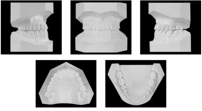
FIGURE 2 - Initial plaster models.
B A
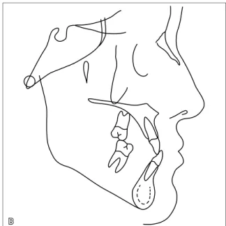
FIGURE 4 - Initial lateral cephalogram (A) and cephalometric tracing (B).
TREATMENT GOALS
The initial goal was to control chronic pain in the left TMJ by referring the patient to a specialist in temporomandibular disorders (TMD).2,3,6 After this issue had been success-fully addressed, orthodontic treatment was ad-ministered with the consent of the specialist. At the patient’s request, combined surgical-orthodontic treatment was ruled out.
Thus, to correct the anterior crossbite, the difference between MIC and CR6 had to be ad-dressed through axial protrusion of the maxillary incisors and retroclination of the mandibular in-cisors, thereby achieving normal occlusion and slightly improving the profile.1
The transverse problem was resolved by cor-recting the left posterior crossbite, which re-quired expanding the upper dental arch4,7 and contracting the lower. Moreover, the purpose of eliminating the difference between MIC and CR was to correct the lower midline and reduce mandibular deviation.
TREATMENT PLAN
The first step would be to refer the patient to a TMD specialist2,3,6 and then have her third mo-lars (38 and 48) extracted, since these teeth were extruded (Figs 1 and 3A).
After TMD treatment a Hyrax-type palatal expansion appliance would be installed (for six months) with bands on all maxillary molars and premolars (eight bands) to expand the upper arch and increase intermolar width.4,7 After expander removal, a palatal bar fabricated from 0.032-in stainless steel would be inserted, with bands on the first molars and palatal extension as far as the first premolars. In the lower arch, a 0.032-in stain-less steel lingual arch would be placed, with bands on the lower first molars.
A B Class III malocclusion with unilateral posterior crossbite and facial asymmetry
induce retroclination of lower incisors and fin-ish the case. In the phase of anterior crossbite correction it would be necessary to use Class III intermaxillary elastic mechanics.
During the finishing stage, the patient would be referred to a speech therapist for evaluation of her oral functions.
After the active treatment phase, an upper wraparound-type retention plate would be used, and on the lower arch a stainless steel 0.028-in lingual canine-to-canine arch (retainer).
TREATMENT PROGRESS
Treatment of the chronic pain in the left TMJ lasted four months under the TMD specialist’s supervision. In addition, the patient was peri-odically evaluated throughout the orthodontic treatment. Extraction of the third molars was performed after this period.
For maxillary expansion, a Hyrax-type ex-pander was installed with bands on all molars and premolars, and 1/4 turn activation once a day for 28 days. The patient wore the appli-ance for six months.
After expander removal, a 0.032-in stainless steel palatal bar was installed, welded to bands
on the first molars and palatal extension as far as the first premolars. The appliance was removed in the early finishing stage and the bands re-placed with bonded brackets.
On the lower arch, a 0.032-in stainless steel lingual arch was placed with bands on the lower first molars. The lingual arch was also removed in the early finishing stage and the bands replaced with bonded brackets.
Upper fixed appliance set-up was performed after removal of the palatal expansion appliance at the same time that the palatal bar was in-stalled. The lower fixed appliance was set up three months after lingual arch installation. All second molars were also included in the treatment, with orthodontic bands. Next, a sequence of 0.014-in to 0.020-in diameter stainless steel alignment and leveling archwires was used. Stainless steel 0.019 X 0.025-in archwires were used to increase the axial inclination of upper incisors and retroclina-tion of lower incisors. At this stage, Class III elastic mechanics was introduced. After crossbite cor-rection, occlusal adjustments were performed by compensatory grinding in some consultations un-til the end of treatment to improve dental inter-cuspation quality. Stainless steel 0.019 X 0.025-in
FIGURE 6 - Final facial and intraoral photographs.
nitely. The patient underwent speech therapy for eight months.
A
B C
Class III malocclusion with unilateral posterior crossbite and facial asymmetry
FIGURE 7 - Final plaster models.
B A
B A
FIGURE 9 - Final lateral cephalogram (A) and cephalometric tracing (B).
FIGURE 10 - Total and partial superimposition of initial (black) and final (red) cephalometric tracings.
occurred in the premolar and molar regions with a 5 mm increase in intermolar width (Table 2), con-tributing to posterior crossbite correction while eliminating a functional shift which had been de-tected and resulted from premature torque in the maxillary left canine4,7 (Figs 6 and 7).
In the lower arch, some improvement was achieved in tooth alignment and a 9º decrease,
also deliberate, in incisor axial inclination (Fig 9 and Table 1).1 In the posterior region, a slight 2 mm contraction was noted at molar level (Table 2), which also contributed to posterior crossbite correction (Figs 6 and 7).
Class III malocclusion with unilateral posterior crossbite and facial asymmetry
MEASUREMENTS A B Difference
A/B
Intercanine Width:
Upper / Lower (mm) 35 / 28 35 / 26 0 / 2
Intermolar Width:
Upper / Lower (mm) 50 / 50 55 / 48 5 / 2 TABLE 2 - Intermolar and intercanine widths (in mm).
adequate intercuspation and crossbite correction in the anterior and left regions6 (Figs 6 and 7).
Facial profile remained concave with a slight improvement in the relationship between the upper and lower lips. In frontal view, a slight de-crease occurred in mandibular deviation (Fig 6).
MEASUREMENTS Standard
values A B
Difference A/B
Skeletal Pattern
SNA(Steiner) 82° 80° 81° 1
SNB(Steiner) 80° 82.5° 84° 1.5
ANB(Steiner) 2° - 2.5° - 3° 0.5
Convexity Angle (Downs) 0° - 8° - 9° 1
Y-Axis (Downs) 59° 61° 60° 1
Facial Angle (Downs) 87° 87° 88° 1
SN – GoGn (Steiner) 32° 29° 29° 0
FMA(Tweed) 25° 28° 27° 1
Dental Pattern
IMPA(Tweed) 90° 91° 81° 10
–1 – NA (degrees)(Steiner) 22° 29° 39° 10
–1 – NA (mm)(Steiner) 4 mm 2 mm 5.5 mm 3.5
–
1 – NB (degrees)(Steiner) 25° 25° 16° 9°
–
1 – NB (mm)(Steiner) 4 mm 5 mm 3 mm 2
–1
1 – Interincisal Angle (Downs) 130° 128º 128° 0
–
1 – APo (mm)(Ricketts) 1 mm 6.5 mm 5 mm 1.5
Profile
Upper Lip – S Line (Steiner) 0 mm -2 mm -2 mm 0
Lower Lip – S Line (Steiner) 0 mm 0 mm 0 mm 0
TABLE 1 - Summary of cephalometric measurements.
The analysis of panoramic and periapical ra-diographs (Fig 8), showed good root parallelism with no significant morphological changes. The lateral cephalometric radiograph (Fig 9, A), clear-ly shows that the anterior crossbite was corrected.
FINAL CONSIDERATIONS
1. Araújo EA, Araújo CV. Abordagem clínica não cirúrgica no tratamento da má oclusão de Classe III. Rev Dental Press Ortod Ortop Facial. 2008 nov-dez;13(6):128-57.
2. Barbosa MC, Araújo EA. Tratamento ortodôntico em pacientes adultos. J CEO. 1999 abr;2(6):3.
3. Conti PC. Ortodontia e disfunções temporomandibulares: o estado da arte. Rev Dental Press Ortod Ortop Facial. 2009 nov-dez;14(6):12-3.
4. Haldelman CS. Nonsurgical rapid maxillary alveolar expansion in adults: a clinical evaluation. Angle Orthod. 1997;67(4):291-305.
5. Locks A, Weissheimer A, Ritter DE, Ribeiro GLU, Menezes LM, Derech CD, et al. Mordida cruzada posterior: uma classiicação mais didática. Rev Dental Press Ortod Ortop Facial. 2008 mar-abr;13(2):146-58.
6. Okeson JP. Critérios para uma oclusão funcional ideal. In. Okeson JP. Tratamento das desordens temporomandibulares e oclusão. 4ª ed. São Paulo: Artes Médicas; 2000. p. 87-100.
REFERENCES
7. Rossi RRP, Araújo MT, Bolognese AM. Expansão maxilar em adultos e adolescentes com maturação esquelética avançada. Rev Dental Press Ortod Ortop Facial. 2009 set-out; 14(5):43-51.
Contact address
Silvio Rosan de Oliveira
Av. Plínio de Castro Prado n. 190 – Jardim Macedo CEP: 14.091-170 – Ribeirão Preto / SP, Brazil E-mail: [email protected]
Submitted: July 2010