Cop
yright
© ABE&M t
odos os dir
eit
os r
eser
vados
.
Severe congenital non-autoimmune
hyperthyroidism associated to a
mutation in the extracellular domain
of thyrotropin receptor gene
Hipertireoidismo congênito não autoimune grave associado com uma mutação no domínio extracelular do gene receptor da tirotroina
Paula A. Scaglia1, Ana Chiesa2, Gabriela Bastida3, Mirta Pacin3,
Horacio M. Domené1, Laura Gruñeiro-Papendieck2
SUMMARY
Activating mutations in the TSH Receptor (TSHR) gene have been identiied as the molecular basis for congenital non-autoimmune hyperthyroidism. We describe the clinical indings and molecular characterization in a girl who presented severe non-autoimmune hyperthyroidism since birth, born to a mother with autoimmune thyroid disease. She was treated with meth-ylmercaptoimidazol and β-blockers, but remained hyperthyroid and required total thyroid-ectomy. To characterize the presence of an activating mutation, the whole coding sequence and intron-exon boundaries of TSHR gene were analyzed. The patient was heterozygous for p.Ser281Asn mutation and p.Asp727Glu polymorphism. This recurrent mutation, p.Ser281Asn, characterized in vitro by increased basal production of cAMP, is the unique germline activating gene variant described so far in the extracellular domain of TSH receptor. Interestingly, the patient’s mother presented hyperthyroidism but without any TSHR gene activating mutation. Although congenital non-autoimmune hyperthyroidism is a rare condition, it should be inves-tigated when severe disease persists, even in a newborn from an autoimmune hyperthyroid mother, in order to differentiate it from the more common congenital autoimmune disease. Arq Bras Endocrinol Metab. 2012;56(8):513-8
SUMÁRIO
Mutações ativadoras no gene receptor de TSH (TSHR) foram identiicadas como a base mo-lecular do hipertireoidismo congênito não autoimune. Descrevemos os achados clínicos e a caracterização molecular de uma menina que apresentou hipertireoidismo autoimune grave desde o nascimento, nascida de uma mãe com desordem tiroidiana autoimune. Tratada com metilmercaptoimidazol e β-bloqueadores, ela permaneceu com hipertiroidismo e necessitou de tiroidectomia total. A im de caracterizar a presença da mutação ativadora, foram anali-sados toda a sequência codiicadora assim como os extremos dos íntrons e éxons do gene TSHR. A paciente era heterozigota para a mutação p.Ser281Asn e para o polimorismo
p.Asp-727Glu. Essa mutação recorrente, p.Ser281Asn, caracterizada in vitro pelo aumento da pro-dução basal de cAMP, é a única variante genética ativadora da linhagem germinativa descrita até hoje no domínio extracelular do receptor de TSH. Interessantemente, a mãe da paciente apresentou hipertireoidismo sem qualquer mutação ativadora do gene TSHR. Embora o hi-pertireoidismo congênito não autoimune seja uma condição rara, ele deve ser investigado quando há persistência da doença grave, mesmo em um recém-nascido de uma mãe com hipertireoidismo autoimune, a im de se diferenciar o caso da forma mais comum da doença autoimune congênita. Arq Bras Endocrinol Metab. 2012;56(8):513-8
1 Centro de Investigaciones Endocrinológicas (CEDIE, CONICET), Buenos Aires, Argentina 2 Hospital de Niños “Ricardo Gutiérrez”, Endocrinology Division, Buenos Aires, Argentina 3 Hospital “E. Castro Rendon”, Pediatrics, Neuquén, Argentina
Correspondence to:
Paula A. Scaglia Centro de Investigaciones Endocrinológicas (CEDIE, CONICET), Hospital de Niños “Ricardo Gutiérrez”, Gallo 1330 C1425EFD – Ciudad Autónoma de Buenos Aires, Argentina [email protected]
Cop
yright
© ABE&M t
odos os dir
eit
os r
eser
vados
.
INTRODUCTION
N
eonatal hyperthyroidism is a rare disease affect-ing about one out of 50,000 neonates (1). It is usually caused by transplacental passage of thyrotropin receptor stimulating antibodies (TRAb) in women with history of Graves’ disease or, more rarely, Hashimoto thyroiditis (2). The neonatal disease is observed within the irst days of life, requiring immediate treatment be-cause of its high morbimortality. Although severe, this condition is usually transient, with spontaneous reso-lution when maternal antibodies are cleared from the neonate’s circulation, within the irst four months of life (3,4).More recently, an even more uncommon type of neonatal hyperthyroidism has been described, result-ing from activatresult-ing mutations of the TSH receptor (TSHR) gene and causing constitutive activation of the
intracellular signaling cascade (5-9). Neonates with this uncommon disorder share the same clinical manifesta-tions of severe neonatal thyrotoxicosis, but with more prolonged symptoms of hyperthyroidism.
The mutations in the TSHR gene leading to
non-autoimmune hyperthyroidism (NAH) may be inherited in an autosomal dominant manner (familial or heredi-tary, HNAH), or may occur sporadically as a de novo
condition (SNAH). To date, at least 20 different muta-tions have been identiied as the cause of HNAH in 27 families with more than a hundred affected indivi duals, while only 15 subjects with 10 different mutations leading to SNAH have been described (10,11).
We report the clinical indings and molecular char-acterization of a girl who presented a persistent and se-vere congenital hyperthyroidism due to an activating mutation of the TSHR gene, born to a mother with
autoimmune thyroid disease.
CASE REPORT
The study was conducted in accordance with the Hel-sinki Declaration (12). Written informed consent for molecular studies was obtained from the mother. The study was approved by the “E. Castro Rendon” Hospi-tal ethics committee.
The girl is the only daughter of a 31-year-old moth-er, born at 38 weeks of gestational age after a poorly controlled pregnancy, with no evaluation of thyroid function. She was delivered by cesarean section because of fetal tachycardia and suffering. Her APGAR score
was 6/8 and she required monitoring in an Intensive Care Unit. Her birth weight was 2410 g (-1.7 SDS); birth length, 47 cm (-0.87 SDS); and head circum-ference, 33.3 cm (< 3rd percentile). Tachycardia (220
beats/min) was present at birth, and thyroid proile conirmed hyperthyroidism (Table 1).
At 8 days of life she had persistent tachycardia (160-200 beats/min), easy sweating, irritability, and an altered thyroid proile (Table 1). Thyroid gland ul-trasound showed a normal sized eutopic gland. Bone age, assessed by hand and wrist maturation (13), was advanced by 1 year.
With diagnosis of hyperthyroidism, she began treat-ment with methylmercaptoimidazol (MMI) and car-diac β-blockers. Nevertheless, she persisted hyperthy-roid with relapses, after an attempt to reduce the MMI dose. Her follow-up showed weight always below the 3rd percentile, normal height around the 50th
percen-tile, while bone age, advanced during the irst 3 years, approached chronological age at the age of 4.
At 6.9 years of age, she underwent a total thyroidec-tomy (Table 1). Histological examination revealed fol-licular cell hyperplasia, with no signs of either malignan-cy or lymphomalignan-cytic iniltration (Figure 1). After surgery, hypothyroidism was conirmed and LT4treatment was indicated (2.5 µg/kg.day). The neuropsychological outcome was normal, without craniosynostosis.
Hyperthyroidism had been diagnosed in her mother at 14 years of age, when she presented goiter and high
131I uptake (64% at 24h) (Table 2). She was initially
treated with MMI, but because of poor compliance she received two doses of 131I. She remained
uncon-trolled and without treatment until delivery. With signs of overt hypothyroidism and positive anti-thyroperox-idase antibodies (ATPO), she began thyroid hormone replacement (LT4 200 µg/day) (Table 2). Data from the father were unavailable.
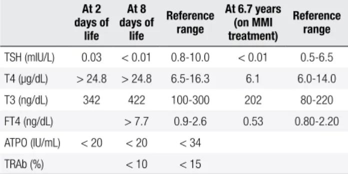
Table 1. Thyroid proile of the patient
At 2 days of
life At 8 days of
life
Reference range
At 6.7 years (on MMI treatment)
Reference range
TSH (mIU/L) 0.03 < 0.01 0.8-10.0 < 0.01 0.5-6.5
T4 (µg/dL) > 24.8 > 24.8 6.5-16.3 6.1 6.0-14.0
T3 (ng/dL) 342 422 100-300 202 80-220
FT4 (ng/dL) > 7.7 0.9-2.6 0.53 0.80-2.20
ATPO (IU/mL) < 20 < 20 < 34
Cop
yright
© ABE&M t
odos os dir
eit
os r
eser
vados
.
RESULTS
Sequencing of TSHR gene in the patient revealed a
het-erozygous G to A transition in exon 9 (rs121908878, c.842G>A), predicted to result in a missense muta-tion changing serine 281 to asparagine, (p.Ser281Asn, p.S281N) in the extracellular domain of TSH receptor. Also, a heterozygous C to G transversion was found in codon 727 (rs1991517, c.2181C>G, exon 10), re-sulting in the substitution of aspartic acid by glutamic acid, a known single nucleotide polymorphism (SNP) (p.Asp727Glu, p.D727E) (Figure 2).
In the mother, no genetic variants were detected in codons 281 or 727 of the TSHR gene, and she was
homozygous for the wild type allele in both positions. However, we found a heterozygous G>A transition in codon 459 (rs113951800, c.1377G>A, exon 10), pre-dicted to result in a silent polymorphism (p.Ala459Ala), not present in her daughter’s sample.
DISCUSSION
The SNAH phenotype was irst described by Kopp and cols. in a patient with the p.Phe631Leu mutation in the TSHR gene (6). Most of the patients with SNAH
reported to date (10 out of 14), were born prematurely and, when compared to HNAH, presented earlier and more severe symptoms in the neonatal period (10,11). Our patient was born at term with normal size, and was severely hyperthyroid since birth, with tachycardia and fetal suffering.
Although congenital NAH is a rare condition, dif-ferential diagnosis with autoimmune disease should be carried out when severe neonatal disease persists, even when the mother presents autoimmune hyperthyroid-ism, in order to differentiate it from the more com-mon congenital autoimmune disorder. In contrast to patients with neonatal Graves’ disease, patients with se-vere and persistent non-autoimmune hyperthyroidism should be treated more aggressively, requiring either total thyroidectomy or radiation to control the disease.
Reported thyroid size is variable in patients with SNAH, with diffuse neonatal goiter observed in 9 out of 14 described patients. Our patient had a normal sized eutopic gland.
Regarding clinical features in NAH, hyperthyroidism persists and commonly relapses following withdrawal of antithyroid drugs, and even after subtotal thyroidecto-my. In the present case, the patient had multiple relapses
Figure 1. Histological inding in the removed thyroid gland: follicular cell hyperplasia without signs of malignancy or lymphocytic iniltration.
Table 2. Thyroid proile of the patient’s mother
14 years At delivery Reference range
TSH (mIU/L) 8.0 0.5-5.0
T4 (µg/dL) 17.0 4.4 4.5-12.5
T3 (ng/dL) 670 66 80-220
FT4 (ng/dL) 0.36 0.80-2.20
ATPO (IU/mL) 58 < 34
ATG (IU/mL) 24.5 < 115
TRAb (%) < 10 < 15
Genomic DNA from the patient and her mother was extracted from peripheral blood based on the use of cetyltrimethylammonium bromide (CTAB) lysis buffer and isoamyl alcohol-chloroform extraction (14). The whole coding sequence (exons 1 to 10) and intron-ex-on boundaries correspintron-ex-onding to TSHR gene (GeneID:
7253; Genebank: NG_009206.1, NM_000369.2, NP_000360.2) were ampliied by PCR using genomic DNA both from the patient and her mother as tem-plate, and automatically sequenced with Applied Bio-systems 3730xl DNA analyzer (Macrogen Inc. Seoul, Korea) with speciic oligonucleotide primers (PCR protocols and primers sequences are available upon re-quest).
Cop
yright
© ABE&M t
odos os dir
eit
os r
eser
vados
.
when withdrawal was attempted, persisting hyperthy-roid until almost 7 years of age, when thyhyperthy-roidectomy was performed. Antithyroid drugs were administered in all previously reported cases without good response, and radioiodine therapy was indicated in 6 cases, but near total thyroidectomy seems to be the treatment of choice for these patients (10,15-17). Various consequences of prolonged neonatal hyperthyroidism, including multi-nodular goiter, microcephaly, craniosynostosis, psycho-motor disturbances, mental retardation, intrauterine growth retardation, prematurity and low birth weight, have been reported in the literature, but none of these complications developed in our patient.
In this patient, we identiied the p.Ser281Asn muta-tion in the TSHR gene, as well as the p.Asp727Glu
poly-morphism. Her mother, who harbors the p.Ala459Ala polymorphism, but not the p.Ser281Asn mutation, was hyperthyroid since adolescence without TRAb antibo-dies, but with low titers of ATPO antiboantibo-dies, indicating an autoimmune thyroid disorder. Although the father could not be studied, no history suggesting hyperthy-roidism was reported. Thus, we might suggest that this is a case of SNAH, resulting from a de novo mutation.
In the patient reported by Grüters and cols. (9), together with the p.Ser281Asn mutation, the authors found the p.Arg528His gene variant, a polymorphism
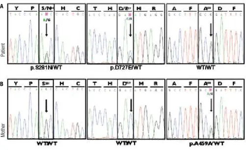
Figure 2. Electropherograms showing partial sequencing results of TSH receptor (TSHR) gene from the patient (A) and her mother (B).
Patient’s genomic DNA sequencing shows a heterozygous G to A transition at the 2nd base of codon 281 (exon 9) leading to p.Ser281Asn substitution (A,
left panel), and a heterozygous transversion C to G in codon 727 (exon 10), resulting in p.Asp727Glu substitution (A, middle panel), while her mother is homozygous for the wild type (WT) allele in both codons (B, left and middle panels). The right panel of Figure B displays a heterozygous A to G transition in the 3rd base of codon 459 (exon 10), presumably resulting in a silent polymorphism (p.Ala459Ala).
that failed to increase cAMP production in functional studies. Some paternal relatives of the affected child, who presented the p.Arg528His polymorphism but did not harbor the p.Ser281Asn mutation, developed hyperthyroidism with negative TRAb but low positive titers of ATPO antibodies. A similar situation is seen in the present case, as the mother, who is negative for the mutation, presented hyperthyroidism with positive ATPO and negative TRAb.
As observed in other conditions caused by gain-of-function mutations, all patients with congenital NAH reported to date (including the present case) have only one affected allele, and are heterozygous for the germ-line mutation.
The p.Ser281Asn TSHR gene variant found in this
patient has already been described as germline muta-tion in 3 patients with SNAH (9,18,19), and as somatic mutation in autonomous thyroid nodules (20). In vi-tro experiments have demonstrated that the resulting
mutant TSH receptor displays signiicantly higher con-stitutive activity when compared with the wild type re-ceptor, with higher basal production of cAMP (17,18). Interestingly, p.Ser281Asn is the unique germline acti-vating mutation described in the extracellular domain, suggesting that residue 281 is a key position for TSH receptor structure and function.
Mother
Patient
Cop
yright
© ABE&M t
odos os dir
eit
os r
eser
vados
.
Alignment analysis of TSHr, FSHr and LH/CGr amino acid sequences from different animal species re-veals that Serine 281 is highly conserved in all three gly-coprotein hormone receptors, suggesting that this resi-due is crucial in maintaining proper three-dimensional structure for receptor function. Serine 281 is located in a highly speciic and conserved motif (SHCCAF) within the hinge domain of the receptor, between the extracellular leucine-rich repeat motif and the trans-membrane helices (17,20-22). The TSH receptor dis-plays signiicant constitutive activity, and it has been hy-pothesized that its extracellular domain in the unbound state would exert a negative constraint maintaining re-ceptor quiescence. Consequently, mutations within the ectodomain would disrupt the three-dimensional struc-ture necessary for receptor silencing, and might lead to increased constitutive activity (9,17-22).
The p.Asp727Glu (p.D727E) polymorphism found in our patient is widely distributed among subjects from different populations. According to the SNP da-tabase (dbSNP) of the National Center for Biotechnol-ogy Information (NCBI), the minor allele frequency (MAF) is 0.108 for the G allele (MAF corresponds to 1,000 Genome phase 1 genotype data from 1,094 worldwide individuals, released in the May 2011 data-set). Gabriel and cols. (23) reported higher frequency of p.Asp727Glu in patients with toxic multinodular goiter than in normal controls, and exaggerated cAMP production in response to TSH stimulation when this variant was studied in vitro, suggesting that the variant
might play a role in the pathogenesis of thyroid disease in patients with toxic adenoma. However, Mühlberg and cols. (24) were unable to detect any signiicant dif-ferences in p.Asp727Glu gene variant frequency among healthy controls and patients with either NAH or Graves’ disease. Similarly, Nogueira and cols. (25) and Sykiotis and cols. (26) also demonstrated, by in vitro
expression studies, that the E727 variant receptor was not signiicantly different from the wild type D727 re-ceptor in terms of basal and stimulated cAMP produc-tion. Therefore, it would seem that the p.Asp727Glu variant is unlikely to play a role in the genesis of NAH or toxic adenoma (25,26).
The heterozygous c.1377G>A transition in exon 10, present only in the mother, is predicted to result in the silent polymorphism p.Ala459Ala. This variant (rs113951800), included in the NCBI dbSNP with low frequency (MAF: 0.008), has been previously re-ported in a hyperfunctioning thyroid nodule (23). Even
though this is presumably a benign change, in vitro
ex-pression and RNA stability studies will be necessary to establish its effect on receptor expression and function, and its potential involvement in thyroid disorders.
Recently, Lueblinghoff and cols. (27) analyzed a possible genotype-phenotype correlation in a system-atic review of the SNAH cases reported to date. They could not ind any consistent association between the degree of in vitro activity of each TSHR mutant and
the clinical course of patients with SNAH, and con-cluded that this may be due, at least in part, to the re-stricted number of case reports and limited follow-up. However, the lack of genotype-phenotype correlation in patients with NAH might also relect the inluence of iodine intake and/or other genetic, epigenetic, or environmental factors.
The molecular characterization of THSR gene
mu-tations in patients with NAH enables both adequate treatment decision and appropriate genetic counseling. Moreover, recognizing NAH will help to avoid the ir-reversible consequences of inadequate treatment of this disorder, which probably affects children since their early fetal development.
Acknowledgments: A. Chiesa is a research associate member of the Gobierno de la Ciudad de Buenos Aires. We would like to thank Mrs. Perla Rossano for her technical assistance. This study did not receive any speciic grant from any funding agency in the public, commercial or non-proit sector.
Disclosure: no potential conlict of interest relevant to this article was reported.
REFERENCES
1. Polak M, Legac I, Vuillard E, Guibourdenache J, Castanet M, Luton D. Congenital hyperthyroidism: the fetus as a patient. Horm Res. 2006;65(5):235-42.
2. Becks GP, Burrow GN. Thyroid disease and pregnancy. Med Clin North Am. 1991;75(1):121-50.
3. Ogilvy-Stuart AL. Neonatal thyroid disorders. Arch Dis Child Fetal Neonatal Ed. 2002;87(3):F165-71.
4. Polak M. Hyperthyroidism in early infancy: pathogenesis, clinical features and diagnosis with a focus on neonatal hyperthyroidism. Thyroid. 1998;8(12):1171-7.
5. Duprez L, Parma J, Van Sande J, Allgeier A, Leclère J, Schvartzm C, et al. Germline mutations in the thyrotropin receptor gene cause non-autoimmune autosomal dominant hyperthyroidism. Nat Genet. 1994;7(3):396-401.
6. Kopp P, van Sande J, Parma J, Duprez L, Gerber H, Joss E, et al. Brief report: congenital hyperthyroidism caused by a mutation in the thyrotropin-receptor gene. N Engl J Med. 1995;332(3):150-4. 7. de Roux N, Polak M, Couet J, Leger J, Czernichow P, Milgrom E,
Cop
yright
© ABE&M t
odos os dir
eit
os r
eser
vados
.
8. Holzapfel HP, Wonerow P, von Petrykowski W, Henschen M, Scherbaum WA, Paschke R. Sporadic congenital hyperthyroidism due to a spontaneous germline mutation in the thyrotropin re-ceptor gene. J Clin Endocrinol Metab. 1997;82(11):3879-84. 9. Grüters A, Schöneberg T, Biebemann H, Krude H, Krohn HP, Dralle
H, et al. Severe congenital hyperthyroidism caused by a germ-line neo mutation in the extracellular portion of the thyrotropin receptor. J Clin Endocrinol Metab. 1998;83(5):1431-6.
10. Gozu HI, Lublingoff J, Bircan R, Paschke R. Genetics and phenom-ics of inherited and sporadic non-autoimmune hyperthyroidism. Mol Cell Endocrinol. 2010;322(1-2):125-34.
11. Hébrant A, van Staveren WC, Maenhaut C, Dumont JE, Leclère J. Genetic hyperthyroidism: hyperthyroidism due to activating TSHR mutations. Eur J Endocrinol. 2011;164(1):1-9.
12. Helsinki, Finland, June 1964, and amended by the 29th WMA Gen-eral Assembly, Tokio, Japan, October 1975, 35th WMA GenGen-eral As-sembly, Venice, Italy, October 1983, 41st WMA General AsAs-sembly, Hong Kong, September 1989, 48th General Assembly, Somerset West, Republic of South Africa, October 1996, 52nd WMA General Assembly, Edinburg, Scotland, October 2000, 53rd WMA General Assembly, Washington 2002 (note of clariication on paragraph 29 added), 55th WMA General Assembly, Tokyo 2004 (note of clar-iication on paragraph 30 added), 59th WMA General Assembly, Seoul, October 2008.
13. Greulich WW, Pyle SI. Radiographic atlas of skeletal development of the hand and wrist. California: Stanford University Press; 1950. 14. Del Sal G, Monioletti G, Schneider C. The CTAB-DNA precipita-tion method a common mini-scale preparaprecipita-tion of template DNA from phagemids, phages or plasmids suitable for sequencing. Biotechniques. 1989;7(5):514-20.
15. Esapa CT, Duprez L, Ludgate M, Mustafa MS, Kendall-Taylor P, Vassart G, et al. A novel thyrotropin receptor mutation in an infant with severe thyrotoxicosis. Thyroid. 1999;9(10):1005-10.
16. Bertalan R, Sallai A, Sólyom J, Lotz G, Szabó I, Kovács B, et al. Hyperthyroidism caused by a gemline activating mutation of the thyrotropin receptor gene: dificulties in diagnosis and therapy. Thyroid. 2010;20(3):327-32.
17. Duprez L, Parma J, Costagliola S, Hermans J, van Sande J, Du-mont JE, et al. Constitutive activation of the TSH receptor by spontaneous mutations affecting the N-terminal extracellular do-main. FEBS lett. 1997;409(3):469-74.
18. Chester J, Rotenstein D, Ringkananont U, Steuer G, Carlin B, Stewart L, et al. Congenital neonatal hypertyroidism caused by germline mutations in the TSH receptor gene. J Pediatr Endocri-nol Metab. 2008;21(5):479-86.
19. Biebermann H, Schöneberg T, Krude H, Gudermann T, Grüters A. Constitutively activating TSH-receptor mutations as a molecular cause of non-autoimmune hyperthyroidism in childhood. Lan-genbecks Arch Surg. 2000;385(6):390-2.
20. Ho SC, van Sande J, Lefort A, Vassart G, Costagliola S. Effects of mutations involoving the highly conserved S281HCC mo-tif in the extracellular domain of the thyrotropin (TSH) recep-tor on TSH binding and constitutive activity. Endocrinology. 2001;142(7):2760-7.
21. Jaeschke H, Neumann S, Kleinau G, Mueller S, Claus M, Krause G, et al. An aromatic environment in the vicinity of serine 281 is a structural requirement for thyrotropin receptor function. Endocri-nology. 2006;147(4):1753-60.
22. Farid NR, Kascur V, Balazs C. The human thyrotropin receptor is highly mutable: a review of gain-of-function mutations. Eur J En-docrinol. 2000;143(1):25-30.
23. Gabriel EM, Bergert ER, Grant CS, van Heerden JA, Thomp-son GB, Morris JC. Germline polymorphism of codon 727 of human thyroid-stimulating hormone receptor is associ-ated with toxic multinodular goiter. J Clin Endocrinol Metab. 1999;84(9):3328-35.
24. Mühlberg T, Herrmann K, Joba W, Kirchberger M, Heberling HJ, Heufelder AE. Lack of association of nonautoimmune hyperfunc-tioning thyroid disorders and a germline polymorphism of codon 727 of the human thyrotropin receptor in a European Caucasian population. J Clin Endocrinol Metab. 2000;85(8):2640-3. 25. Nogueira CR, Kopp P, Arseven OK, Santos CL, Jameson JL,
Me-deiros-Neto G. Thyrotropin receptor mutations in hyperfunction-ing thyroid adenomas from Brazil. Thyroid. 1999;9(11):1063-8. 26. Sykiotis GP, Neumann S, Georgopoulos NA, Sgourou A,
Papa-chatzopoulou A, Markou KB, et al. Functional signiicance of the thyrotropin receptor germline polymorphism D727E. Biochem Biophys Res Commun. 2003;301(4):1051-6.