385
Rev Bras Med Esporte – Vol. 17, No 6 – Nov/Dec, 2011
Positive Diabetes Family History Alters Chronotropic
Response to Acute Exercise
Michelle Sartori1 Marcelo Velloso Hereen1 Juliana Valente1 Márcio Tubaldini1 Maria Cláudia Irigoyen2 Kátia De Angelis1,3
1. Laboratory of Human Movement São Judas Tadeu University – São Paulo, SP. 2. Laboratory of Experimental Hyper-tension, (InCor), Medicine College, University of São Paulo – São Paulo, SP. 3. Laboratory of Translational Physiol-ogy, Nove de Julho University, São Paulo, SP.
Mailing address:
Kátia De Angelis
Av Francisco Matarazzo, 612 1 andar, Diretoria do Mestrado em Ciências da Reabilitação
05001-100 – São Paulo, SP E-mail: [email protected]
Original Article
EXERCISE AND SPORTS MEDICINE CLINIC
ABSTRACT
The aim of this study was to verify the effects of one aerobic exercise session (AES) on the metabolic and hemodynamic profile in diabetes type II offspring. Healthy young males were assigned into 2 groups: diabetic offspring (DO, n=7) and euglycemic offspring (EO, n=7). Meta-bolic (triglycerides and glucose levels) and cardiovascular (arterial pressure, AP, heart rate, HR) measures were realized before, during and after the AES. The AES was realized on a treadmill during 30 minutes, with progressive intensity. The groups showed similar triglycerides, AP and HR values at rest. The glucose level was higher in DO group when compared to the ED group (99±2 vs. 89±2 mg/dL). The DO group showed higher chronotropic response at the beginning of AES when compared to the EO group (86±4 vs. 125±8 bpm), however, the HR was similar between the groups at the other exercise intensities. At the fifth minute of the recovery, the HR was higher in the DO group when compared to the EO group (FN: 88 ± 3 vs FD: 97± 4 bpm). The AP was similar between groups during and after the AES. The results showed that young diabetic offspring presented metabolic alterations at rest, and exacerbated HR response at the beginning and in the recovery period of the AES, suggesting a higher cardiovascular risk in this population.
Keywords: heritage, autonomic control, physical exercise, risk factors.
INTRODUCTION
Diabetes mellitus (DM) is a chronic disease in which the genetic and auto-immune elements, leading to alterations in the metabolism of carbohydrates, lipids and proteins. Type II DM has a dominant autosomal genetic transmission pattern which can be hereditary. Moreover, many environmental factors play an important role in the development of the disease(1).
Studies demonstrate that the DM doubles the risk of developing cardiocirculatory diseases in men and triples in women(2). It is
interesting to note that the cardiocirculatory complications represent the greatest cause of morbimortality among diabetic patients. Additionally, individuals with positive type II DM family history present increase of 40% in the risk of developing cardiovascular diseases. It is known that cardiovascular complications in diabetic patients are related to autonomous neuropathy, which is initially characterized by dysfunction of the parasympathetic nervous system(3).
Studies have currently related hemodynamic alterations at rest during or after physical exercise with higher risk of developing cardiovascular diseases(3-6)such as hypertension(3-7)and
diabetes(6),higher risk of besides being a predictor of higher risk of
mortality(8). Specifically concerning DM, the chronotropic response
to physical exercise has been associated to the disease diagnosis as well as mortality in patients who have already been diagnosed(9).
Epidemiological studies have shown that increase of the development of MD risk related to positive family history of this disease is strongly connected to sedentary lifestyle. Therefore, it is suggested that increase of the disease development related to genetic factors may be decreased and even eliminated with increase in the physical activity level(10,11).
Moreover, there is strong and consistent evidence that a single exercise session may, acutely, improve insulin sensitivity and glucose homeostasis, besides improving the sympatovagal balance in cardiac or diabetic patients(12). However, little is known
about the metabolic, hemodynamic and autonomic alterations in offspring of diabetic patients at rest or in response to a single session of dynamic exercises. Early detection of metabolic and/or cardiovascular alterations at rest and in response to exercise may be very important to prevent DM in young individuals with positive family history for this disease. Thus, the aim of this study was to verify the effects of a session of aerobic exercise in hemodynamic and metabolic parameters in diabetic offspring.
MATERIALS AND METHODS
14 male subjects were selected and divided in two groups (n= 7 in each group: euglycemic offspring(EO) and diabetic offspring (DO). The evaluations were carried out in three distinct phases, respecting a 48-hour interval between them.
In order to be apt to participate in the research, the subjects had to meet the following inclusion criteria: age range of 18 and 35 years; sedentarianism;blood pressure (BP) below 140/90mmHg obtained in two different occasions; absence of smoking; fast glycemia below 120mg/dl; BMI up to 27kg/m2. The participants who presented
injury, pain or recent orthopedic surgery in upper or lower limbs, any other pathologies, such as cardiologic, metabolic, neurological or rheumatic nature; participants who had hypertensive parents and/or coronary arterial disease and/or cardiac insufficiency.
386 Rev Bras Med Esporte – Vol. 17, No 6 – Nov/Dec, 2011
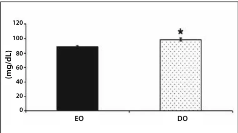
Figure 1. Glycemia at rest of the euglycemic offspring (EO) and diabetic offspring (DO) groups. *p < 0.05 versus DO.
120
100
80
60
40
20
0
(mg/dL)
EO DO
Biodinamics). Subsequently, using reagent bands, triglycerides and
total cholesterol were measured with the Accutrend GCT equipment and glycemia with the Accu-Chek Advantage equipment. Finally, weight and height were measured to obtain the body mass index values(BMI).
Part 2 – Ergospirometric test on treadmill was performed (Johnson brand name, model Jet 2000), in which the ventilatory thresholds and O2 peak values (VO2000 gas analyzer) were determined. HR
was monitored during the test through the electrocardiogram. BP was checked at each load increment. This phase was performed to determine the suitable intensity of the acute exercise session.
Ventilatory threshold 1 (or anaerobic threshold) was determined from the occurrence of at least two of the following variables: loss of linearity of the CO2 production and O2 consumption ratio by
the lowest value of the O2 alveolar concentration (PetO2/ O2partial
pressure at the end of the expiration); and the loss of linearity of the E (pulmonary ventilation) and the O2ratio. Ventilatory threshold
2 (or respiratory decompensation point) was determined in the occurrence of the highest PetCO2 value (CO2partial pressure at the
end of the expiration) preceding its sudden decrease and/or from the loss of linearity of the E and CO2 ratio.
Part 3 – This part consisted of the aerobic exercise session. Prior to the exercise, the individual remained at 30-minute rest and lactate (Accutrend Lactate), BP by the auscultatory method, and HR were measured with a frequency meter (Polar S-810). The instruments used as reference pattern for BP and HR measurement in this study had been previously inspected by INMETRO and were calibrated accordingly. The conventional and validated methods use were an aneroid sphygmomanometer with a cuff brand name Missouri®
(according to specifications by the British Society of Hypertension, 2004) and a stethoscope in perfect condition for BP measurement. All hemodynamic measurements were performed by the same evaluator and according to the guidelines by the Brazilian Society of Hypertension (2000) and IV Brazilian Guidelines for Hypertension (2002) for the measurements at rest and according to the guidelines by the American College of Sports Medicine (2003) for BP and HR check during exercise. All measurements of the volunteers were performed at sitting position and the upper limb was kept at the heart level.
The acute exercise was performed during 30 minutes with progressive load increase: five minutes of warm-up, five minutes of with HR at 30% below the one obtained at the second ventilatory threshold, five minutes with HR 20% below the one obtained at the second ventilator threshold, 10 minutes with HR at 10% below the one obtained at the second ventilator threshold and five minutes back to calmness.The lactate was measured at rest, at the 25th
minute of exercise and at recovery. BP was checked immediately after each load increment during the exercise (30min) and the HR monitored during the entire exercise through the frequency meter. The individuals then remained sitting during 30 minutes of recovery. In that period the HR was continuously monitored and at the end the BP and glycemia measurements were taken.
Results were statistically analyzed through the SPSS software forWindows 12.0. Means and means standard error (MSE) of the evaluated variables were calculated. In order to have the obtained
results compared, ttest followed by Student-Newman-Keuls complementary test were applied accordingly. The diferences were considered significant for p< 0.05 values.
RESULTS
The blood triglycerides and total cholesterol values were similar between groups after a four-hour fast period. Glycemia, though, despite being within the normality range, was higher in the diabetic offspring group (9.0 ± 1.50mg/dL) compared to the control offspring group (89.0 ± 2.30mg/dL) (figure 1).
The body composition measurements (lean mass and fat mass) and BMI were similar between the two groups. The BP and HR were similar between the groups at rest. The O2 peak, an important aspect related to physical capacity, was also similar between groups. These results demonstrate the sample’s homogeneity (table 1).
Lactate presented normal behavior and similar between groups at rest (EO: 3.3 ± 0.46 versusDO: 2.8 ± 1.10mmol/dL), at the end of the exercise (EO: 4.4 ± 0.83 versusDO: 4.5 ± 1.70mmol/dL) and at recovery (EO: 3.0 ± 0.96 versusDO: 3.7 ± 1.65mmol/dL).
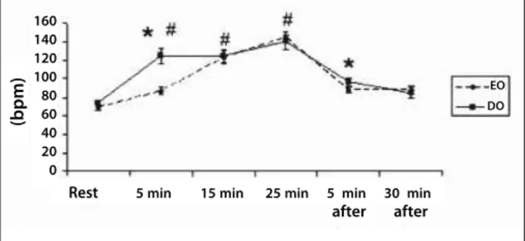
The SBP and DBP and HR presented normal behavior and similar between groups during the exercise session. However, the DO group demonstrated exacerbated increase of HR at the first five minutes of physical exercise when compared to the EO group (EO: 87 ± 4 versusDO: 125 ± 8bpm) (figure 2).
At the fifth minute of recovery, the EO group presented HR values lower when compared to the DO group (EO: 88 ± 3 versus
DO: 97 ± 4bpm). After the total recovery period (30min), there was not difference in the HR (EO: 88 ± 5 versusDO: 85 ± 6bpm), in the SBP or DBP (table 2).
Table 1. Anthropometric, metabolic ad hemodynamic measurements of the euglyce-mic offspring (EO) and diabetic offspring (DO) groups at rest and after four-hour rest .
EO DO
BMI (kg/m2) 23 ± 1.0 22 ± 0.8
Lean mass (%) 81 ± 2.0 81 ± 1.5
Fat mass (%) 18 ± 2.0 19 ± 1.4
TGL (mg/dL) 121 ± 22 137 ± 19
Cholesterol (mg/dL) 166 ± 5.5 160 ± 3.5
SBP (mmHg) 110 ± 3.5 107 ± 3.2
DBP (mmHg) 77 ± 3.7 73 ± 2.2
HR (bpm) 69 ± 3.4 74 ± 3.0
387
Rev Bras Med Esporte – Vol. 17, No 6 – Nov/Dec, 2011
Figure 2. Heart rate at rest, during and after exercise (recovery) in the euglycemic offspring (EO) and diabetic offspring (DO) groups. *p < 0.05 versus EO, #p < 0.05
versus rest.
DISCUSSION
The aim of the present study was to verify the effects of a session of aerobic exercise on the hemodynamic and metabolic parameters in offspring of diabetic individuals.The results of the present study demonstrate that offspring of diabetic patients present metabolic (glycemia increase) and cardiovascular alterations (higher HR response at the beginning, at the fifth minute of exercise and at the fifth minute of recovery) compared to offspring of euglycemic individuals.
DM is a disease with severe endocrine alterations which interfere in the metabolism of carbohydrates, lipids and proteins. These alterations result in the glucose plasmatic increase, triggering a fasting hyperglycemia status, caused mainly by the insulin deficient or absent secretion, associated to varied degrees of resistance to the activity of this hormone(13,14). Having the guidelines of the Brazilian
Society of Diabetes (2007) as reference, it was observed that both groups presented glycemia values within the normality range. However, the offspring of diabetic individuals presented higher glycemia values when compared to the offspring ofeuglycemic individuals. Conversely, further studies did not observe difference in glycemia at fast in offspring of diabetic subjects(15,16); however,
the insulin at rest levels were higher in offspring of diabetic ones(16).
It is worth mentioning that in these studies already published the sample was predominately composed of women.
It is known that besides environmental factors such as sedentarism and bad eating habit, it is currently clear that the development of diabetes is also related to the genetic characteristics of each individual(13,14). Thus, considering that the sample of the
present study presented similar and within healthy limits metabolic, (glycemia, triglyderides and cholesterol), hemodynamic (BP and HR) and body composition (BMI, fat and lean mass percentage) parameters, it is suggested that genetic factors justifies, at least
partly, the higher glycemia at fast observed in the group of offspring of diabetic individuals.
The interest for evaluating alterations in the HR during rest, in exercise and recovery after exercise has been increasing for some time now(6). When we start a physical activity, one of the
earliest effects on the cardiovascular system is the increase of the chronotropic response. This increase occurred linearly and proportionally to the increase in the exercise intensity(17). In fact,
in a work conducted by our group, linear HR increase in women during physical exercise performance(18). HR increase during exercise
is firstly a combination of vagal removal, and subsequently, of increasing activation of the sympathetic nervous system(6). Although
the HR presents normal behavior in the offspring of euglycemic parents, exacerbated increase was verified at the first five minutes of exercise in the DO group compared to the EO group. This result presents important clinical relevance in the stratification of the cardiovascular risk in this population, since there is a direct correlation between HR at rest or submaximal and development of cardiovascular diseases. Thus, HR abnormality during exercise is directly associated to mortality due to cardiovascular diseases and other causes of diseases(19).Therefore, individuals with lower HR
at rest or lower tachycardia during submaximal physical exercise present lower probability to develop cardiopathies(3). Interestingly,
individuals who present higher HR at rest or higher chronotropic response to exercise have higher chances to develop diseases such as hypertension or diabetes(6). It is important to highlight that the
HR increase in diabetic offspring was observed in the exercise phase characterized as warm-up, in which all individuals were submitted to the same absolute exertion intensity, suggesting hence that offspring of diabetic individuals present increased initial reactivity to physical exertion when compared to offspring of euglycemic parents. During the rest of the exercise session the HR was similar between groups, while the systolic and diastolic blood pressure presented normal behavior during the entire exercise(20)and in the
recovery period, being similar between groups.
It is worth mentioning that HR has been related to higher risk of cardiovascular diseases and episodes not only during restor exercise, but there is also high correlation between HR reduction at the first minutes of recovery after exercise and cardiovascular risk(4). Concerning this aspect, in the present study, at the end of
the recovery period, HR was similar between groups. However, at the fifth minute of recovery from exercise, DO group presented higher values compared to EO group. The reduced HR return to rest values during the recovery beginning is associated to increase in mortality risk due to cardiovascular diseases(21,22), sudden death(5)
and other mortality causes in asymptomatic patients(21,22), besides
being associated with diseases such as hypertension (3-7). In fact,
in an interesting study developed with healthy individuals, the HR profile during the recovery period was associated with the sudden death event(3).
Moreover, HR has been associated with diabetes diagnosis as well as increase of mortality in diabetic patients(9). Therefore, one of the
aspects which can answer for this increase in cardiovascular risk in these individuals is possible misadjustment of the parasympathetic nervous system. It is worth remembering that the HR decrease immediately after physical exercise mainly occurs in response to the reestablishment of the activity of the parasympathetic nervous system(6).Therefore, both the exacerbated HR increase
Table 2. Systolic blood pressure (SBP) and diastolic blood pressure (DBP) values at rest, during and after recovery from exercise in the euglycemic offspring (EO) and diabetic offspring (DO) groups.
Group Rest 5min 15min 25min Recovery
EO SBP (mmHg) 110 ± 3 120 ± 5 144 ± 6# 159 ± 9# 101 ± 4 DBP (mmHg) 77 ± 4 81 ± 2 81 ± 2 80 ± 3 77 ± 4 DO SBP (mmHg) 107 ± 3 120 ± 4 136 ± 5# 152 ± 7# 110 ± 6
DBP (mmHg) 73 ± 2 76 ± 3 77 ± 4 75 ± 4 76 ± 3
Values represent mean ± mean standard error. #p < 0.05 versus rest.
(bpm)
160 140 120 100 80 60 40 20 0
Rest 5 min 15 min 25 min 5 min 30 min after after
EO
388 Rev Bras Med Esporte – Vol. 17, No 6 – Nov/Dec, 2011 in the beginning of the exercise and the attenuation of the HR
return to baseline values may be associated with dysfunction on the cardiac autonomic control manly characterized by parasympathetic reduction, according to what has been observed in diabetic subjects.
CONCLUSION
The main finding of the present study was to demonstrate that young adults with positive diabetes family history present an exacerbated chronotropic response at the beginning of a physical exercise session and slower heart rate recovery in the period after exercise when compared to the group of offspring of euglycemic
subjects, Thus, it is suggested that these individuals, despite being considered healthy due to their evaluation at rest, already present glycemia at rest alteration, as well as exacerbated response to exercise which can be considered as predicting factors to the development of diabetes and cardiovascular disease. Considering this evidence of higher disposition to diseases, it becomes important that diabetic offspring adopt a healthy lifestyle in order to prevent, treat or delay the disorders to which they are genetically prone.
All authors have declared there is not any potential conflict of interests concerning this article.
REFERENCES
1. Zecchin HG, Carvalheira JBC, Abdalla MJ. Bases genéticas da resistência à insulina, da síndrome metabólica e do diabetes melito tipo 2. Rev SOCESP 2002:3:508-20.
2. Muir A, Schatz DA, Maclaren NK. The Pathogenesis, Prediction, and Prevention of Insulin-Dependent Diabetes Mellitus.EndocrinolMetabClin North Am 1992:21:199-21.
3. Jouven X, Empana JP, Schwartz PJ, Desnos m, Courbon D, Ducimetière P. Heart-rate Profile During Exercise as a Predictor of Sudden Death. N Engl J Med 2005:352:1951-8.
4. Ellestad MH, Wan MK. Predictive Implications of Stress Testing: followup of 2700 subjects after maxi-mum treadmill stress testing. Circulation 1975:51:363-9.
5. Sandivik L, Erikssen J, Ellestad MH, Erikssen G, Thaulow E, Mundal R, et al. Heart rate increase and maximal heart rate during exercise as predictors of cardiovascular mortality: a 16 years follow-up study of 1960 healthy men. Coron Artery Dis 1995:6:667-79.
6. Cole CR, Blackstone EH, Pashkow FJ, Snader CE, Lauer MS. Heart-rate recovery Immediately After Exercise as a Predictor of Mortality. N Engl J Med 1999:341:1351-7.
7. Singh JP, Larson MG, Manolio TA, O’donnell CJ, Lauer M, Evans JC, et al. Blood pressure response during treadmill testing as a risk factor for new-onset hypertension. The Framingham Heart study. Circulation 1999:99:1831-6.
8. Imai K, Sato H, Hori M, Kusuoka H, Ozaki H, Yokoyama H, et al. Vagally mediated heart rate recovery after exercise is accelerated in athletes but blunted in patients with chronic heart failure. J Am Col-lCardiol 1994:24:1529-35.
9. Carnethon MR, Yan L, Greenland P, Garside DB, Dyer AR, Metzger B, et al. Resting Heart Rate in Middle Age and Diabetes Development in Older Age. Diabetes Care 2008:31:335-9.
10. Hu FB, Sigal RJ, Rich-Edwards JW, Colditz GA, Solomon CG, Willett WC, et al. Walking compared with vigorous physical activity and Risk of type 2 diabetes in women: a prospective study. JAMA 1999:282:1433-9.
11. Sargeant LA, Wareham NJ, Khaw KT. Family history of diabetes identifies a group at increase risk for the metabolic consequences of obesity and physical inactivity in EPIC-Norfolk: a population-based study. The European Prospective Investigation into Cancer.Int J ObesRelatMetabDisord 2000:24:1333-9.
12. Thompson PD, Crouse SF, Goodpaster B, Kelley D, Moyna N, Pescatello L. The acute versus the chronic response to exercise. Med Sci Sports Exerc 2001:33:438-45.
13. Yagihashi S. Pathology and pathogenetic mechanisms of diabetic neuropathy. Diabetes Metab Rev 1995:11:193-225.
14. Forjaz CLM, Santella DF, Rezende LO. A duração do exercício determina a magnitude e a duração da hipotensão pós-exercício. Arq Bras Cardiol 1998:70:99-104.
15. Barwell ND, Malkova D, Moran CN, Clelan SJ, Packard CJ, Zammit VA, et al. Exercise Training has Greater Effects on Insulin Sensitivity in Daugheters of Patients With Type 2 Diabetes Than in Women With no Family History of Diabetes. Diabetologia 2008:51:1912-19.
16. Giannattasio C, Failla M, Capra A, Scanziani E, Amigoni M, Boffi L, et al. Increased Arterial Stiffnes in Normotensive Offspring of Type 2 Diabetic Parent. Hypertension 2008:51:182-7.
17. Alonso DO, Forjaz CLM, Rezende LO, Braga AMFW, Barreto ACP, Negrão CE, et al. Comportamento da freqüência cardíaca e sua variabilidade durante as diferentes fases do exercício progressivo máximo. Arq Bras Cardiol 1998:71:787-92.
18. Pureza DY, Sargentini L, Laterza R, Flores LJF, Irigoyen MC, De Angelis K. Efeitos cardiovasculares da abstinência do fumo no repouso e durante o exercício submáximo em mulheres jovens fumantes. Rev Bras Med Esporte 2007:13:292-6.
19. Gillum RF, Mussolino ME, Madans JH. Diabetes Mellitus, Coronary Heart Disease incidence, and Death From All Causes in African American and European American Woman: The NHANES I Epidemiologic Follow-up Study. J ClinEpidemiol 2000:53:511-8.
20. American College of Sports Medicini. Position stand: progression models in resistance training for healthy adults. Med Sci Sports Exerc 2002:34:364-80.
21. Kurl S, Laukkanen JA, Rauramma R, Lakka TA, Sivenius J, Salonen JT. Systolic blood pressure response to exercise stress test and risk of stroke. Stroke 2001:32:2036-41.