Tensile bond strength of glass fiber posts
luted with different cements
Resistência à tração de pinos de fibra de vidro
cimentados com diferentes materiais
Abstract: Proper selection of the luting agent is fundamental to avoid failure due to lack of retention in post-retained crowns. The objective of this study was to investigate the tensile bond strength and failure mode of glass iber posts luted with different cements. Glass iber posts were luted in 40 mandibular premolars, divided into 4 groups (n = 10): Group 1 – resin-modiied glass ionomer RelyX Luting; Group 2 – resin-modiied glass ionomer Fuji Plus; Group 3 – resin cement RelyX ARC; Group 4 – resin cement Enforce. Specimens were assessed by tensile strength testing and light microscopy analysis for ob-servation of failure mode. The tensile bond strength values of each group were compared by ANOVA and Tukey test. The signiicance level was set at 5%. The failure modes were described as percentages. The following tensile strength values were obtained: Group 1 – 247.6 N; Group 2 – 256.7 N; Group 3 – 502.1 N; Group 4 – 477.3 N. There was no sta-tistically signiicant difference between Groups 1 and 2 or between Groups 3 and 4, yet the resin cements presented signiicantly higher tensile bond strength values than those presented by the glass ionomer cements. Group 1 displayed 70% of cohesive failures, whereas Groups 2, 3 and 4 exhibited 70% to 80% of adhesive failures at the dentin-ce-ment interface. We concluded that resin cedentin-ce-ments and glass ionomer cedentin-ce-ments are able to provide clinically suficient retention of glass iber posts, and that glass ionomer cements may be especially indicated when the application of adhesive techniques is dificult. Descriptors: Post and core technique; Tensile strength; Resin cements; Glass ionomer cements.
Resumo: A seleção adequada do agente cimentante é essencial para evitar falhas por per-da de retenção em coroas retiper-das por núcleos. O objetivo deste estudo foi investigar a resistência à tração e o tipo de falha de pinos de ibra de vidro cimentados com diferentes materiais. Cimentaram-se pinos de ibra de vidro em 40 pré-molares inferiores, divididos em 4 grupos (n = 10): Grupo 1 – ionômero de vidro modiicado por resina RelyX Luting; Grupo 2 – ionômero de vidro modiicado por resina Fuji Plus; Grupo 3 – cimento resinoso RelyX ARC; Grupo 4 – cimento resinoso Enforce. Avaliaram-se os espécimes por teste de resistência à tração e análise por microscopia óptica para observação do tipo de falha. Compararam-se os valores de resistência à tração de cada grupo por análise de variância e teste de Tukey, sendo que o nível de signiicância estabelecido foi de 5%. Descreveram-se os tipos de falha na forma de porcentagens. Os valores de resistência à tração obtidos foram: Grupo 1 – 247,6 N; Grupo 2 – 256,7 N; Grupo 3 – 502,1 N; Grupo 4 – 477,3 N. Não houve diferença estatisticamente signiicativa entre os Grupos 1 e 2 ou entre os Gru-pos 3 e 4, porém os cimentos resinosos apresentaram valores de resistência à tração sig-niicativamente maiores do que os apresentados pelos cimentos ionoméricos. O Grupo 1 exibiu 70% de falhas coesivas, enquanto os Grupos 2, 3 e 4 apresentaram 70% a 80% de falhas adesivas na interface dentina-cimento. Concluiu-se que os cimentos resinosos e ionoméricos são capazes de proporcionar retenção clinicamente suiciente de pinos de i-bras de vidro, e que os cimentos ionoméricos podem ser indicados principalmente quando houver diiculdades de aplicar técnicas adesivas.
Descritores: Técnica para retentor intra-radicular; Resistência à tração; Cimentos de resina; Cimentos de ionômeros de vidro.
Gerson Bonfante(a) Osvaldo Bazzan Kaizer(b) Luiz Fernando Pegoraro(a) Accácio Lins do Valle(a)
(a) PhDs, Professors, Department of
Prosthodontics, School of Dentistry of Bauru, University of São Paulo.
(b) PhD, Professor, Department of Restorative
Dentistry, School of Dentistry of Santa Maria, Federal University of Santa Maria.
Corresponding author:
Osvaldo Bazzan Kaizer Rua Tuiuti, 2121, apto. 902 Santa Maria - RS - Brazil CEP: 97015-000
E-mail: [email protected]
Introduction
Post-retained crowns may present mechanical or biological failures,11 commonly due to loss of
reten-tion.20 Thus, root canal posts should have enough
tensile bond strength to avoid displacement during function.3,19 The main function of posts is to aid in
crown retention,8 especially when 50% or more of
the remaining coronal structure have been lost.8,13
The quality of the cement is fundamental for post retention; however, there is no consensus in the literature as to the superiority of one cement com-pared to others, since the outcomes of tensile bond strength studies are conlicting.5,15,16 Moreover, no
luting agents have all ideal properties.5
Zinc phosphate is still the luting agent most often employed,16 and it is the irst choice for posts with
adequate mechanical retention when luoride release is not essential since it presents a long history of reli-ability and clinical success.5,16 It is used as standard
in comparative studies.21 Its main disadvantages are
high clinical solubility and lack of adhesion to the tooth structure.16
Resin cements are indicated only when post re-tention is severely impaired.5 However, these
ce-ments are highly technique-sensitive, are affected by moisture and require extended chair time.13
More-over, the shorter working time, high viscosity and possibility of accumulation of adhesive13 preclude its
application in narrow root canals, with risk of in-complete itting of the post.2
Bonding of resin cements is mainly impaired by unfavorable root canal coniguration, related to a high C factor (cavity coniguration factor), which may be up to 40 times higher compared to direct intracoronal restorations with similar cement thick-ness.26 The C factor is the ratio between the bonded
and non-bonded surface areas. The latter allows res-in low with consequent decrease res-in polymerization shrinkage stress.22 When the non-bonded area is
minimum, as inside the root canals, stress decrease is not enough and polymerization shrinkage may be higher than the bond strength, leading to the forma-tion of gaps at the cement-dentin interface.26
Since the bonding of root canal posts with resin cements is highly unpredictable,26 conventional glass
ionomer cements (GIC) or resin-modiied glass
iono-mer cements (RMGIC)3 may be alternatively
indicat-ed for the luting of iber posts. Both cements present bonding to dentin by micromechanical mechanisms and chemical bonding,6 and, despite the
polymer-ization shrinkage they present, their more favor-able viscoelastic properties (viscosity and modulus of elasticity), compared to those of resin cements (which are more rigid), and their longer setting time allow a better maintenance of the bonding.3,6
More-over, the hygroscopic expansion occurring after maturation of GIC e RMGIC partially compensates for shrinkage, thus reducing the stress and provid-ing a closer adaptation between cement and dentin at completion of maturation.27 The RMGIC intend
to keep the advantages of GIC and minimize their most signiicant disadvantages: They are less sensi-tive to moisture, present higher dimensional stabil-ity and higher bonding to the tooth structure than GIC. They also release luoride, are easily prepared and are more resistant to compression than zinc phosphate cement.5,21
A recent study10 with the push-out technique
suggested that the resistance to displacement of iber posts is more related to friction than to true bonding to the root canal. Therefore, GIC and RMGIC may be used for iber post luting since their hygroscopic expansion6 increases the frictional resistance to post
displacement. Also, no signiicant differences in mi-croleakage have been observed between carbon iber posts luted with resin cement Panavia 21 or glass ionomer cement Fuji-I.21
This study investigated the tensile bond strength of glass iber posts luted with different cements and the failure mode occurring for each cement. The null hypothesis was that there would be no differences in tensile bond strength or failure mode between luting agents.
Materials and Methods
Maillefer, Ballaigues, Switzerland), as recommended by the manufacturer of the glass iber posts (Refor-post n. 2, Odonto-Lógika Ltda., Londrina, Paraná, Brazil). The posts had a cylindrical shape (1.25 mm in diameter) and a tapered apex (0.9 mm in diam-eter in the inal 3 mm), in addition to large mechani-cal undercuts throughout their extension.
The roots were ixed to plastic cylinders with self-curing acrylic resin, keeping 3 mm of root ex-posed. Before luting, the root canals were cleaned, rinsed with distilled water for 60 seconds and dried with suction and paper points. The specimens were randomly divided into 4 groups (n = 10), as follows:
Group 1 – Posts luted with resin-modiied glass ionomer cement RelyX Luting (3M ESPE, St. Paul, MN, USA). The cement was prepared fol-lowing the manufacturer’s instructions and mixed for 30 seconds. The mixture was applied on the post and inserted into the root canal with a Lentulo spiral. The posts were itted under in-ger pressure and submitted to a 2-kg static load for 10 minutes.
Group 2 – Posts luted with resin-modiied glass ionomer cement Fuji Plus (GC America, Alsip, IL, USA). The root canal dentin was previously etched with Fuji Plus Conditioner for 20 seconds, followed by thorough rinsing with distilled water and drying with paper points. The cement was prepared following the manufacturer’s instruc-tions and mixed for 20 seconds. Luting was then performed as described for Group 1.
Group 3 and Group 4 – Posts luted with dual-cure resin cements. Group 3 was luted with resin cement RelyX ARC (3M ESPE, St. Paul, MN, USA) and Group 4 with the cement En-force (Dentsply Ind. e Com., Rio de Janeiro, RJ, Brazil). Acid etching of the root canal walls was performed with 37% phosphoric acid for 15 seconds, followed by thorough rinsing and dry-ing with endodontic canula and paper points. The adhesive system Scotchbond Multi-Purpose Plus (3M ESPE, St. Paul, MN, USA) was applied on the root canal walls, carefully following the manufacturer’s instructions to achieve dual cure. The post was cleaned with ethyl alcohol and a coat of silane (Scotchbond Ceramic Primer, 3M
•
•
•
ESPE, St. Paul, MN, USA) was applied for one minute, gently air-dried, and the catalyst of the adhesive system Scotchbond Multi-Purpose Plus was applied.
After cement preparation following the manu-facturer’s instructions for 10 to 20 seconds, luting was performed as described for Group 1, followed by light curing (XL 2500, 3M ESPE, St. Paul, MN, USA) for 40 seconds.
Tensile bond strength testing
The coronal portion of the post was fabricated with light cured composite resin Filtek Z250 (3M ESPE, St. Paul, MN, USA), applied in 2-mm in-crements. To avoid adhesion of resin to the root, two sheets of tin foil (Alumileste Ind., Cajamar, SP, Brazil) were placed on the dentin for isolation. A U-shaped loop cast in Ni-Cr alloy was inserted to connect the specimen to the testing machine. Af-ter storage of the specimens in distilled waAf-ter for 24 hours at 37°C, they were submitted to tensile bond strength testing in a universal testing machine (Model K-2000MP, Dinamômetros Kratos Ltda., Taboão da Serra, SP, Brazil). A metallic pin was placed through a perforation at the lower third of the acrylic resin cylinder for adaptation to the lower articulation of the testing machine. The crosshead speed applied to the specimen (through the hook connected to the upper portion of the testing ma-chine and the loop at the coronal portion of the post) was 0.5 mm/min.
The failure mode was established by analysis un-der a light microscope Mitutoyo series 164 (Mitu-toyo Corporation, Tokyo, Tokyo, Japan) at a 30 X magniication and classiied as follows:17
adhesive failure at the cement-post interface; adhesive failure at the dentin-cement interface; cohesive failure;
combined failure.
The tensile bond strength values of each group were compared by one-way ANOVA and Tukey’s test (p = 0.05).
Results
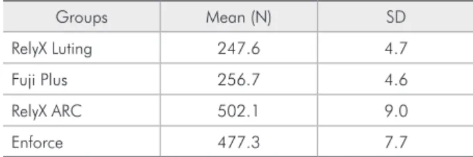
Table 1 presents the mean tensile bond strength values and standard deviations. The 1-way ANOVA 1.
revealed statistically signiicant differences between groups (p < 0.05). The Tukey test for multiple com-parisons revealed that RelyX ARC and Enforce presented signiicantly higher tensile bond strength values than those presented by the other cements (p < 0.05), without statistically signiicant differenc-es between RMGIC or between rdifferenc-esin cements.
Discussion
Fiber-reinforced resin posts are the most recent option for the reconstruction of endodontically treated teeth. Even though clinical and laboratory studies on iber posts are limited, the low number of failures and nearly total absence of root fractures should be highlighted.9,12,14 Theoretically, these posts
would be able to reinforce weakened roots, without increasing the risk of fracture.
In this study, even though the bond strength val-ues obtained for the RMGIC (Groups 1 and 2) were signiicantly lower compared to those obtained for Groups 3 and 4 (resin cements), all cements showed proper retention considering that posts should have a minimum tensile bond strength of 200 N to allow clinical success.17 The post coniguration (parallel
with the tapered apex and undercuts throughout its extension) probably contributed to the retention
val-ues of 247.6 N and 256.7 N respectively for Groups 1 and 2. Moreover, the retention of posts luted with RMGIC may be related to the frictional retention provided by hygroscopic expansion occurring after cement maturation,27 which also aids the
self-seal-ing at the dentin-cement interface.
The highest bond strength values were observed for the resin cements RelyX ARC (502.1 N) and En-force (477.3 N). Considering that polymerization of chemically-cured or dual-cured resin cements is im-paired by the association with simple (2-step) conven-tional adhesives or single-step self-etching adhesives, due to the high acidity of these systems, a 3-step dual-cured conventional adhesive was used for both resin cements in the present study. This enhanced the polymerization of the adhesive considering that the access for light curing at the most apical regions is limited. Scotchbond Multi-Purpose Plus has been successfully used since 1992 and has a record of less than 2% of loss of direct restorations.25 Also, the
ten-sile bond strength of self-curing or dual-cured resin cements obtained with this system is higher than the polymerization shrinkage stress produced by the po-lymerization of thin layers of resin cements in closed spaces, which is nearly 20 MPa.
When resin cements were employed, 70 to 80% of failures occurred at the dentin-cement interface (Table 2), which is in agreement with previous ind-ings1 that adhesive failures occurred between the
adhesive system and dentin. Bonding to the root canal dentin may be reduced by several unfavorable conditions, such as the materials employed during root canal preparation, eugenol-based endodontic sealers, dificulty to achieve an ideal degree of mois-ture, insuficient impregnation by the adhesive, and a high C factor, which is related to root canal con-iguration. When resin cements were employed, no specimen presented complete failure at the post-res-in cement post-res-interface, suggestpost-res-ing that the application of silane combined with the mechanical undercuts of the posts was effective to aid the cement against displacement and also suggesting a good bonding between the resin matrix of the iber posts and the resin cements.18 Finally, the lack of observation of
cohesive failures in the resin cements is related to their high resistance.23
Table 1 - Means and standard deviations of tensile bond strength (N) for the cements.
Groups Mean (N) SD
RelyX Luting 247.6 4.7
Fuji Plus 256.7 4.6
RelyX ARC 502.1 9.0
Enforce 477.3 7.7
Table 2 - Failure modes (%) observed for each group.
Failure mode RelyX
Luting Fuji Plus RelyX
ARC Enforce
Adhesive at the
dentin-cement interface 1 (10%) 7 (70%) 8 (80%) 7 (70%)
Adhesive at the
post-cement interface – – – –
Combined 2 (20%) 2 (20%) 2 (20%) 3 (30%)
Both RMGICs presented different failure modes, despite their similar bond strengths: RelyX presented 70% of cohesive failures, whereas Fuji Plus exhibited 70% of adhesive failures between dentin and cement (similarly to the resin cements). Some glass ionomer cements have increased bond strength due to the for-mation of a hybrid layer in dentin, and HEMA is able to penetrate into the dentinal tubules up to a depth of 1.5 mm.4,7 The failure mode observed for Fuji Plus
suggests the formation of a hybrid layer, and dentin pretreatment with a weak acid may have helped in the removal of the smear layer and increased dentin wettability, thereby enhancing adhesion. The low number of cohesive failures observed for Fuji Plus suggests that its resin components (HEMA, TEG-DMA) provide it with good mechanical resistance. The failure mode observed for RelyX Luting indi-cates that retention of this cement probably is more dependent on mechanical retention than on bonding
to dentin, and the high number of cohesive failures may be associated with its low intrinsic resistance and presence of bubbles within the cement.
All cements investigated in this study provided clinically suficient retention of glass iber posts.17,24
The results suggest that RMGIC may be indicated for iber post luting, especially when application of adhesive techniques is dificult.
Conclusions
Within the limitations of the present study, the following could be concluded:
Resin-modiied glass ionomer cements may be indicated for luting of glass iber posts.
Posts luted with RelyX Luting presented predom-inance of cohesive failures, whereas posts luted with Fuji Plus and resin cements exhibited pre-dominance of adhesive failures at the dentin-ce-ment interface.
1. 2.
References
1. Alster D, Feilzer AJ, de Gee AJ, Davidson CL. Polymerization contraction stress in thin resin composite layers as a function of layer thickness. Dent Mater. 1997;13(3):146-50. 2. Bachicha WS, Difiore PM, Miller DA, Lautenschlager EP,
Pashley DH. Microleakage of endodontically treated teeth restored with posts. J Endod. 1998;24(11):703-8.
3. Bouillaguet S, Troesch S, Wataha JC, Krejci I, Meyer JM, Pash-ley DH. Microtensile bond strength between adhesive cements and root canal dentin. Dent Mater. 2003;19(3):199-205. 4. Carvalho RM, Yoshiyama M, Horner JA, Pashley DH.
Bonding mechanism of Variglass to dentin. Am J Dent. 1995;8(5):253-8.
5. Creugers NH, Kayser AF, van’t Hof MA. A meta-analysis of durability data on conventional fixed bridges. Community Dent Oral Epidemiol. 1994;22(6):448-52.
6. Dauvillier BS, Feilzer A, de Gee AJ, Davidson CL. Viscoelastic parameters of dental restorative materials during setting. J Dent Res. 2000;79(3):818-23.
7. Ferrari M, Davidson CL. Interdiffusion of traditional glass ionomer cement into conditioned dentin. Am J Dent. 1997;10(6):295-7.
8. Ferrari M, Vichi A, Grandini S, Goracci C. Efficacy of a self-curing adhesive-resin cement system on luting glass-fiber posts into root canals: a SEM investigation. Int J Prosthodont. 2001;149(9):543-9.
9. Fredriksson M, Astback J, Pamenius M, Arvidson K. A retrospective study of 236 patients with teeth restored by
carbon fiber-reinforced epoxy resin posts. J Prosthet Dent. 1998;80(2):151-7.
10. Goracci C, Fabianelli A, Sadek FT, Papacchini F, Tay FR, Ferrari M. The contribution of friction to the dislocation re-sistance of bonded fiber posts. J Endod. 2005;31(8):608-12. 11. Hatzikyriakos AH, Reisis GI, Tsingos N. A 3-year postopera-tive clinical evaluation of posts and cores beneath existing crowns. J Prosthet Dent. 1992;67(4):454-8.
12. Heydecke G, Peters MC. The restoration of endodontically treated, single-rooted teeth with cast or direct posts and cores: a systematic review. J Prosthet Dent. 2002;87(4):380-6. 13. Kimmel SS. Restoration and reinforcement of endodontically
treated teeth with a polyethylene ribbon and prefabricated fiberglass post. Gen Dent. 2000;48(6):700-6.
14. Love RM, Purton DG. Retention of posts with resin, glass ionomer and hybrid cements. J Dent. 1998;26(7):599-602. 15. Mendoza DB, Eakle S. Retention of posts cemented with various
dentinal bonding cements. J Prosthet Dent. 1994;72(6):591-4.
16. Mitchell CA. Selection of materials for post cementation. Dent Update. 2000;27(7):350-4.
17. Monticelli F, Grandini S, Goracci C, Ferrari M. Clinical be-havior of translucent-fiber posts: a 2-year prospective study. Int J Prosthodont. 2003;16(6):593-6.
19. Purton DG, Love RM. Rigidity and retention of carbon fibre versus stainless steel root canal posts. Int Endod J. 1996;29(4):262-5.
20. Sorensen JA, Martinoff JT. Intracoronal reinforcement and coronal coverage: a study of endodontically treated teeth. J Prosthet Dent. 1984;51(6):780-4.
21. Stockton LW. Factors affecting retention of post systems: a literature review. J Prosthet Dent. 1999;81(4):380-5. 22. Tay FR, Loushine RJ, Lambrechts P, Weller RN, Pashley DH.
Geometric factors affecting dentin bonding in root canals: a theoretical modeling approach. J Endod. 2005;31(2):584-9. 23. Tay FR, Pashley DG, Yiu CKY, Sanares AME, Wei SHY.
Factors contributing to the incompatibility between
simpli-fied-step adhesives and chemical-cured or dual-cured com-posites. Part I. Single-step, self-etch adhesive. J Adhes Dent. 2003;5(1):27-40.
24. Turner CH. The retention of dental posts. J Dent. 1982;10(3):154-65.
25. van Meerbeek B, Perdigao J, Lambrechts P, Vanherle G. The clinical performance of adhesives. J Dent. 1998;26(1):1-20. 26. Watanabe F, Powers JM, Lorey RE. In vitro bonding of
prostho-dontic adhesives to dental alloys. J Dent Res. 1998;67(2):479-83.