Study of the areas and thicknesses of
mesiobucal root canals prepared by
three endodontic techniques
Estudo das áreas e espessuras de canais
radiculares mésio-vestibulares preparados por
três técnicas endodônticas
Abstract: The purpose of this study was to compare, in vitro, by means of computerized analysis of digital radiographic images, the anatomic alterations produced in the mandibu-lar momandibu-lar tooth dentinal walls of mesiobucal canals with severe curvature by three different endodontic techniques: Progressive Preparation, Staged and Serial Preparation. A selection was made of 45 extracted, human, mandibular molars, with root curvatures greater than 25°. They were divided into three groups for every technique studied, which were then sub-divided into three sub-groups in accordance with the position of the curvature along the root: cervical, median or apical. After access surgery and tooth length determination, the canals were illed with 100% Barium Sulphate radiological contrast and the teeth were then radiographed with a direct digital radiography system, using a special apparatus ca-pable of keeping the samples in the same spatial position during the different radiographic takes. After the above-mentioned endodontic techniques had been performed, the teeth were again illed with Barium sulphate and were also radiographed under the same previ-ously mentioned conditions. The pre- and post-operative digital images were then analyzed in two computerized programs, AutoCAD 2004 and CorelDraw 10, to assess, respectively, the areas and the horizontal alterations which occurred in the internal and external walls of the root canals. The results indicated that although no signiicant differences among the techniques were shown in the statistical analysis, in a descriptive analysis the Progressive Preparation technique was shown to be more regular, uniform and effective.
Descriptors: Root canal therapy; Root canal, anatomy & histology; Digital radiography; Contrast media.
Resumo: Objetivou-se comparar, in vitro, através de análise computadorizada de ima-gens radiográicas digitais, as alterações anatômicas promovidas nas paredes dentinárias de canais mésio-vestibulares com curvatura severa de molares inferiores por três técnicas endodônticas diferentes: Preparo Progressivo, Escalonada e Seriada. Foram selecionados 45 molares inferiores humanos extraídos, com curvaturas radiculares superiores a 25°, que foram divididos em três grupos para cada técnica estudada e subdivididos em três subgrupos de acordo com a posição da curvatura ao longo da raiz: cervical, mediana ou apical. Após cirurgia de acesso e odontometria, os canais foram preenchidos com contras-te radiológico de sulfato de Bário a 100% e os dencontras-tes então foram radiografados por um sistema de radiograia digital direta, utilizando-se um aparato capaz de manter as amos-tras na mesma posição espacial nas diferentes tomadas radiográicas. Após a realização das técnicas endodônticas supracitadas, os dentes foram novamente preenchidos com o sulfato de Bário e radiografados nas mesmas condições anteriores. As imagens digitais pré e pós-operatórias foram então analisadas em dois programas computadorizados, o Auto-CAD 2004 e o CorelDraw 10, para veriicar, respectivamente, as áreas e as alterações ho-rizontais ocorridas nas paredes internas e externas dos canais radiculares. Apesar de não ter havido diferenças signiicantes entre as técnicas na análise estatística, em uma análise descritiva a técnica do Preparo Progressivo mostrou-se mais regular, uniforme e eicaz. Descritores: Tratamento do canal radicular; Canal radicular, anatomia & histologia; Radiograia digital; Meios de contraste.
Isa Geralda Teixeira Constante(a)
Harry Davidowicz(b)
Fernando Branco Barletta(c)
Abilio Albuquerque Maranhão de Moura(d)
(a) MSc in Endodontics; (b)Full Professor; (d)Full Professor and Chairman – Department of Endodontics, Paulista University, São Paulo. (c) Professor, Graduate Program in
Endodontics, Lutheran University of Brazil, Canoas.
Corresponding author:
Abilio Albuquerque Maranhão de Moura Rua Barão do Triunfo, 1650, ap. 22, Campo Belo, São Paulo - SP - Brazil CEP: 04602-006
E-mail: [email protected]
Braz Oral Res 2007;21(2):118-26 119
Introduction
The presence of alterations in the internal anat-omy of the canal, such as different angulations of curvatures, isthmus, and apical deltas, among oth-ers, make the maintenance of the original anatomy of the canal a dificult objective to attain. Several studies5,10,16,18 have investigated the ability of endo-dontic techniques to maintain the root canal in its initial position, quantifying the occurrence of api-cal deviations, zips, loss of working length and oth-er undesirable altoth-erations in the inal shape of the preparation.
A great deal of study has been done about the correlation between these deformations and the in-struments used during root canal preparation. Since Endodontics began, the use of manual iles has caused concern both to researchers and clinicians, who endeavored to diminish the risks of undesirable occurrences caused by manual instrumentation. The use of Nickel-Titanium iles led to a lower possibil-ity of these deviations occurring.7,13,19
The Serial Technique was the irst technique introduced for the treatment of either straight or curved root canals.8 The Staged Technique appeared to attempt to avoid the undesirable alterations of the previous technique, proposing a withdrawal in the working length for instruments with a higher cali-ber. It was indicated for the treatment of canals with a curvature.20
The “Progressive Preparation Technique”, based on the crown-apex principle, recommended a differ-entiated use of manual iles, combining the use of K, Flex and NiTi type iles depending on the root curvature. Thus, it offered a safe alternative for the treatment of canals with accentuated curvature.11
The aim of this study was to compare the Pro-gressive Preparation, Staged and Serial techniques as regards greater eficacy and less risk of undesirable alterations in the original anatomy of root canals with severe curvatures, located in different positions along the root, by means of computerized analyses of digital radiographic images.
Material and Methods
A selection was made of 45 teeth according to the Berbert & Nishiyama method1 for the root curvature
position, and according to the Schneider method15 for the degree of curvature. The selected teeth were divided into three groups: one for the Serial Tech-nique, one for the Staged Technique and one for the Progressive Preparation Technique. Each group was sub-divided into three sub-groups, depending on the curvature: Sub-group A – cervical curvature, sub-group B – median curvature and sub-sub-group C – api-cal curvature. All the samples had a root curvature angulation greater than 25 degrees.
The mesiobucal canals of each sample were illed with a radiological contrast of 100% Barium sul-phate and radiographed by a direct digital radio-graphic system with the aid of an apparatus that guaranteed that the radiographs before and after instrumentation would be in the same spatial posi-tion.
The Serial Technique was performed with instru-ments worked along the working length, up to in-strument number 30.8
The Staged Technique was performed sequen-tially up to instrument #25 (Dentsply Maillefer, Bal-laigues, Switzerland), which was denominated the “memory instrument”. The staged preparation was performed next with a 1 mm reduction of the work-ing length for each increase in instrument caliber, at all times reviewing the total working length with the memory instrument.20
The next instruments were of successively smaller calibers and reached an increasing apical depth of the root canal, at all times interspersed with the memory instrument, up to the working length, in this greater to smaller sequence, until the memory instrument number was attained. The memory in-strument was then replaced by a NiTi inin-strument (Dentsply Maillefer, Ballaigues, Switzerland) of the same number, which was now worked actively in the apical region, followed by another NiTi instrument of the immediately higher diameter, number 30, in the working length to perform apical preparation. The apical preparations were performed with NiTi instruments, as all the teeth had curvatures greater than 25 degrees, i.e. severe curvatures.11
The root canal preparations of all the techniques were performed with 1% NaOCl.
New, inal radiographs were taken after the en-dodontic techniques had been performed, which were superimposed on the initial ones in order to analyze widening.
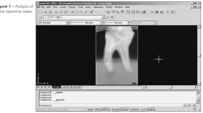
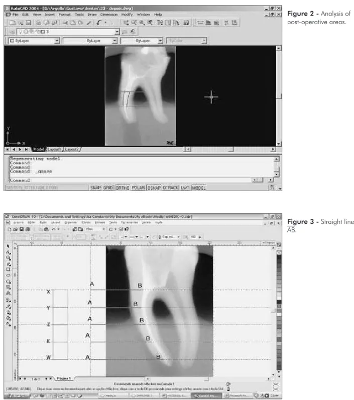
Analyses were made using the AutoCAD 2004 program (Autodesc, Inc., USA) to determine the partial and total areas of images before and after the preparations. The program imported the images and, by means of multiple points (Polyline tool), it
outlined the image of the canal thus making an ir-regular polygon. The program offered the areas of the polygons relative to the cervical, median and apical thirds. The sum of the partial areas resulted in the value of the total area (Figures 1 and 2). Wid-ening was assessed by the difference between the i-nal and initial areas.
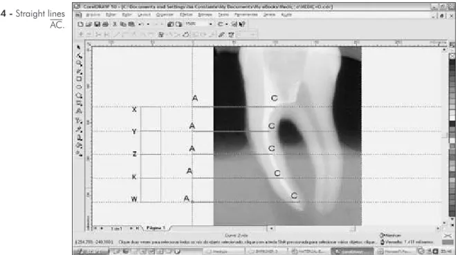
In the CorelDraw 10 program (MicroSafe, RJ, Brazil) the canal thicknesses before and after the preparations were assessed by analyzing the hori-zontal alterations observed in the internal and exter-nal walls of the mesiobuccal caexter-nal of each sample.
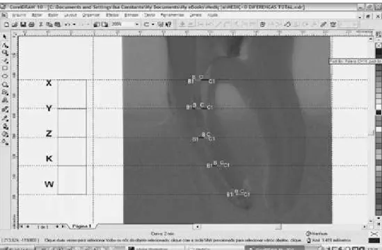
Straight lines were traced at ive equidistant points along the canal, starting from a reference guide line up to the external wall of the canal. They were denominated straight lines AB (Figure 3). New straight lines were traced in relation to the internal wall, and they were denominated straight lines AC (Figure 4).
The difference between straight lines AC and straight lines AB revealed the thickness of the canal (BC) (Figure 5).
The thicknesses of the canals before and after preparations were then determined and superim-posed. Superimposition of an image of the canal thickness before preparation on an image of the
Figure 1 - Analysis of
Braz Oral Res 2007;21(2):118-26 121 canal thickness after preparation is able to show
the canal wall at whose expense the widening oc-curred.
In order to do image superimposition, some of the tools available in CorelDraw10 were applied: “Effects” and “invert”, to transform light into dark
and vice-versa; “Interactive Transparency”, to en-able visualization of the two superimposed images; and “Brightness-contrast-intensity”, for small ad-justments. By doing so, a better visualization was obtained of the two superimposed thicknesses (the pre-operative images were inverted and seen in
Figure 2 - Analysis of
post-operative areas.
Figure 3 - Straight lines
black, and the post-operative ones became semi-transparent and seen in white) in the different tech-niques studied (Figure 6).
In this case, widening was assessed by the dif-ference between the inal and initial thicknesses (straight lines BC).
Results
A descriptive analysis of the areas showed that in all the locations considered, the Progressive Prepa-ration Technique presented greater widening when compared with the other techniques. In greater de-tail, considering the total tooth area, in spite of the
Figure 4 - Straight lines
AC.
Figure 5 - Initial canal
Braz Oral Res 2007;21(2):118-26 123 Progressive Preparation presenting 0.420 for the
min-imum value and 2.100 for the maxmin-imum value, the mean and median values were higher and the stand-ard deviation was the lowest obtained, indicating a greater precision of the results found when compared with the results of the other techniques (Table 1).
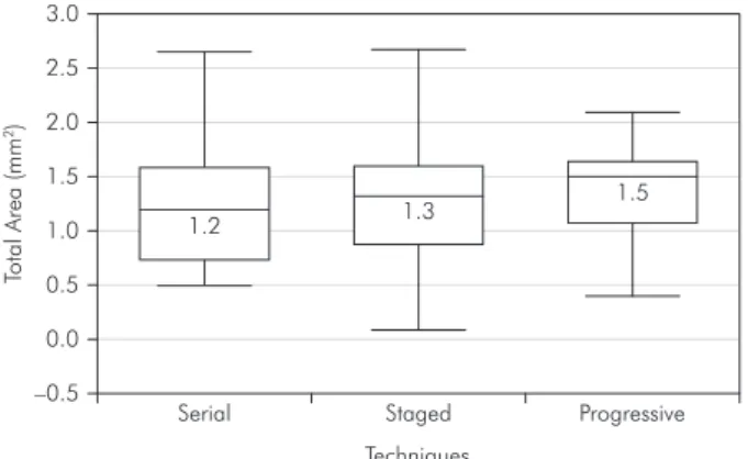
Graph 1 demonstrates that the results were closer to each other for the Progressive Preparation
tech-nique, that is to say, the variation among the results obtained was smaller (this same result is conirmed by the standard deviation, which was the lowest for this technique). The greatest widening was found through the median (line traced inside the rectangles) which was shown to be in a superior position to the others.
With regard to the thicknesses, the Serial Tech-nique presented the best results at point X, whereas
Table 1 - Difference between initial and final areas in mm², according to location and technique.
Location Technique Descriptive Measurements
No. of Observations Minimum Mean Median Standard Deviation Maximum
Total
Serial 15 0.510 1.278 1.210 0.580 2.670 Staged 15 0.100 1.223 1.330 0.661 2.680 Progressive 15 0.420 1.349 1.510 0.466 2.100
Cervical
Serial 15 –0.120 0.556 0.390 0.297 1.120 Staged 15 0.100 0.419 0.530 0.257 0.790 Progressive 15 0.030 0.530 0.650 0.255 0.910
Median
Serial 15 0.190 0.466 0.440 0.234 1.080 Staged 15 0.030 0.509 0.440 0.322 1.350 Progressive 15 0.080 0.497 0.520 1.888 0.860
Apical
Serial 15 0.000 0.265 0.240 0.193 0.710 Staged 15 0.000 0.299 0.310 0.227 0.700 Progressive 15 0.005 0.327 0.330 0.151 0.630
Figure 6 - Initial thickness
the Progressive Preparation technique presented the best results at points Y, Z and K, which demon-strates a greater regularity of the latter technique along the canal. At point W, the Staged technique presented the greatest widening (Table 2).
Negative values in the inal thicknesses indicated the occurrence of deformations (negative deviations) in the prepared canals. These were observed in six samples in the Serial technique.
Table 2 - Widening (in mm) of the canals of 45 teeth, measured after the application of three techniques, in different
loca-tions.
Location Technique Descriptive Measurements
No. of Observations Minimum Mean Median Standard Deviation Maximum
X
Serial 15 –3.520 4.200 4.122 3.555 11.800 Staged 15 0.803 3.696 3.307 2.246 7.880 Progressive 15 0.001 4.148 4.819 2.538 7.920
Y
Serial 15 0.341 3.446 2.790 2.181 7.472 Staged 15 0.010 3.607 3.570 2.199 8.330 Progressive 15 0.994 4.902 5.310 2.123 9.678
Z
Serial 15 0.640 3.148 3.010 2.255 7.126 Staged 15 1.040 3.968 4.110 1.930 8.600 Progressive 15 0.415 4.232 3.892 1.729 6.979
K
Serial 15 –2.440 2.571 2.460 2.517 8.340 Staged 15 0.417 2.939 2.450 1.658 5.860 Progressive 15 0.339 3.953 3.953 1.607 5.511
W
Serial 15 0.020 1.293 0.574 1.524 5.070 Staged 15 0.110 1.859 1.090 1.667 5.320 Progressive 15 0.059 1.790 1.190 1.622 5.530
Graph 1 - Comparison between the standard deviations
and medians between the techniques in accordance with the initial and final differences.
Serial 3.0
2.5
2.0
1.5
1.0 1.2 1.3
1.5
0.5
0.0
–0.5
Staged
Techniques
To
tal
A
rea
(m
m
2)
Progressive
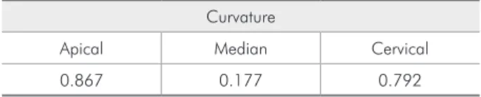
However, statistical analysis of the area and thick-ness data, performed by means of the Kruskal-Wallis test, did not lead to statistically signiicant differenc-es among the techniqudifferenc-es, considering the curvaturdifferenc-es (p-value > 0.05 in the three groups) (Table 3).
Discussion
With regard to the descriptive analysis of the difference between inal and initial areas (Table 1 and Graph 1), the Progressive Preparation technique showed closer results, thus indicating a more regular preparation. As regards the total area, it presented a greater value for the mean and the median, which indicated better performance. As regards the partial areas, the Serial and Staged techniques presented a greater maximum value in the cervical and median regions, respectively. However, the highest standard deviations of these techniques in the regions referred to showed greater irregularity in the preparations.
wid-Braz Oral Res 2007;21(2):118-26 125 ening the canal was to model it, in which the inal
shape respected the original one, but assumed a fun-neled and regular shape.
Although the inal preparations for all the tech-niques would be standardized with a #30 ile, the Progressive Preparation technique presented a better performance in the region for all the groups of teeth, principally for the teeth with apical curvature. This may be explained by the removal of the cervical inter-ferences before apical preparation, as recommended by the technique referred to. This enabled a better control of the instrument in the apical region and, consequent-ly, a greater advantage to be taken of its kinematics.
These results corroborate those of Contreras et al.3 (2001), Goerig et al.6 (1982), Holland et al.9 (1991) and Nishiyama, Garcia12 (1993), who agreed that initial preparation of the cervical region facilitates the subse-quent action of the instruments in the apical region.
Tan, Messer17 (2002), however, disagree. When comparing the manual apex-crown and crown-apex techniques with the LightSpeed techniques, they ob-served that there was no statistically signiicant dif-ference between those techniques in relation to apical deviation and shape of the canal, which was also ob-served by Testa18 (2003) when he compared manual and rotary crown-apex and apex-crown techniques.
As regards the thicknesses, the Progressive Prep-aration technique presented a better performance at three of the ive levels studied, showing greater reg-ularity of the preparation along the canal (Table 2). Negative deviations occurred in six samples in the Serial technique. The fact that both the Staged and the Progressive Preparation techniques did not present negative deviations indicated that the use of instruments of different lexibilities for inal prepa-ration (stainless steel for the Staged technique and Nickel-Titanium for the Progressive Preparation) was not a factor that determined whether or not devia-tions occurred.
This is in agreement with the work of Testa18 (2003), Pereira et al.13 (2001) and Chan, Cheung2 (1996), who did not obtain statistically signiicant differences when they compared the results of prepa-rations with stainless steel or Nickel-Titanium iles. It is, however, in disagreement with the work of Vanni et al.19 (2004) and Esposito, Cunning-ham4 (1995), who studied the alterations that vari-ous endodontic techniques produced in the curved canals of extracted teeth, and concluded that those that used Nickel-Titanium iles were more capable of maintaining the original anatomy; and of Souza et al.16 (2001), who compared the result of manual techniques with conventional iles with that of automated Nickel-Titanium systems, and observed that the manual technique presented bet-ter results.
Although the descriptive analysis points towards a better performance of the Progressive Prepara-tion technique in comparison with that of the other preparations, statistical analysis did not present niicant statistical difference at the 5% level of sig-niicance for decision making.
Conclusions
Based on an analysis of the results, it was con-cluded that:
The Progressive Preparation technique tended to be shown as more effective, precise and regular in a descriptive analysis that compared the Serial, Staged and Progressive Preparation techniques, as it presented more uniform preparations in all of the groups of teeth with root curvatures in different positions.
Deformations (negative deviations) occurred in 40% of the samples only in the Serial technique, and did not occur in the other two techniques. The use of instruments of different lexibilities in the Progressive Preparation and Staged tech-niques was not a preponderant factor for deter-mining the occurrence of deviations.
Although there were no statistically signiicant differences, the descriptive analysis indicates that, clinically, the Progressive Preparation technique presented a better performance in relation to the other preparations.
1.
2.
3.
Table 3 - Result of the Kruskal-Wallis Test for analyzing the
areas.
Curvature
References
1. Berbert A, Nishiyama CK. Curvaturas radiculares: uma nova metodologia para a mensuração e localização. Rev Gaúcha Odont. 1994;42(6):356-8.
2. Chan AW, Cheung GS. A comparison of stainless steel and nickel-titanium K-files in curved root canals. Int Endod J. 1996;29(6):370-5.
3. Contreras MA, Zinman EH, Kaplan SK. Comparison of the first file that fits at the apex, before and after early flaring. J Endod. 2001;27(2):113-6.
4. Esposito PT, Cunningham CJ. A comparison of canal prepa-ration with nickel-titanium and stainless steel instruments. J Endod. 1995;21(4):173-6.
5. Gambil JM, Alder M, Del Rio CE. Comparison of Nickel-Titanium and Stainless Steel hand-file instrumentation using computed tomography. J Endod. 1996;22(7):369-75. 6. Goerig AC, Michelich RJ, Schultz HH. Instrumentation of
root canals in molar using the step-down technique. J Endod. 1982;8(12):550-4.
7. Heck AR, Garcia RB. Avaliação radiográfica do desvio apical do canal radicular após a instrumentação manual com limas Flexofile, Flex-R, Onyx-R e o sistema mecânico rotatório Pro-file. Rev Fac Odont Bauru. 1999;7(3/4):27-32.
8. Heuer MA. The biomechanics of endodontic therapy. Dent Clin North Am. 1963;13(1):341-59.
9. Holland R, Souza V, Otoboni Filho JA, Nery MJ, Bernabé PFE, Mello W. Técnicas mistas de preparo do canal radicular. Rev Paul Odont. 1991;13(4):17-23.
10. Iqbal MK, Firic S, Tulcan J, Karabucak B, Kim S. Comparison of apical transportation between Profile and Protaper Niti rotary instruments. Int Endod J. 2004;37(6):359-64.
11. Moura AAM, Moura Netto C, Carvalho CF. Técnica do preparo progressivo do canal radicular. ACDC em Ação. 2003;15:22. 12. Nishiyama CK, Garcia RB. Estudo comparativo entre as técnicas
de instrumentação escalonada regressiva, oregon modificada, Sistema Canal Finder e Canal máster “U” na limpeza de canais radiculares. Rev Odontol Univ São Paulo. 1993;7(12):173-9. 13. Pereira AJA, Fidel RAS, Fidel SR, Santa Cecília M, Duarte
MAH. Comportamento das limas manuais de aço inoxidável e de Níquel-Titânio em relação ao transporte apical. Rev Bras Odontol. 2001;58(4):266-9.
14. Schilder H. Cleaning and shaping the root canal. Dent Clin North Am. 1974;18(2):269-96.
15. Schneider SW. A comparison of canal preparations in straight and curved root canals. Oral Surg Oral Med Oral Pathol. 1971;32(2):271-5.
16. Souza V, Otoboni Filho JA, Holland R, Nery MJ, Bernabé PFE, Dezan Junior E. Avaliação de alguns aspectos relacionados ao preparo manual e automatizado do canal radicular. Rev Paul Odont. 2001;23(5):22-4.
17. Tan BT, Messer HH. The quality of apical canal prepara-tion using hand and rotary instruments with specific crite-ria for enlargement based on initial apical file size. J Endod. 2002;28(9):658-64.
18. Testa FM. Influência das Técnicas de instrumentação no desvio apical de canais radiculares [Dissertação de Mestrado]. Bauru: Faculdade de Odontologia de Bauru da USP; 2003.
19. Vanni JR, Albuquerque DS, Reiss C, Barato Filho F, Limongi O, Della Bona A. Apical displacement produced by rotary nickel-titanium instruments and stainless steel files. Appl Oral Sci. 2004;12(1):51-5.