Neuromuscular/Neurology Division, Internal Medicine Departament, Hospital de Clínicas da Universidade Federal do Paraná, Curitiba PR, Brazil (UFPR). Supported: Fundação Araucária and CAPES.
Received 7 June 2004, received in final form 10 November. Accepted 25 November 2004.
Dr. Lineu Cesar Werneck - Hospital de Clínicas da UFPR - Rua Gal. Carneiro 181/3º andar - 80060-900 Curitiba PR - Brazil. E-mail: [email protected]
The cytogenetic localization of the gene of the Duchenne muscular dystrophy (DMD) in the short arm of chromosome X1, locus Xp212, with posteri-or cloning of the DNA3, codification4and identifica-tion of the gene product, dystrophin5and subse-quent characterization of the dystrophin
glycopro-tein complex (DGC)6 , 7have brought great advances in the briefing of molecular pathogeneses of mus-cular dystrophies. The DGC is a multisubunit com-plex of proteins, which form a structural linkage between the cytoskeleton (F-actin) and the extracel-lular matrix (laminin-α2 )8. The integral proteins
LIMB-GIRDLE MUSCULAR DYSTROPHY
An immunohistochemical diagnostic approach
Enio Alberto Comerlato, Rosana Hermínia Scola, Lineu César Werneck
ABSTRACT - The limb-girdle muscle dystrophy (LGMD) represents a heterogeneous group of muscular dis-eases with dominant and recessive inheritance, individualized by gene mutation. A group of 56 patients, 32 males and 24 females, with suggestive LGMD diagnosis were submitted to clinical evaluation, serum muscle enzymes, electromyography, muscle biopsy, and the immunoidentification (ID) of sarcoglycans (SG) α, β, γand δ, dysferlin and western blot for calpain-3. All the patients had normal ID for dystrophin (rod domain, carboxyl and amine terminal). The α-SG was normal in 42 patients, β-SG in 28, β-SG in 45, δ-SG in 32, dysferlin in 37 and calpain-3 in 9. There was a reduction in the α-SG in 7 patients, β-SG in 4, γ-SG in 2, and δ-SG in 8. There was deficiency of α-SG in 7 patients, β-SG in 6, γ-SG in 9, δ-SG in 5, dysferlin in 8, and calpain-3 in 5. The patients were grouped according the ID as sarcoglycans deficiency 18 cases, dysferlin deficiency 8 cases and calpain-3 deficiency 5 cases. Only the sarcoglycans deficiency group showed calf hyper-trophy. The dysferlin deficiency group was more frequent in females and the onset was later than sarco-glycan and calpain-3 deficiency groups. The calpain-3 deficiency group occurred only in males and showed an earlier onset and weaker muscular strength.
KEY WORDS: limb-girdle muscular dystrophy, immunoidentification, sarcoglycans, dysferlin, calpain-3.
Distrofias musculares de cinturas: uma abordagem diagnóstica imuno-histoquímica
RESUMO - As distrofias musculares de cinturas (DMC) representam grupo heterogêneo de doenças mus-culares com heranças autossômicas dominante ou recessivas, caracterizadas geneticamente por mutações gênicas específicas. Cinqüenta e seis pacientes, 32 masculinos e 24 femininos, com diagnóstico sugestivo de DMC, foram submetidos a avaliação clínica, dosagem séricas das enzimas musculares, eletromiografia, biópsia muscular e imunoidentificação (ID) das proteínas sarcoglicanas (SG) α, β, γeδ, disferlina e calpaí-na-3. A ID da distrofina (domínio rod e terminais carboxila e amino) era normal em todos os pacientes. Apresentaram ID normal para α-SG 42 casos, β-SG 28, γ,-SG 45, δ-SG 32, disferlina 37 e calpaína-3 9. Foi observada redução de α-SG em 7 pacientes, β-SG em 4, γ-SG em 2 e δ-SG em 8. Houve deficiência de α-SG em 7 pacientes, β-SG em 6, γ-SG 9, δ-SG em 5, disferlina em 8 e calpaína-3 em 5. Os pacientes foram classi-ficados de acordo com a ID em deficiência de SG em 18 casos, disferlina em 8 e calpaína-3 em 5. A hipertrofia de panturrilhas foi observada apenas no grupo com deficiência de SG. O grupo com deficiência de disfer-lina teve maior número de mulheres acometidas e a idade de início dos sintomas foi mais tardio em relação aos grupos com deficiência de SG e calpaína-3. O grupo com deficiência de calpaína-3 ocorreu apenas em pacientes do sexo masculino, a idade do início dos sintomas foi menor e teve maior fraqueza muscular.
that comprise the DGC are structurally organized into sub-complexes, formed by the dystrophin, the dystroglycan complex (αand βsubunits), the sarcoglycan (SG) complex (α, β, γ,δand εsubunits), α-dystrobrevin, syntrophins and sarcospan9. At least six forms of muscular dystrophy arise from pri-mary mutations in genes encoding components of this complex10,11. With the identification of these genes and their product, the limb-girdle muscular dystrophies (LGMD) where classified in autosomal dominant (LGMD1) and recessive (LGMD2). Patho-genic mutations in the SG complex components de-termine a group of autosomal recessive limb gir-dle muscular dystrophies (LGMD2) known as sarco-glycanopathies: the γ, α, βand δs a r c o g l y c a n o p a t h y, genetically classified as LGMD2C12, 2D13, 2E14and 2 F1 5respectively .The sarcoglycanopathies present a variable clinical features and is characterized by the biochemical deficiency of its subunits, independ-ently of any primary gene defects16.
Among the LGMD where expression of sarcogly-cans is normal, other genes are involved, that can cause defects or deficiencies in sarcolemmal pro-teins: dysferlin (LGMD2B)1 7, caveolin-3 (autosomal dominant limb girdle muscular dystrophy -L G M D 1 C )1 8; cytoplasmatic proteases: calpain-3 ( L G M D 2 A )1 9; cytoplasmatic proteins associated with organelles: TRIM32 (LGMD2H)2 0, fukutin relat-ed protein (FKRP) (LGMD2I)2 1; sarcomeric proteins: telethonin (LGMD2G)22, titin (LGMD2J)23, myotilin ( L G M D 1 A )2 4, filamin C (LGMD1F)2 5; and nuclear membrane proteins: lamin A/B (LGMD1B)26.
Therefore, the limb-girdle muscular dystrophy (LGMD) becomes a clinically and genetically hetero-geneous group of degenerative muscular diseases where the clinical, laboratory, electromyographic, histopathological and immunohistochemical have turned to be of great importance in the guideline of the specific genetic study. These made us to carry through this work, with the intention to im-prove the diagnosis in a heterogeneous group of patients with LGMD.
METHOD
We selected 56 patients with LGMD diagnostic and normal dystrophin by immunofluorescence (rod domain, carboxy and amino terminal) admitted to the Neuromus-cular Unit, from January 1976 to May 2001. The patients were submit to clinical evaluation, serum muscle enzymes, e l e c t r o m y o g r a p h y, muscle biopsy, and the immunoiden-tification (ID) of α-sarcoglycan, β- sarcoglycan, γ -sarco-glycan, δ-sarcoglycan, dysferlin and calpain-3.
Clinical evaluation – We collected data regarding gen-der distribution, family history, age and mode of onset, muscle strength, muscle atrophy and hypertrophy, func-tional abilities, and progression of the disease. To assess muscle strength we used a manually muscle testing of the British Medical Research Council (MRC) scale converted to 0-7 point system as follows: 0=0, 1=1, 2=2, 3=3, 4(-)=4, 4=5, 4(+)=6, 5=72 7. The proximal and distal muscles of the upper and lower limbs were tested. The functional grade was classified the Vignos and Archibald scale2 8.
Muscle enzymes – The serum muscle enzymes activ-ity to creatine kinase (CK) was performed in 49 cases, lactic dehydrogenase (LDH) in 25 cases, alanine amino-transferase (ALT) in 40 cases, aspartate aminoamino-transferase (AST) in 25 cases and aldolase in 19 cases. The plasmat-ic levels were registered as time fold increased above the normal limit.
Electromyography – The electromyography (EMG) was performed in 50 patients and was classified as nor-mal, myopathic, and mixed.
Muscle biopsy – Open muscle biopsies were taken from deltoid, biceps or quadriceps. All samples were fro-zen in liquid nitrogen and cryostat sections stained his-tologically and histochemically according to standard pro-cedures29. The following features were assessed: varia-tion in muscle fiber diameter, the distribuvaria-tion of atro-phied and hypertroatro-phied fibers; fiber degeneration and regeneration processes, architectural changes, connec-tive and fat tissue increase and inflammatory changes.
im-munofluorescence to dysferlin was deficient only when the fluorescence was absent.
Western blot – The western blot was performed in patients with normal labeling to α- S G ,β- S G ,γ- S G ,δ- S G , and dysferlin. Only thirteen muscles samples were avail-able. The muscle proteins were extracted in treatment b u ffer containing 0.125 mol/l Tris-HCL buffer pH 6.4, 10% glycerol, 4% SDS, 4 mol/l urea, 10% mercaptoethanol and 0.001% bromophenol blue (final pH of the treatment b u ffer was 6.8). Soluble proteins were separated using a SDS-PAGE gel 10% and the transferred into nitro-cellulose membrane. The visualization of blotted proteins nitrocellulose strips were blocked in 5% milk powder in a pH 8 buffer containing 10 mmol/l Tris-HCL, 0.15mol/l NaCl and 0.05% Tween 20 (TBST). Blots were probed with antibodies against to calpain-3 diluted 1:100 (Novo-castra/NCL-12A2, Newcastle upon Tyne, UK) and visual-ized using peroxidase-conjugated anti-mouse secondary antibody diluted 1:1000 (Amersham/NA931, Little Chalfont, UK) followed by exposure to freshly prepared 0.05% diaminobenzidine and 0.1% H2O23 1. Only the absence of band to calpain-3 was considered deficient.
Statistical analysis – Chi-square and Mann-Whitney tests were used to analyze the relation between the pres-ences of abnormalities in the ID groups.
RESULTS
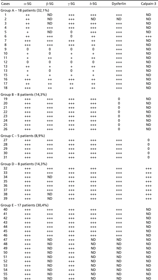
The α-SG was normal in 42 patients, β-SG in 28, γ-SG in 45, δ-SG in 32, dysferlin in 37 and calpain-3 in 9. There was a reduction in the α-SG in 7 pa-tients, β-SG in 4, γ-SG in 2, and δ-SG in 8. There was deficiency of α-SG in 7 patients, β-SG in 6, γ-SG in 9, δ-SG in 5, and dysferlin in 8. The calpain-3 was absent in 5 patients (Table 1).
The patients were classified according to the type of protein deficiency. The Group A, characte-rized by reduction or deficiency of one or several the SG-complex, was reported in 18 patients (Fig 1); group B, characterized by dysferlin deficiency, in 8 (Fig 2); group C with calpain-3 deficiency in 5 (Figs 3 and 4). The group D, not classified, did not show any deficiency. The group E with 17 cases had a non-conclusive evaluation due to insuff i c i e n t material for the tests and was not included in the statistical analysis (Table 1).
Clinical evaluation – Both male and female pa-tients were affected, with a preponderance of ma-le patients. There was a significant statistical rel-evance among the groups with dysferlin and cal-pain-3 deficiency (p=0.005).
Most of patients were sporadic cases. Patients with family history and, or consanguinity of the par-ents were more common in the dysferlin deficien-cy group (Table 2), but without statistical signifi-cance (p>0.05). An autosomal dominant pattern was observed only in a female patient of group D. The mean age of onset and at evaluation was significantly higher in dysferlin deficiency group (group B) than among sarcoglycans (p= 0.014) and calpain-3 (p = 0.010) deficiency groups (Table 3). The diseases duration do not showed statistical sig-nificance among the ID groups (p>0.05).
The symptoms at presentation occurred by we-akness of the muscles of the lower limbs in 29 pa-tients, upper limbs in 6 and both in 4 (Table 3). No statistical difference occurred among the ID groups (p>0.05).
The proximal muscle atrophy was observed in all ID groups. Only the dysferlin deficiency group (group B) does not showed distal muscle atrophy ( Table 4). Facial weakness occurred in all the groups of ID. In the sarcoglycanopathy group it was ob-served only in the cases with specific deficiency of γ-SG. The calf hypertrophy was observed in 4 pa-tients with SG complex deficiency. The calpain-3 deficiency group (group C) presented the lowest muscle strength tests, but without statistical signifi-cance. The waddling gait and Gowers sign were commons features in all ID groups. The loss of gait occurred only in SG complex deficiency (group A) and the non-classified (group D) patients. Most of patients showed at evaluation gait impairment, dif-ficulties in running or climbing stairs (Table 4).
Muscle enzymes – The mean elevation of CK and AST in the serum was more expressive in dysferlin deficiency patients; and of LDH, ALT and aldolase in SG complex deficiency patients, but without statistical significance (Table 5).
Electromyography – The myopathic electromyo-graphic pattern was a common finding, observed in 31 patients, but the neuromyopathic pattern oc-curred in all ID groups (Table 6).
Table 1. Immunocytochemical and western blot analysis.
Cases α-SG β-SG γ-SG δ-SG Dysferlin Calpain-3
Group A – 18 patients (32,1%)
1 ++ ND +++ +++ +++ ND
2 ++ ND +++ ND ND ND
3 ++ ND +++ +++ +++ ND
4 ++ +++ +++ +++ +++ ND
5 + ND 0 +++ +++ ND
6 ++ +++ 0 ++ +++ +++
7 +++ +++ +++ ++ +++ ND
8 +++ +++ +++ ++ +++ ND
9 0 0 0 0 +++ ND
10 + 0 + + +++ ND
11 + ++ + ++ +++ ND
12 0 0 0 0 +++ ND
13 ++ + + ++ +++ ND
13 + 0 0 + +++ ND
15 + + + + +++ ND
16 +++ ++ +++ ++ +++ ND
17 ++ ++ ++ ++ +++ ND
18 +++ ++ ++ ++ +++ ND
Group B – 8 patients (14,3%)
19 +++ +++ +++ +++ 0 ND
20 +++ +++ +++ +++ 0 ND
21 +++ +++ +++ +++ 0 ND
22 +++ +++ +++ +++ 0 ND
23 +++ +++ +++ +++ 0 ND
24 +++ +++ +++ +++ 0 ND
25 +++ +++ +++ +++ 0 ND
26 +++ +++ +++ +++ 0 ND
Group C – 5 patients (8,9%)
27 +++ +++ +++ +++ +++ 0
28 +++ +++ +++ +++ +++ 0
29 +++ +++ +++ +++ +++ 0
30 +++ +++ +++ +++ +++ 0
31 +++ +++ +++ +++ +++ 0
Group D – 8 patients (14,3%)
32 +++ +++ +++ +++ +++ +++
33 +++ +++ +++ +++ +++ +++
34 +++ ND +++ +++ +++ +++
35 +++ +++ +++ +++ +++ +++
36 +++ +++ +++ +++ +++ +++
37 +++ +++ +++ +++ +++ +++
38 +++ ND +++ +++ +++ +++
39 +++ ND +++ +++ +++ +++
Group E – 17 patients (30,4%)
40 +++ +++ +++ +++ +++ ND
41 +++ +++ +++ +++ +++ ND
42 +++ +++ +++ +++ +++ ND
43 +++ +++ +++ +++ +++ ND
44 +++ +++ +++ +++ +++ ND
45 +++ +++ +++ +++ +++ ND
46 +++ +++ +++ +++ +++ ND
47 +++ ND +++ ND ND ND
48 +++ ND +++ ND ND ND
49 +++ ND +++ ND ND ND
50 +++ ND +++ ND ND ND
51 +++ ND +++ ND ND ND
52 +++ ND +++ ND ND ND
53 +++ ND +++ ND ND ND
54 +++ ND +++ ND ND ND
55 +++ ND +++ ND ND ND
56 +++ ND +++ ND ND ND
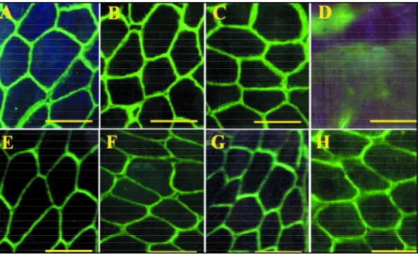
Fig 2. Dyspherlin deficiency (Case 22, immunofluorescence). A, Dystrophin carboxyl terminal; B, Dystrophin amino terminal; C, Dystrophin Rod domain; D, Dyspherlin; E, α-Sarcoglycan; F, β-Sarcoglycan; G, γ-Sarcoglycan; H, δ -Sarcoglycan. (Bar 100µin A,B,C,E,F,G,H; 25µin D).
Fig 1. Sarcoglycan deficiency (Case 12, immunofluorescence). A, Dystrophin carboxyl terminal; B, Dystrophin amino terminal; C, Dystrophin Rod domain; D, Dyspherlin; E, α-Sarcoglycan; F, β-Sarcoglycan; G, γ- S a r c o g l y c a n ; H, δ-Sarcoglycan. (Bar 100µin A,B,C,D; 25µin E,F,G,H).
Table 2. Gender and family history by immunoidentification groups.
Immunoidentification groups A B C D Total
Number of patients 18 8 5 8 39
Female 8 7 – 3 18
Male 10 1 5 5 21
Family history 7 5 2 4 18
Fig 4. Calpain-3 deficiency (Case 28, Western blot). A, Normal control; B, Absent band (Patient).
Fig 3. Calpain-3 deficiency (Case 28, immunofluorescence). A, Dystrophin carboxyl terminal; B, Dystrophin amino terminal; C, Dystrophin Rod domain; D, Dyspherlin; E, α-Sarcoglycan; F,β-Sarcoglycan; G, γ-Sarcoglycan; H, δ -Sarcoglycan. (Bar 100µin A,B,C,E,F,G,H; 25µin D).
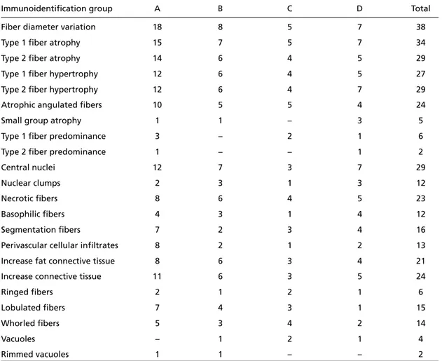
Small group atrophy was found only in 5 specimens. Type 1 and 2 fibers predominance and vacuoles we-re uncommon findings. Central nuclei and nuclear clumps were found more frequently in the groups with dysferlin deficiency and also in the non-classi-fied group. Fibers with necrosis were found more frequently in the calpain-3 deficiency group. Ba-sophilic and segmented fibers were more frequent-ly reported in the group with dysferlin deficiency and in the non-classified group. The perivascular inflammatory infiltrates was more common in the group with SG complex deficiency. The increase of fat and fibrous connective tissues was more fre-quent in dysferlin deficiency patients (group B). The ring fibers, lobulated fibers, and whorled fibers were more common in the group with calpain-3 deficiency. No statistically relevance were found among the ID groups and the several abnormal spe-cific histological findings (p>0.05) (Table 7).
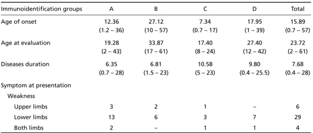
Table 3. Mean age of onset of the symptoms and at evaluation, symptom at presentation by immunoidentifica -tion group.
Immunoidentification groups A B C D Total
Age of onset 12.36 27.12 7.34 17.95 15.89
(1.2 – 36) (10 – 57) (0.7 – 17) (1 – 39) (0.7 – 57)
Age at evaluation 19.28 33.87 17.40 27.40 23.72
(2 – 43) (17 – 61) (8 – 24) (12 – 42) (2 – 61)
Diseases duration 6.35 6.81 10.58 9.80 7.68
(0.7 – 28) (1.5 – 23) (5 – 23) (0.4 – 25.5) (0.4 – 28) Symptom at presentation
Weakness
Upper limbs 3 2 1 – 6
Lower limbs 13 6 3 7 29
Both limbs 2 – 1 1 4
A, sarcoglycanopathy; B, dysferlinopathy; C, calpainopathy; D, not classified.
Table 4. Clinical findings in the neurological examination according the immunoidentification groups.
Immunoidentification groups A B C D Total
Muscular atrophy Upper limbs
Proximal 10 5 5 7 27
Distal 4 – 2 3 9
Lower limbs
Proximal 11 3 4 6 24
Distal 3 – 2 3 8
Calf Hypertrophy 4 – – – 4
Muscle force graduation (mean) Upper limbs
Proximal 4.94 5.13 4.00 4.88 4.85
(2 – 7) (3 – 7) (3 – 5) (3 – 6) (2 – 7)
Distal 6.61 6.75 5.80 6.13 6.44
(4 – 7) (5 – 7) (4 – 7) (5 – 7) (4 – 7) Lower limbs
Proximal 4.83 4.63 4.20 4.50 4.64
(2 – 7) (3 – 7) (3 – 6) (4 – 5) (2 – 7)
Distal 6.78 6.63 5.00 6.25 6.41
(5 – 7) (5 – 7) (4 – 7) (4 – 7) (4 – 7)
Facial weakness 2 1 1 2 6
Gait type
Normal 2 2 1 1
Waddling 15 6 4 6 31
Unable to walk 1 – – 1 2
Gowers sign
Present 13 6 5 7 31
Unable to perform 2 – – 1 3
Vignos functional scale
1 – 4 17 8 5 7 37
7 – – – 1 1
9 1 – – – 1
Table 5. Mean muscular enzymes by immunoidentification group.
Immunoidentification group A B C D Total
Muscular enzymes
CK 17.39 23.85 10.45 12.26 17.41
(0 – 66) (0.8 – 46) (0.4 – 20.5) (2.6 – 23) (0 – 66)
LDH 1.86 1.38 0.10 0.52 1.39
(0 – 11) (0 – 3.3) (0 – 0.1) (0 – 0.2) (0 – 11)
AST 1.13 16.01 0.70 4.08 5.45
(0 – 4) (0 – 80) (0 – 1.4) (0 – 20) (0 – 1)
ALT 0.68 0.37 1.00 0.16 0.55
(0 – 2) (0 – 1.3) (0 – 2) (0 – 0.5) (0 – 2)
Aldolase 3.21 – 0.10 – 2.49
(0 – 12) (0 – 0,2) (0 – 12)
A, sarcoglycanopathy; B, dysferlinopathy; C, calpainopathy; D, not classified; CK, Creatine kinase; LDH, Lactic dehydrogenase; AST, Aspartate aminotransferase; ALT, Alanine aminotransferase.
Table 6. Electromyographic pattern by immunoidentificatoin group.
Immunoidentification group A B C D Total
Electromyography pattern
Myopathic 15 7 2 7 31
Mixed 1 1 1 1 4
A, sarcoglycanopathy; B, dysferlinopathy; C, calpainopathy; D, not classified.
Table 7. Immunoidentification groups and histopathology.
Immunoidentification group A B C D Total
Fiber diameter variation 18 8 5 7 38
Type 1 fiber atrophy 15 7 5 7 34
Type 2 fiber atrophy 14 6 4 5 29
Type 1 fiber hypertrophy 12 6 4 5 27
Type 2 fiber hypertrophy 12 6 4 7 29
Atrophic angulated fibers 10 5 5 4 24
Small group atrophy 1 1 – 3 5
Type 1 fiber predominance 3 – 2 1 6
Type 2 fiber predominance 1 – – 1 2
Central nuclei 12 7 3 7 29
Nuclear clumps 2 3 1 3 12
Necrotic fibers 8 6 4 5 23
Basophilic fibers 4 3 1 4 12
Segmentation fibers 7 2 3 4 16
Perivascular cellular infiltrates 8 2 1 2 13
Increase fat connective tissue 8 6 3 4 21
Increase connective tissue 11 6 3 5 24
Ringed fibers 2 1 2 1 6
Lobulated fibers 7 4 3 1 15
Whorled fibers 5 3 4 2 14
Vacuoles – 1 2 1 4
Rimmed vacuoles 1 1 – – 2
generally important in others recessive forms2 2 , 3 4 , 4 2. Important variations can occur independent of the stage of the disease43. The mean serum levels of muscular enzymes were similar for all groups, not being possible to differentiate them.
The progressive loss of the muscle fibers results in the generation of myopathic motor unit poten-tials in the electromyography. However, neuro-genic motor unit potentials can also be observed in areas with clustering or fiber hypertrophy, owing to motor unit remodeling caused by segmentary necrosis process, which can isolate the distal por-tions of the muscular fibers from the myoneural e n d p l a t e4 3. The presence of neurogenic motor unit potentials has described in calpainopathy, dysferli-nopathy and sarcoglycanopathies3 9 , 4 4 , 4 5. The dystro-phic changes at muscle biopsy, characterized by vari-ation in muscle fiber size, necrotic/regenerating pro-cess, and increase of the endomysial and perimysial connective tissue, is the landmark of LGMD46.
The variation in muscle fiber size is generally li-ght to moderate degree in the calpainopathy and d y s-f e r l i n o p a t h y, and more intense in the sarcoglycano-p a t h i e s3 3 , 4 5, in the telethoninopathy (LGMD2G)4 7a n d L G M D 2 H4 8. Type 1 fiber predominance can be mo-re intense in the sarcoglycanopathies (LGMD2C-2F)4 7 and calpainopathy (LGMD2A)4 2 , 4 9. The degenerat-ing and regeneratdegenerat-ing process do not characterize any specific form in LGMD. The proliferation of the conjunctive and fat connective proliferation usual-ly tends to be more intense in the final stages of the d i s e a s e4 6. The cellular reactions in the muscular dys-trophies are unspecific, and generally vary accord-ing to the degree of muscular necrosis, because of the activation and release of the complement4 6. So-me perivascular inflammatory reaction can be very similar to inflammatory myopathy, as observed in the sarcoglycanopathies4 7 and dysferlinopathy5 0. The structural alterations in the majority of the au-tosomal recessive forms of the LGMD are of little in-tensity and unspecific, with exception of the teletho-ninopathy (LGMD2G) and the titinopathy (LGMD2I), where there have been reported the formation of rimmed vacuoles2 3 , 4 7. Notwithstanding, the exis-tence of rimmed vacuoles is a non-specific finding and has been reported in many neuromuscular di-seases, like the inclusion body myositis, spinal mus-cular atrophies and peripheral neuropathies5 1. The segmentary necrosis processes, can isolate the dis-tal portions of the muscular fibers from the myo-neural endplate, and remodeled the motor unit4 3.
DISCUSSION
The identification of the forms of LGMD with autosomal recessive inheritance is often difficult to be established, considering the great number of sporadic cases, the lack of convincing data in fam-ily history and its great clinical similarity with the Duchenne and Becker muscular dystrophies3 2. These cases had no correlation among the groups of ID and family history, but it was more frequent in the group of the dysferlinopathy3 3. The lack of changes in the ID of the cases of the autosomal dominant trait corroborates the literature data34. The great variability in the age of onset difficult the characte-rization of a typical pattern to each type of LGMD, and the mean age of onset can be common to more than one. We have verified that the cases with dys-ferlin deficiency showed a later onset33,35and the degrees of deficiency of the sarcoglycan protein complex proteins did not interfere in a meaning-ful way in the age at onset either36.
The classic presentation of the symptoms has been the weakness of the hip-girdle muscles, but can also present as involvement of the shoulder gir-dle or lower-limb distal muscles, muscular pain, and exercise intolerance20,36,37. Some specific patterns can be observed in the calpainopathy (LGMD2A), by involvement of posterior limb-girdle and trunk muscles; in the dysferlinopathy (LGMD2B), by in-volvement of the posterior compartment of the legs33,38,39; in the telethoninopathy (LGMD2G) and titinopathy (LGMD2J), by involvement of the ante-rior compartment of leg22,34. The facial weakness can be observed in advanced stages of the illness in the sarcoglycanopathies (LGMD2C-2F)4 0, and oc-casionally in the calpainopathy (LGMD2A) and telethoninopathy (LGMD2G)2 2 , 3 7. The calf hypertro-phy is a common finding among the sarcoglycano-pathies but can occasionally be observed in the ear-ly stages of the calpainopathy and dysferlinopa-thy35. In our cases the muscular involvement was unspecific, the facial musculature took place in intermediate stages of the disease in all groups of ID, and the calf hypertrophy was observed in the group of SG-complex deficiency.
H o w e v e r, secondary reductions had been also observed in the cases with mutations in the gene of the dysferlin (LGMD2B) and titin (LGMD2J)23. Other alterations also can be caused due to the muscle biopsy storage time, the amount of pres-ent protein and by the process of homogenization of muscular tissue. Distortions of the lane of blot difficulties the characterization of the calpain-3 band reduction, probably caused by contamination with the mountant medium. The mutations in the calpain-3 gene have been observed between 9 to 40% LGMD cases3 5 , 5 3 , 6 0 , 6 1. However, is estimated that 10% of the mutations of the gene of calpain-3 are not detected by the molecular techniques cur-rently used62.
The immunocytochemical and western blot ana-lysis were useful methods to classify the LGMD pa-tients. The sarcoglycan deficiency was more fre-quent, followed of the dysferlinopathy and calpain-o p a t h y. Heaven the clinical and labcalpain-oratcalpain-ory findings were very similar between the ID groups, the dys-ferlin deficiency patients had more delayed onset, and occurred more in female, and the calpain-3 de-ficiency patients occurred only in males and had greater impairment in the muscle strength.
REFERENCES
1. Greenstein RM, Reardon MP, Chan TS. An X/ autosomal translocation in a girl with Duchenne muscular dystrophy (DMD): evidence for DMD gene localization. Pediatr Res 1977;11:457.
2. Francke U, Ochs HD, De Martinville B, et al. Minor XP21 chromosome deletion in a male associated with expression of Duchenne muscular dystrophy, chronic granulomatous disease, retinitis pigmentosa, and McLeod syndrome. Am J Hum Genet 1985;37:250-267.
3. Kunkel LM, Monaco AP, Middleswoth W, Ochs HD, Latt AS. Specific cloning of DNA fragments absents from the DNA of male patient with an X chromosome deletion. Proc Natl Acad Sci 1985;82:4778-4782. 4. Koenig M, Hoffmann EP, Beterlson CJ, Monaco AP, Feener C, Kunkel
LM. Complete cloning of Duchenne muscular dystrophy (DMD) cDNA and preliminary genomic organization of the DMD gene in normal and affected individuals. Cell 1987;50:509-517.
5. Hoffman EP, Brown RH, Kunkel LM. Dystrophin: the protein product of the Duchenne muscular dystrophy locus. Cell 1987;51:919-928. 6. Campbell KP, Khal SD. Association of dystrophin and an integral
mem-brane glycoprotein. Nature 1989;338:259-262.
7. Ervasti JM, Campbell KP. Membrane organization of the dystrophin-glycoprotein complex. Cell 1991;66:1121-1131.
8. Yoshida M, Ozawa E. Glycoprotein complex anchoring dystrophin to sarcolemma. J Bichem 1990;108:748-752
9. Tinsley AM, Blake DJ, Roche A, et al. Primary structure of dystrophin-related protein. Nature 1992;360:591-593.
10. Campbell KP. Three muscular dystrophies: loss of cytoskeleton-extracelu-lar matrix linkage. Cell 1995;80:675-679.
11. Matsumura K, Campbell KP. Deficiency of dystrophin-associated pro-teins: a common mechanism leading to muscle cell necrosis in severe childhood muscular dystrophies. Neuromusc Disord 1993;3:109-118. 12. Noguchi S, McNally EM, Ben Othmane K, et al. Mutations in the
dys-trophin-associated protein ?-sarcoglycan in chromosome 13 muscular dystrophy. Science 1995;270:819-822.
13. Roberds S, Leturcq F, Allamand V, et al. Missense mutations in the adhalin gene linked to autosomal recessive muscular dystrophy. Cell 1994;78:625-633.
Therefore, angulated atrophic fibers, small groups of atrophic fibers, and nuclear clumps can occasio-nally be observed in the muscular dystrophies, espe-cially in facioescapulohumeral dystrophy and the LGMD syndromes4 6.
In the sarcoglycanopathies (LGMD2C-2F), the modifications of the SG-complex may also cause a secondary dystrophin deficiency, making the sep-aration from the dystrophinopathies very diff i-cult. Therefore, we chose to include only the cas-es with normal dystrophin52,53. The ID of α-SG has been used as the main mark the sarcoglycano-pathies, because of the structural alterations of complex SG caused for the mutations in the genes of these proteins52. The α-sarcoglycan deficiency varies of 9% to 30% of the cases, depending on the studied population1 6 , 5 3 , 5 4. Considering the possi-bility of the preservation of this marker in the form LGMD2C, we used the remaining ID markers to the proteins of the SG complex. This choice must have contributed to the identification of a larger number of cases when compared to the lit-erature data1 6 , 5 4. The alterations of complex SG must be analyzed with caution; therefore they are normally not followed by mutations of the genes of the proteins of complex SG1 6. This fact make pos-sible the occurrence of a secondary deficiency of the complex by other types of mutations, as ob-served in LGMD2I, where it occur secondary defi-ciency of α-distroglycan and merosin55. In βand γ -sarcoglycanopathies (LGMD2E and 2F), the deficien-cy of proteins of complex SG occurs in a much mo-re uniform way, and does not pmo-resent a specific pat-tern16,56,57. In the α-sarcoglycanopathy (LGMD2D), also a larger correlation with the degree of defi-ciency of the α-sarcoglycan can exist1 6 , 5 8. Only in the γ-sarcoglycanopathy (LGMD2C) the deficiency iso-lated of protein γ-SG seems to be a specific result, which suggests a strong correlation of this dis-e a s dis-e5 9. However, there is a better degree of correla-tion with mutacorrela-tions of complex SG concerning the cases with important deficiency exist16,52.
14. Bönnemann CG, Modi R, Noguchi S, et al. Beta sarcoglycan (A3b) mutations cause autosomal recessive muscular dystrophy with loss of the sarcoglycan complex. Nature 1995;11:266-273.
1 5 . Passos-Bueno MR, Moreira ES, Vainzof M, Marie SK, Zatz M. Linkage analy-sis in autosomal recessive limb-girdle muscular dystrophy (AR LGMD) maps a sixth form to 5q33-34 (LGMD2F) and indicates that there is at least one more subtype of AR LGMD. Hum Mol Genet 1996;5:818-820. 16. Duggan DJ, Gorospe R, Fanin M, Hoffman EP, Angelini C. Mutations
in the sarcoglycan genes in patients with myopathy. N Engl J Med 1997;336:618-624.
17. Bashir R, Strachan T, Keers S, et al. A gene for autosomal recessive limb-girdle muscular dystrophy maps to chromosome 2p. Hum Mol Genet 1994;3:455-457.
1 8 . Speer MC, Yamoaka LH, Gilchrist JH, et al. Confirmation of genetic het-e roghet-enhet-eity in limb-girdlhet-e muscular dystrophy: linkaghet-e of an autosomal dominant form to chromosome 5q. Am J Hum Genet 1992;50:1211 - 1 2 1 7. 19. Richard I, Broux O, Allamand V, et al. Mutations in the proteolytic enzyme calpain 3 cause limb-girdle muscular dystrophy type 2A. Cell 1995;81:27-40.
20. Weiler T, Gre e n b e rg CR, Zelinski T. A gene for autosomal recessive limb-g i rdle muscular dystrophy in Manitoba Hutterites maps to chro m o s o m e region 9q31-q33: evidence for another limb-girdle muscular dystro p h y locus. Am J Hum Genet 1998;63:140-147.
2 1 . Driss A, Amouri C, Hamida CB, et al. A new locus for autosomal recessive limb-girdle muscular dystrophy in a large consanguineous Tunisian family maps to chromosome 19q13.3. Neuromusc Disord 2 0 0 0 ; 1 0 : 2 4 0 - 2 4 6 .
22. Moreira ES, Wiltshire TJ, Faulkner G, et al. Limb-girdle muscular dys-t rophy dys-type 2G is caused by mudys-tadys-tion in dys-the gene encoding dys-the sarc o m-eric protein telethonin. Nat Gene 2000;24:163-166.
23. Haravuori H, Vihola A, Straub V, et al. Secondary calpain3 deficiency in 2q-linked muscular dystrophy: Titin is the candidate gene. Neuro l o g y 2001;56:869-877.
24. Hauser MA, Horrigan SK, Salamikangas P, et al. Myotilin is mutated in limb-girdle muscular dystrophy 1A. Hum Mol Genet 2000;9:2141-2147.
25. Palenzuela L, Andreu AL, Gamez J, et al. A novel autosomal dominant l i m b - g i rdle musclar dystrophy (LGMD 1F) maps to 7q32.1-32.2. Neurology 2003;61:404-495.
26. Muchir A, Bonne G, van der Kooi AJ, et al. Identification of mutations in the gene encoding lamins A/C in autosomal dominant limb girdle muscular dystrophy with atrioventricular conduction disturbances. Hum Mol Genet 2000;9:1453-1459.
2 7 . Mendell JR, Florence J, Manual muscle testing. Muscle Nerve 1 9 9 0 ; 1 3 : 1 6 - 2 0 .
28. Vignos PJ, Spencer GE, A rchibald KC. Management of pro g re s s i v e muscular dystrophy of childhood. JAMA 1963;184:89-96.
29. Dubowitz V. Muscle biopsy: a practical approach. London: Bailliüre Tindall, 1985.
3 0 . Werneck LC, Bonilla E. Immunohistochemical alterations of dystro p h i n in congenital muscular dystro p h y. A rq Neuropsiquiatr 1995;53:416-423. 31. Spencer MJ, Tidball JG, Anderson LVB, et al. Absence of calpain 3 in a form of limb-girdle muscular dystrophy (LGMD2A). J Neurol Sci 1997;146:173-178.
32. Dinçer P, Leturcq F, Richard I, et al. A biochemical, genetic, and clini-cal survey of autosomal recessive limb girdle muscular dystrophies in Turkey. Ann Neurol 1997;42:222-229.
33. Soares CN, Freitas MR, Nascimento OJ, et al. Myopathy of the distal lower limbs: the clinical variant of Miyoshi. A rq Neuro p s i q u i a t r 2003;61:946-949.
34. Bushby KMD. The limb-girdle muscular dystrophies: multiple genes, multiple mechanisms. Hum Mol Genet 1999;10:1875-1882.
35. Passos-Bueno MR, Vainzof M, Moreira ES, Zatz M. Seven autosomal recessive limb-girdle muscular dystrophies in the Brazilian population: from LGMD2A to LGMD2G. Am J Med Genet 1999;82:392-398. 36. Morandi L, Barresi R, Di Blasi C, et al. Clinical heterogeneity of adhalin
deficiency. Ann Neurol 1996;39;196-202.
37. Chou FL, Angelini C, Daentl D, et al. Calpain III mutation analysis of a heterogeneous limb-girdle muscular dystrophy muscular dystrophy population. Neurology 1999;52:1015-1020.
38. Weiler T, Greenberg CR, Nylen E, et al. Limb-girdle muscular dystro-phy and Miyoshi myopathy in an aboriginal Canadian kindred map to LGMD2B and segregate with the same haplotype. Am J Hum Genet 1996;59:872-878.
39. Fardeau M, Hillaire D, Mignard C, et al. Juvenil limb-girdle muscular d y s t rophy: clinical, histopathological and genetic data from a small com-munity living in Reunion Island. Brain 1996;119:295-308.
40. Duggan DJ, Hoffman EP. Autosomal recessive muscular dystro p h y and mutations of the sarcoglycan complex. Neuromusc Disord 1996;6:475-482.
41. Emery AEH. Duchenne muscular dystro p h y, 2n ded. Oxford: Oxford Univ
Press, 1993.
42. Urtasun M, Sáenz A, Roudant C, et al. Limb-girdle muscular dystro-phy in Guipúzcoa (Basque Contry, Spain). Brain 1998;121:1735-1747. 43. Sonoo M. New attempts to quantify concentric needle
electromyogra-phy. Muscle Nerve. 2002;S11:S98-S102.
44. Mahjneh I, Bushby K, Pizzi A, Bashir R, Marconi G. Limb-girdle mus-cular dystrophy: a follow-up study of 79 patients. Acta Neurol Scand 1996;94:177-189.
45. Ben Jelloun-Dellagi S, Chaffey P, Ben Hamida CH, et al. Presence of nor-mal dystrophin in Tunisian severe childhood autosonor-mal recessive mus-cular dystrophy. Neurology 1990;40:1903.
46. Walton JN, Nattrass FJ. On the classification, natural history and treat-ment of myopathies. Brain 1954 ;77:169-231.
47. M o reira ES, Vainzof M, Marie SK, Sertié AL, Zatz M, Passos-Bueno MR. The seventh form of autosomal recessive limb-girdle muscular dystro-phy is mapped to 17q11-12. Am J Hum Genet 1997;61:151-159. 48. Shokeir MHK, Kobrinsky NL. Autosomal recessive muscular
dystro-phy in Manitoba Hutterites. Clin Genet 1976;9:197-202.
49. Chae J, Minami N, Jin Y, et al. Calpain 3 gene mutations: genetic and clinical-pathologic findings in limb-girdle muscular dystro p h y. Neuromusc Disord 2001;11:547-555.
50. McNally EM, Chantal TL, Rosenmann H, et al. Splicing mutation in dys-ferlin produces limb-girdle muscular dystrophy with inflammation. A m J Med Genet 2000;91:305-312.
51. Scola RH, Werneck LC, Franco CRC. Cytoplasmic inclusion bodies: a study in several diseases and a review of the literature. A rq Neuropsiquiatr 1996;54:245-259.
52. Vainzof M, Passos-Bueno MR, Canovas M, et al. The sarcoglycan com-plex in the six autosomal recessive limb-girdle muscular dystrophies. Hum Mol Genet 1996;5:1963-1969.
53. Shilling CJ, Wicklund MP, Moore SA, et al. Establishing the prevalence of LGMD genotypes in North America: an ongoing multi-center col-laborative study. Neurology 2004;7(S5):A412.
54. Hayashi YK, Mizuto Y, Yoshida M, Nonaka I, Ozawa E, Arahata K. The frequency of patients with 50-kd dystrophin-associated glycoprotein (50DAG or adhalin) deficiency in a muscular dystrophy patient pop-ulation in Japan: immunocytochemical analysis of 50DAG, 43DAG, dys-trophin, and utrophin. Neurology 1995;45:551-554.
55. Brockington M, Blake DJ, Prandini P, et al. Mutations in the fukutin-related protein gene (FKRP) cause a form of congenital muscular dys-trophy with secondary laminin ?2 deficiency and abnormal glycosyla-tion of ?-dystroglycan. Am J Hum Genet 2001;69:1198-1209. 56. Bönnemann CG, Passos-Bueno MR, McNally EM, et al. Genomic scre e
n-ing for beta-sarcoglycan gene mutations: missense mutations may cause severe limb-girdle muscular dystrophy type 2E (LGMD 2E). Hum Mol Genet 1996;5:1956-1961.
57. Moreira ES, Vainzof M, Marie SK, Nigro V, Zatz M, Passos-Bueno MR. A first missense mutation in the delta sarcoglycan gene associated with a severe phenotype and frequency of limb-girdle muscular dystrophy type 2F (LGMD2F) in Brazilian sarcoglycanopathies. J Med Genet 1998;35:951-953.
58. E y m a rd B, Romero NB, Leturcq F, et al. Primary adhalinopathy (?-sarc o-glycanopathy): clinical, pathologic, and genetic correlation in 20 patients with autosomal recessive muscular dystro p h y. Neurology 1997;48:1227-1234.
59. Vo rg e rd M, Gencik M, Mortier J, Epplen JT, Malin JP, Mortier W. Isolated
loss of γ- s a rcoglycan: diagnostic implications in autosomal recessive
limb-girdle muscular dystrophies. Muscle Nerve 2001;24:421-424. 60. Argov Z, Sadeh M, Mazor K, et al. Muscular dystrophy due to
dysfer-lin deficiency in Libyan Jews: cdysfer-linical and genetic features. Brain 2000;123:1229-1237.
61. Minami N, Nishino I, Kobayashi, Ikezoe K, Yu-ichi G, Nonaka I. Mu-tation of calpain 3 gene in patients with sporadic limb-girdle muscu-lar dystrophy in Japan. J Neurol Sci 1999;171:31-37.