Joana Rolo
Dissertation presented to obtain the Ph.D degree in Molecular Biology
Instituto de Tecnologia Química e Biológica António Xavier | Universidade Nova de Lisboaresistance determinant in staphylococci
Joana Rolo
Dissertation presented to obtain the Ph.D degree in Molecular Biology
Instituto de Tecnologia Química e Biológica António Xavier | Universidade Nova de LisboaOeiras, April, 2016
Origin and evolution of the ß-lactam
resistance determinant in
Second edition, © Joana Rolo
Cover by Hélio Mateus
iii
Supervisors: Dr. Maria Miragaia
Professor Hermínia de Lencastre
Dr. Rita Sobral
Chairman of examiners: Professor Miguel Teixeira
Examiners: Dr. Matthew Holden
Dr. Stefan Schwarsz
v
ACKNOWLEDGMENTS
To Dr. Maria Miragaia, my supervisor and Head of the Laboratory of Bacterial Evolution and Molecular Epidemiology, at Instituto de Tecnologia Química e Biológica António Xavier (ITQB-AX) da Universidade Nova de Lisboa. Maria was one of my very first scientific supervisors, who very patiently taught me how to work in a lab. I thank her for all the patience and guidance in those first years. I also thank her for her criticism, for pushing me forward and for sharing my enthusiasm about Science. Finally, I thank her for her kindness and endless support, for never letting me give up and most importantly for trusting and believing in my abilities.
To Professor Hermínia de Lencastre, my co-supervisor and Head of the Laboratory of Molecular Genetics, at the Instituto de Tecnologia Química e Biológica António Xavier (ITQB-AX) da Universidade Nova de Lisboa, for accepting me in her lab as an undergraduate and for introducing me to a scientific career. I thank her also for her criticism, her careful revision of my writing and her guidance in my scientific career. I learned a lot from her, both scientifically and personally.
To Dr. Rita Sobral, also my co-supervisor and Head of the Laboratory of Molecular Microbiology of Bacterial Pathogens at UCIBIO-REQUIMTE, Faculdade de Ciências e Tecnologia da Universidade Nova de Lisboa, for teaching me some methodologies at the bench, for her valuable suggestions that have always helped this thesis to move forward and for her detailed revisions of my writing. A word of thanks also for her friendship and support.
To Dr. Henrik Westh, my supervisor at the Department of Microbiology, at the Hvidovre Hospital, Denmark, where part of the studies were performed. I felt very welcomed at Copenhagen; I thank him for his kindness and thoughtfulness. A word of thanks also for his interest in my work and helpful critics that many times helped to move my research forward while in Copenhagen.
To Professor Alexander Tomasz, head of the Laboratory of Microbiology and Infectious Diseases at The Rockefeller University, New York, USA, for the valuable critique and thorough revisions of my manuscripts. I also thank him for the unique opportunity to discuss my results and to be able to share my enthusiasm with my work.
group at Instituto Gulbenkian de Ciência, for their helpful critics and suggestions. A special additional acknowledgment to Dr. Isabel Couto, for her previous work with S. sciuri strains that helped to select a representative collection for the studies that are part of this thesis.
To Fundação para a Ciência e Tecnologia (FCT, Portugal), for my PhD fellowship SFRH/BD/72675/2010 and for partially funding the studies in this thesis. To the Federation of European Societies of Microbiology (FEMS) for the Research Grant FEMS-RG-2014-0078, that supported one of my visits to Copenhagen.
To Instituto de Tecnologia e Química Biológica António Xavier (ITQB-AX), for providing excellent work facilities.
To the Department of Microbiology, at the Hvidovre Hospital, Copenhagen, Denmark, for accepting me as a foreign student and for providing financial support during my visits to Copenhagen.
To all current and former members of the Laboratory of Molecular Genetics, the Laboratory of Bacterial Evolution and Molecular Epidemiology and the Laboratory of Microbiology of Human Pathogens, for all the help and suggestions that made this thesis go forward. Thank you also for the great relaxing moments during coffee breaks and lunches (especially the ones involving my beloved grilled sardines!). To Sra D. Isilda Gueifão and Sra D. Manuela Nogueira, for the precious technical and administrative support, respectively. I also thank their friendship, kindness and care. A special word of thanks to Professor Ana Madalena Ludovice, who introduced me to the field of molecular genetics, and Dr. Raquel Sá-Leão, head of the Laboratory of Microbiology of Human Pathogens, for their helpful critics.
To my fellow PhD students, Carina Valente, Inês Grilo and Débora Tavares, for walking with me through this long and whining road! We did it!
For all the support at the lab and outside of it, I thank my friends Catarina Milheiriço, Nelson Frazão and Ana Tavares. Thank you for tirelessly and patiently supporting me all the way. You accompanied my scientific growth and are definitely a part of it – and a part of me. I am truly thankful to you and will never forget all the small (and not so small) things you did for me.
vii Part of this thesis was performed in Denmark, in the incredible city of Copenhagen. A huge “tusind tak” to Peder Worning and Jesper Boye Nielsen, who introduced me to the field of bioinformatics and very kindly gave me a hand whenever I needed. A special thanks to Frederik Hertz, Jesper Boye Nielsen and Klaus Gamst Hansen, who very patiently put up with me all day long during my three stays in Copenhagen. I am proud to say (despite all the complaints and longing, especially for Portugal’s sunny weather) that I enjoyed every single minute in your company and we eventually became friends. I must thank all the people at the Department of Microbiology, at the Hvidovre Hospital, and especially Louise Christensen, Mette Pinholt, Mette Bartels and Kit Boye, for your kindness and for your efforts in making me feel integrated and part of the team. Finally, I thank Torkil Svensgaard, who very patiently solved all the hardware issues. Of course outside the hospital, life went on and for making it so enjoyable and stimulating, I have to thank my friends and fellow PhD students “the mob” Lara Marcos da Silva, Diana Campos and Rita Pinto. Thank you for always remembering to bring me along in all adventures - I am truly blessed to have had the opportunity to share this experience with you! A special thanks also to João Martins and Daniel Carvalho with whom I had the pleasure of sharing housing with. You had the power of cheering me up every single day, whenever I came home, and you were always there for anything that I needed. Finally, I would like to thank all the people I met in Copenhagen and made my stay there truly unforgettable: Thor, Alex, Natalia, Timo and Claudio. Thank you all for your incredible support and stimulating discussions!
I could never have pulled this off without a lot of help from my friends. A special thanks to Bruno Guerra and Catarina Rodrigues (and Lara, of course!). You were always there and kept pushing me forward. I will never forget that. Thank you so much for your friendship!
For keeping me sane and with my feet on the ground, I thank my dearest friends Inês Vieira and Liliana Amaral. You never let me gave up and supported me blindly all the way! I truly could never have made it without you. Thank you for your love and support.
To the great women that were part of my life and helped this thesis move forward: my grandmother, my sister, my mother and my baby daughter. To my grandmother, Ermelinda, for teaching me the most valuable lessons ever. You are missed every day. To my sister, Margarida, for being such an inspiration. You have always been the wise one amongst us two – thank you for all the advice. For having proud in me and pushing me forward in a way that only you know how to do. Finally, to my incredible mother, Teresa. For having always a word of confidence and friendship, even in the darkest of moments. For teaching me to be strong. A
me forward to finish this thesis.
Finally, I dedicate this thesis to the two most important men in my life and the ones that most inspired me: my father and my husband.
To my father, Carlos, I can only say thank you. You did everything right. As a child, you inspired me to be curious. As an adult, you inspired me to be ambitious. Thank you for all the good advice you have always gave me, for all the interest you have always demonstrated in my career, and for all the inspiration you provided. The truth is I feel I am never truly alone - you are with me every minute of my life, guiding me as you always did.
To my husband, Hélio, for pushing me forward and supporting me all the way. For patiently listening to me and always having a kind word, even through a computer screen. For being so understanding and helpful. And of course, for walking this path with me – you were there in every single step, every curve, every bump on the road.
This thesis is as much yours as it is mine.
A todos os restantes familiares e amigos que me apoiaram ao longo deste anos, um grande obrigado e bem-hajam! Estão no meu coração!
ix
ABSTRACT
In staphylococci, resistance to methicillin and to all β-lactam antibiotics is provided by the mecA gene, which encodes a penicillin-binding protein with low affinity to β-lactams (PBP2a). The mecA is carried by a mobile genetic element, the staphylococcal cassette chromosome mec (SCCmec), one of the most widely studied bacterial pathogenicity islands. SCCmec carries mecA and its regulators (the mec complex), as well as cassette chromosome recombinases encoded by ccr genes that form the ccr complex. These recombinases assure the mobility of the cassette. In addition, SCCmec cassettes carry joining regions (J regions) that link the orfX to the
mec complex (J3); the mec complex to the ccr complex (J2) and the ccr complex to the
end of the cassette (J1). The J regions can carry additional antibiotic resistance determinants, transposons, insertion sequences and plasmids. The SCCmec element always inserts at the same site in the bacterial chromosome, downstream orfX (which encodes a RNA methyltransferase), located 500 kb downstream the origin of replication. SCCmec is a very diverse element; so far eleven different types have been identified in Staphylococcus aureus and many more are probably carried by coagulase-negative staphylococcal species. SCCmec is transferred horizontally among strains and species of Staphylococcus, through an unknown molecular mechanism.
The crucial event leading to the emergence and dissemination of methicillin-resistant S. aureus (MRSA) and methicillin-methicillin-resistant Staphylococcus epidermidis (MRSE) was the acquisition of SCCmec in the genetic background of these bacteria, shortly after the introduction of methicillin in clinical practice. However little is known regarding the origin of this element. In this Thesis we aimed to contribute to the understanding of the origin and steps of assembly of SCCmec and to assess its impact in the emergence and evolution of MRSE as a nosocomial pathogen.
Previous studies indicated that mecA has originated in the most primitive group of staphylococcal species, the phylogenetic sciuri group. The sciuri group comprises five species that are widespread in nature and only rarely colonize humans. Noteworthy, mecA homologues with different degrees of nucleotide identity with S.
homologues carried by these species have the same chromosomal location (the native location, 200 kb downstream orfX), suggesting that these native penicillin-binding proteins have evolved from a common ancestor, the most primordial one being
mecA1. In addition, we studied the evolution of the mecA homologues towards the
expression of resistance in their own native host, in species belonging to the sciuri group. We found that the development of resistance in the species of the sciuri group was achieved by distinct mechanisms: diversification of the promoter region of the gene, accumulation of single-nucleotide polymorphisms (SNPs) in the non-binding domain of mecA1-encoded PBP4, and adaptation of the genetic background. Moreover, we found that major diversification of mecA homologues begun during the introduction of β-lactam antibiotics, namely penicillin, in human clinical practice and in animal feeding as additives. We have also described, for the first time, the high frequency and diversity of ccr genes among S. sciuri isolates collected in different time periods, different hosts and different geographic locations; the results indicate that the
ccr complex originated in S. sciuri. We suggest that the most primitive Staphylococcus
species, S. sciuri, is the source of the mecA gene and the ccr complex, the building blocks of SCCmec.
Besides focusing in ccr and mecA, we have also searched for elements carried in the J regions of SCCmec in order to understand the contribution of the three species of the sciuri group to the assembly of this element. Using whole-genome sequencing analysis, we propose for the first time, a model for the assembly of SCCmec. According to our data, the mec complex (mecR2-mecR1-mecI-mecA-IS431mec) in native location evolved in parallel with SCC in the orfX region. The mecR2 and genes within J2-J3 regions originated in S. sciuri and evolved over phylogeny (S. sciuri-> S. vitulinus -> S.
fleurettii), but the remaining regulators were only added in S. fleurettii. Once formed,
the mec complex from S. fleurettii was mobilized to S. vitulinus, probably by recombination. The first SCC in S. sciuri was formed by the creation of direct/inverted repeats (DR/IR) around ccr genes and housekeeping genes resident within the orfX region. The final assembly step of SCCmec occurred in S. sciuri, wherein the ccr
xi complex and the adjacent J1 region located within a resident SCC element incorporated the mec complex with adjacent J2-J3 regions from S. vitulinus. Moreover, we showed that the very first SCCmec type was an ancestral of SCCmec III, a cassette that has spread to a large number of different staphylococcal species and is responsible for the emergence of the MRSA ST239-III clone, associated to one of the most important MRSA pandemics.
Overall our data showed that S. sciuri, the most primitive staphylococcal species and also the most widespread in nature, was the origin of mecA, the ccr complex and of a primordial SCCmec element. Our studies reinforce the importance of animal-associated staphylococcal species as a source of antimicrobial resistance determinants and the use of antibiotics in treatment and animal feeding additives as the driving force for their emergence. We suggest that the detailed study of these primitive staphylococcal species could help to anticipate the emergence of other antibiotic resistance determinants.
Our results have also shown that similarly to S. sciuri, other coagulase-negative staphylococci, were important players in the assembly of SCCmec. This was the case of
S. epidermidis, which we established to be involved in the assembly of SCCmec IV. We
studied a collection of S. epidermidis isolates obtained in Denmark in 1965, and found that a structure resembling SCCmec IV cassettes, a ψSCCmec-IV-like element (a SCC element lacking the ccr complex), was already carried by these early S. epidermidis. This structure did not contain a ccr complex and carried a mecA copy disrupted by an IS431 and thus did not provide resistance to β-lactams. In addition, early methicillin-susceptible S. epidermidis, lacking mecA, carried in the orfX vicinity genes with high homology with genes that are part of SCCmec IV. Overall, the data provided in this Thesis, highly suggest that SCCmec IV emerged in S. epidermidis through a mechanism similar to that described for S. sciuri, wherein a resident SCC containing specific ccr complexes incorporated heterologous mec complexes. In addition, we unraveled a mechanism that can be used by early bacteria to accommodate mecA in the chromosome: the interruption of mecA coding frame by a copy of the insertion sequence IS431. This observation could correspond to a first step in the domestication
Besides being involved in the assembly of SCCmec IV, we showed that S.
epidermidis were also active players in the diversification of SCCmec, when the contact
with the hospital environment increased. Actually, when we compared S. epidermidis strains of the same genetic background collected in the community and the hospital in the same time period and geographic origin, we found that strains collected in the hospital environment presented a higher frequency and diversity of SCCmec than their community counterparts.
The impact of the contact of S. epidermidis with the hospital environment in its development as a pathogen was further evidenced when we compared the genomes of the early S. epidermidis with contemporary isolates collected in Denmark. We found that the core genomes of highly virulent contemporary isolates were related with the ones found among early isolates, although the distribution of mobile genetic elements varied greatly between the two collections. Specifically, contemporary isolates carried insertion sequences and SCCmec in higher frequency and diversity, while prophages were more abundant in early genomes. Of note, we have also found that the number and frequency of biofilm-associated genes, like ica, aap, bap, sdrF and ACME-I, was higher among contemporary genomes. Taking our results together, we suggest that adaptation towards the hospital environment has favored the accumulation of genes involved in biofilm, β-lactam resistance and genome plasticity. Our findings allowed a better understanding of the molecular evolutionary pathways used by opportunistic nosocomial pathogens, like S. epidermidis, during adaptation to the hospital environment and might help to design new strategies for treatment and infection control of S. epidermidis.
In conclusion, in this Thesis we were able to:
i. Identify S. sciuri as the original source of the ccr complex and the mecA determinant;
xiii ii. Identify the molecular mechanisms involved in the development of
-lactam resistance in primitive staphylococcal species;
iii. Propose a model for SCCmec assembly, which occurred in the phylogenetic sciuri group;
iv. Produce evidence that SCCmec IV originated in S. epidermidis;
v. Establish that S. epidermidis adaptation to the hospital environment involved multiple mechanisms, namely acquisition and diversification of SCCmec, acquisition of insertion sequences, loss of phages and accumulation of genes involved in biofilm formation.
xv
RESUMO
No género Staphylococcus, a resistência à meticilina e a todos os antibióticos β-lactâmicos é conferida pelo gene mecA, que codifica uma proteína envolvida na síntese da parede celular, denominada PBP2a (PBP de, penicillin-binding protein) com afinidade reduzida para os antibióticos β-lactâmicos. O gene mecA está incluído no elemento genético móvel SCCmec (de, staphylococcal cassette chromosome mec), considerado uma das mais importantes ilhas de patogenicidade bacterianas. O elemento SCCmec transporta o gene mecA e os seus reguladores (denominado complexo mec), bem como recombinases que asseguram a mobilidade da cassette, denominadas ccr (de, cassette chromosome recombinases) e que formam o complexo
ccr. Adicionalmente, as cassettes SCCmec contém regiões de junção (joining ou regiões
J) que ligam a orfX ao complexo mec (J3); o complexo mec e o complexo ccr (J2); e o ccr ao final da cassette (J1). As regiões de junção podem conter outros determinantes de resistência a antibióticos, inseridos em transposões, sequências de inserção e plasmídeos. O elemento SCCmec insere-se sempre no mesmo local cromosómico, a jusante do gene orfX (que codifica uma metiltransferase de RNA), localizado a 500 kb da origem de replicação. O SCCmec é um elemento genético extremamente diverso; existem onze tipos diferentes descritos em Staphylococcus aureus e numerosos tipos adicionais foram identificados em Staphylococcus coagulase-negativos. A transferência do SCCmec ocorre horizontalmente entre estirpes e espécies de Staphylococcus, por via de um mecanismo molecular ainda desconhecido.
O acontecimento-chave que levou à emergência e disseminação de MRSA (de,
methicillin-resistant Staphylococcus aureus) e de MRSE (de, methicillin-resistant Staphylococcus epidermidis), foi a aquisição do elemento SCCmec, que se pensa possa
ter ocorrido imediatamente após a introdução da meticilina na prática clínica. Contudo, a origem deste elemento é ainda desconhecida. Neste Tese de Doutoramento, os nossos principais objectivos foram o estudo da origem e dos passos que levaram à construção do SCCmec e ainda a avaliação do seu impacto na emergência e evolução de S. epidermidis.
formam um grupo designado, grupo sciuri, que engloba espécies que estão disseminadas na natureza e que apenas raramente colonizam o ser humano. De notar, genes homólogos do mecA com diferentes graus de identidade nucleotídica foram identificados em diversas espécies dentro deste grupo, nomeadamente em
Staphylococcus sciuri (mecA1, 80%), Staphylococcus vitulinus (mecA2, 90%) e Staphylococcus fleurettii (mecA, 99%).
Nos nosso estudos verificámos que todos os genes homólogos de mecA destas espécies têm a mesma vizinhança genética e estão localizados no mesmo locus cromossómico (local nativo, 200 kb a jusante da orfX), o que sugere que evoluíram de um ancestral comum. O estudo da evolução dos genes homólogos do mecA no seu hospedeiro, em espécies pertencentes ao grupo filogenético sciuri permitiu-nos também descrever que a resistência nestas espécies emergiu através de diversos mecanismos: diversificação do promotor, acumulação de mutações pontuais no domínio non-binding das proteínas PBPs nativas (codificada pelos genes nativos homólogos do mecA) e adaptação do património genético. Verificámos também que o momento no qual ocorreu a maior diversificação do gene mecA1 em S. sciuri coincide com a introdução dos antibióticos β-lactâmicos como forma de tratamento de infecções bacterianas em humanos e como aditivos em rações para animais. Identificámos também, pela primeira vez, uma elevada frequência e diversidade de genes ccr numa colecção de isolados de S. sciuri obtidos em diferentes períodos, diversos hospedeiros e países distintos. Os resultados sugerem que a espécie estafilocócica mais primitiva, S. sciuri, foi não só a origem do gene mecA mas também do complexo ccr, que juntos constituem os dois elementos centrais do SCCmec.
Para além de estabelecermos qual a origem dos principais elementos do SCCmec, complexo mec e complexo ccr, estudámos também a origem de elementos presentes nas regiões J do SCCmec e os passos evolutivos que levaram à construção do SCCmec. O estudo da sequência dos genomas de um grande número de isolados de S.
sciuri, S. vitulinus e S. fleurettii permitiu desenhar um modelo para a construção do
xvii nativo ocorreu em paralelo com a construção de elementos SCC na região orfX. O gene
mecR2 e genes pertencentes à região J2/J3 tiveram origem em S. sciuri evoluíram no
local nativo ao longo da filogenia (S. sciuri-> S. vitulinus -> S. fleurettii), mas os restantes genes reguladores do mecA só foram adicionados ao complexo mec mais tarde na evolução filogenética, em S. fleurettii. Uma vez formado, o mec complex terá sido transferido para S. vitulinus onde recombinou com o mecA2 no local nativo. Por outro lado a construção do elemento SCC terá ocorrido na orfX através da criação de DR/IR (de, direct/inverted repeats) à volta de recombinases e genes housekeeping residentes nesta região (pertencentes à região J1). O passo final de construção do elemento SCCmec terá ocorrido em S. sciuri, onde o mec complex de S. vitulinus terá sido introduzido num elemento SCC já previamente formado. Os nossos dados indicaram, também, que o primeiro tipo SCCmec foi um elemento ancestral do SCCmec III, uma cassette que se disseminou num elevado número de espécies estafilocócicas diferentes e é responsável pela emergência do clone MRSA ST239-III, associado a uma das mais importantes pandemias mundiais de MRSA.
Em resumo, os nossos dados revelaram que S. sciuri, a espécie estafilocócica mais primitiva e também a mais distribuída na natureza, foi a origem do mecA, do complexo ccr e de uma estrutura primordial do SCCmec. Os estudos presentes nesta Tese de Doutoramento, vêm reforçar a importância de espécies estafilócocicas associadas aos animais como fontes de genes de resistência a antibióticos, e o uso de antibióticos no tratamento de infecções bacterianas em humanos e como aditivos alimentares em rações, como causas da emergência desta resistência. O estudo detalhado destas espécies poderá ajudar a antecipar a ocorrência de emergência de resistência a outras classes de antibióticos.
Os nossos resultados demonstraram também que, à semelhança de S. sciuri, outras espécies de Staphylococcus coagulase-negativos, estiveram envolvidas na construção de elementos SCCmec. Uma destas bactérias foi S. epidemidis, que verificámos ter contribuído para a construção do SCCmec IV. Em particular verificámos que uma estirpe arcaica de S. epidermidis obtida na Dinamarca em 1965, transportava um elemento muito semelhante ao SCCmec IV, denominada -SCCmec IV. Esta estrutura não contém o complexo ccr, e tem o mecA interrompido por uma sequência
Para além disso, verificou-se que outros isolados de S. epidermidis recolhidos na mesma data e local geográfico continham, na região da orfX, genes com elevada homologia com genes pertencentes à cassete SCCmec IV. Em conclusão, os nossos dados demonstraram que o SCCmec IV surgiu em S. epidermidis, provavelmente por um mecanismo semelhante àquele descrito para S. sciuri, onde um elemento SCC residente terá incorporado o mec complex. Adicionalmente, identificou-se um mecanismo que poderá ter sido utilizado pelos isolados de S. epidermidis de 1965 para acomodar o gene mecA no seu cromossoma: a interrupção do mecA por uma cópia intacta da sequência de inserção IS431. Este fenómeno poderá corresponder a um passo intermédio de domesticação do mecA, que permitirá a adaptação do genoma à expressão da resistência aos antibióticos β-lactâmicos.
Para além de estar envolvido na construção do SCCmec, os nossos estudos indicam que a espécie S. epidermidis contribuiu para a diversificação do SCCmec, principalmente no ambiente hospitalar. Na realidade, a comparação da estrutura populacional de isolados de S. epidermidis obtidos na comunidade e no hospital no mesmo período temporal e com a mesma origem geográfica demonstrou que as estirpes isoladas nos dois ambientes pertenciam à mesma linhagem genética. No entanto verificámos que quando isoladas nos hospitais as estirpes apresentaram uma maior frequência do SCCmec e um maior número de tipos diferentes de SCCmec.
O impacto do hospital na adaptação, evolução e emergência de S. epidermidis como agente patogénico foi adicionalmente evidenciado quando comparámos os genomas dos isolados S. epidermidis obtidos em 1965 com o genoma de isolados S.
epidermidis contemporâneos obtidos também na Dinamarca. Verificou-se que as
estirpes arcaicas pertenciam à mesma linhagem genética que as estirpes contemporâneas; no entanto variavam quanto à distribuição de elementos genéticos móveis e conteúdo genético. Em particular, os isolados contemporâneos apresentaram maior frequência de sequências de inserção, maior frequência e diversidade de SCCmec, e menor conteúdo de profagos. Adicionalmente, verificou-se que os isolados contemporâneos continham um maior número e frequência de genes associados à formação de biofilme, nomeadamente os genes ica, aap, bap e sdrF, bem como uma
xix maior frequência do elemento ACME-I. Assim, os nossos dados sugerem que a adaptação de S. epidermidis ao hospital ocorreu através da acumulação de determinantes genéticos associados à formação de biofilme, resistência aos antibióticos β-lactâmicos e plasticidade genética.
Em conclusão, os nossos estudos sugerem que o contacto de S. epidermidis com o ambiente hospitalar, favoreceu a emergência da resistência aos antibióticos -lactâmicos, a acumulação de genes envolvidos na formação de biofilme e a sua plasticidade genética. Estes resultados contribuíram para o conhecimento das estratégias usadas por microorganismos oportunistas na sua adaptação ao ambiente hospitalar, o que poderá ajudar no desenho de estratégias de tratamento e controlo da infecção.
Resumindo, nesta Tese de Doutoramento:
i. Identificámos S. sciuri como a origem do complexo ccr e o determinante genético mecA;
ii. Identificámos os mecanismos moleculares envolvidos na aquisição da resistência aos antibióticos β-lactâmicos nas espécies estafilocócicas mais primitivas;
iii. Elaborámos um modelo para a construção do SCCmec, que ocorreu no grupo filogenético sciuri;
iv. Produzimos evidências que suportam a origem do SCCmec IV em S.
epidermidis;
v. Estabelecemos que a adaptação de S. epidermidis ao ambiente hospitalar envolveu diversos mecanismos, nomeadamente, a aquisição e diversificação do SCCmec, a aquisição de sequências de inserção, perda de profagos e a acumulação de genes associados à formação de biofilmes.
xxi
THESIS OUTLINE
This Thesis focuses on the origin and assembly of SCCmec, in particular: (i) the role of Staphylococcus sciuri as the origin of the methicillin resistance determinant
mecA and the ccr complex; (ii) the assembly of a primordial SCCmec III structure in the
phylogenetic sciuri group; (iii) the assembly of SCCmec IV in S. epidermidis; (iv) the impact of hospital contact in SCCmec and S. epidermidis evolution.
In Chapter I, a general introduction providing background on the relevant literature regarding SCCmec origin, assembly and impact in the evolution of staphylococcal species, is provided. In addition, the gaps in the current knowledge that we aim to address with the results of this Thesis are highlighted.
The findings and results of this Thesis are organized in two parts. In Part I, the role of the most primitive staphylococcal species in the origin of SCCmec is discussed. Part I comprises three chapters. Chapter II focuses on the evolution of the recombinases responsible for the mobilization of SCCmec (the ccr genes) and the role of S. sciuri in the construction of the first ccr complexes. In Chapter III the evolution of the ancestral form of the central element of methicillin resistance, mecA and the molecular mechanisms that led to the development of β-lactam resistance in primitive staphylococcal species are assessed. Finally, in Chapter IV, the role of S. sciuri and related species in the assembly of a primordial SCCmec is discussed in detail.
Part II focuses on the impact of hospital contact in the evolution of S.
epidermidis and SCCmec. In Chapter V, the study of a collection of S. epidermidis
isolates obtained in the early antibiotic era sheds light on the evolutionary origin of MRSE and SCCmec IV and their comparison with contemporary isolates showed the impact of contact with hospital environment on the genetic content of S. epidermidis. In Chapter VI, the comparison of contemporary S. epidermidis isolates obtained in the hospital and in the community allowed to identify additional strategies developed by this species to adapt to the hospital environment.
Finally, in Chapter VII, the results of this Thesis are discussed; a model for assembly of SCCmec, including several species of the phylogenetic sciuri group, is
xxiii
LIST OF ABBREVIATIONS A
AAP – accumulation-associated protein
ACME – arginine catabolic mobile element
AMP – antimicrobial peptide
Atl – autolysin
B
Bap – biofilm-associated protein
bp – base pairs
BURST – based upon related sequence types
C
CA – community-associated
CC – clonal complex
ccr – cassette chromosome recombinase
CDS – coding sequences
CI – confidence interval
CoNS – coagulase-negative staphylococci
D
DLV – double-locus variant
DNA – deoxyribonucleic acid
dNTP – deoxynucleotide
HA – hospital-associated HVR – hypervariable region I IR – inverted repeat IS – insertion sequence M
MIC – minimum inhibitory concentration
MLST – multilocus sequence typing
MRCoNS – methicillin-resistant coagulase-negative staphylococci
MRS – methicillin-resistant staphylococci
MRSA – methicillin-resistant Staphylococcus aureus
MRSE – methicillin-resistant Staphylococcus epidermidis
MSSA – methicillin-susceptible Staphylococcus aureus
MSSE – methicillin-susceptible Staphylococcus epidermidis
N
NA – not available
ND – not determined
NGS – next generation sequencing
NT – non-typeable
O
xxv
P
PCR – polymerase chain reaction
PBP – penicillin-binding protein
PIA – polyssacharide intercellular adhesin
PFGE – pulsed-field gel electrophoresis
PSM – phenol-soluble modulins
R
RNA – ribonucleic acid
S
SID – Simpson’s index of diversity
SCC – staphylococcal cassette chromosome
SCC – CI – staphylococcal cassette chromosome composite island
SCCmec – staphylococcal cassette chromosome mec
SLV – single-locus variant SNP – single-nucleotide polymorphism ST – sequence type T Tn – transposon U
xxvii
TABLE OF CONTENTS
ACKNOWLEDGMENTS ... v ABSTRACT………. ... ix RESUMO... ... xv THESIS OUTLINE….. ... xxi LIST OF ABBREVIATIONS ... xxiii
Chapter I: General Introduction ... 1
1. Staphylococcus in the clinical setting: a worldwide concern ... 3 1.1. The genus Staphylococcus and clinically relevant species ... 3 1.2. Molecular typing techniques and whole-genome sequencing analysis in
Staphylococcus... ... 5
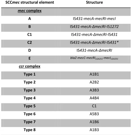
1.2.1. Pulsed-field gel electrophoresis (PFGE) ... 5 1.2.2. Multilocus sequence typing (MLST) ... 7 1.2.3. Whole-genome sequence analysis ... 7 1.2.3.1. Historical prespective ... 7 1.2.3.2. Whole-genome sequencing analysis ... 12 1.2.3.2.1. Closed reference genome-based analysis ... 12 1.2.3.2.2. De novo assembly ... 13 1.2.3.2.3. Downstream analysis ... 14 1.3 The opportunistic pathogen Staphylococcus epidermidis ... 15 1.3.1. Staphylococcus epidermidis as a commensal ... 15 1.3.2. Staphylococcus epidermidis as a pathogen ... 17 1.3.3. Molecular epidemiology and population structure ... 19 1.3.4. Virulence genes ... 23 2. Methicillin resistance: a crucial event in the evolution of Staphylococcus ... 27 2.1. Mechanism of resistance ... 28 2.2. The staphylococcal cassette chromosome mec (SCCmec) ... 29 2.2.1. Historical perspective ... 29 2.2.2. Basic structure and diversity ... 29 2.2.3. Transfer and mobility ... 31 2.2.4. The mec complex ... 32 2.2.5. The ccr complex ... 34
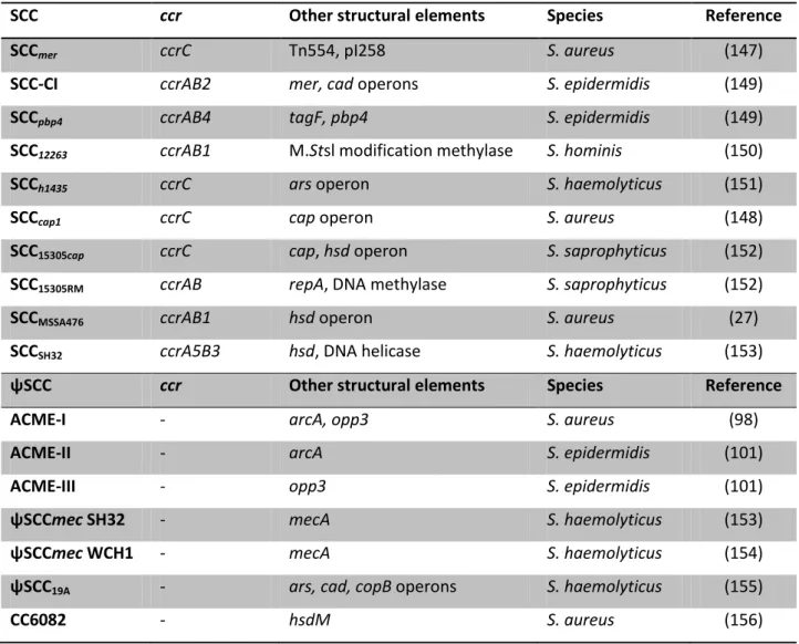
2.2.8. SCCmec typing ... 39 2.2.9. SCCmec distribution in staphylococci: CoNS as the origin of specific SCCmec types... ... 40
2.3. The mec homologues in animal-related species: the origin of mecA... 41 2.3.1. Staphylococcus sciuri mecA1 ... 42 2.3.2. Staphylococcus vitulinus mecA2 ... 43 2.3.3. Staphylococcus fleurettii mecA ... 44 2.3.4. Staphylococcus lentus ... 45 2.3.5. Other mec homologues (mecB, mecC) ... 45 3. The missing links in SCCmec assembly and its impact in Staphylococcus evolution ... 47
Part I: Role of the most primitive staphylococcal species in the origin and assembly of SCCmec
... 49
Chapter II: High frequency and diversity of cassette chromosome recombinases (ccr) in
methicillin-susceptible Staphylococcus sciuri ... 51
Chapter III: Evidence for the evolutionary steps leading to β-lactam resistance in
staphylococci... ... 77
Chapter IV: Role of the primitive Staphylococcus species in the assembly of the primordial
staphylococcal cassette chromosome mec (SCCmec): a key antimicrobial and virulence element in the pathogen Staphylococcus aureus ... 117
Part II: Impact of SCCmec in the evolution and adaptation of S. epidermidis to the hospital
environment ... 165
Chapter V: Evolution of the skin commensal Staphylococcus epidermidis in the hospital
environment: insights from an early collection ... 167
Chapter VI: Strategies of adaptation of Staphylococcus epidermidis to hospital and
community: amplification and diversification of SCCmec... 209
Chapter VII: Concluding Remarks ... 235
Chapter I
General Introduction
Chapter I | 3
1. Staphylococcus in the clinical setting: a worldwide concern
Since their discovery in the late 1800s (1), staphylococci, and particularly
Staphylococcus aureus have been widely studied due to their importance in the clinical
setting. Despite being primarily commensals, staphylococci can cause a wide range of infections that are often difficult to treat, due to their adaptive power to environmental stresses such as the ones in the hospital environment, and increased resistance to antimicrobials. Actually, the burden caused by staphylococcal infections is increasing worldwide (2), and recent advances in molecular typing techniques have revealed some of the secrets behind the success of these highly adaptable pathogens. In the following sections, the available literature on this subject is reviewed, with a focus on the opportunistic pathogen Staphylococcus epidermidis.
1.1. The genus Staphylococcus and clinically relevant species
The genus Staphylococcus is part of the family Staphylococcaceae, that belongs to the order Bacillales of the class Bacilli (3). Bacilli belong to the phylum Firmicutes, which comprises Gram-positive bacteria that typically have a low G+C DNA content (30-40%) (2). The genus Staphylococcus comprises 52 species and 28 subspecies (2, 4) and the bacteria belonging to Staphylococcus have a typical round shape (cocci) and agglomerate as “grape-like” clades (5). Phenotypically, these bacteria are non-motile and facultative anaerobes. They can be easily identified by their biochemical phenotypic properties: non-production of oxidase, production of catalase and tolerance to high concentrations of salt (6). Regarding their ability to produce coagulase the genus can be subdivided into two large groups, one comprising 38 species that do not produce coagulase, so-called coagulase-negative staphylococci, CoNS (2) and 14 which produce coagulase, coagulase-positive staphylococci. Moreover, Staphylococcus cells are enveloped by a characteristic cell wall enriched in O-acetylated peptidoglycan. The peptidoglycan layer in Staphylococcus cell wall is highly crossed-linked, composed by linear glycan chains linked by short peptides, which together with teichoic acids, constitute the cell wall of Staphylococcus (7).
Recently, the genus Staphylococcus has been re-classified, due to the advances and availability of molecular data (in particular whole-genome sequencing data) that allowed the establishment of more accurate phylogenetic relationships. The 52
Staphylococcus species have been organized into distinct cluster groups, which,
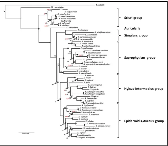
together with their phenotypic and epidemiological characteristics, were divided into six groups of species (Auricularis, Hyicus-Intermedius, Epidermidis-Aureus, Saprophyticus, Simulans and Sciuri) (2, 8)(Figure 1).
Figure 1. Maximum likelihood phylogram based on the nucleotide sequence of 16S ribosomal genes of
46 staphylococcal species. The consensus phylogram was generated from 200 bootstrap replicates with five maximum likelihood search replicates per bootstrap. Adapted from (8).
Sciuri group Auricularis Simulans group Saprophyticus group Hyicus-Intermedius group Epidermidis-Aureus group
General Introduction
Chapter I | 5 The vast majority of the most clinically relevant species belong to the Epidermidis-Aureus group, such as Staphylococcus aureus, Staphylococcus epidermidis,
Staphylococcus haemolyticus, Staphylococcus hominis, Staphylococcus capitis and Staphylococcus lugdunensis. Other clinically relevant species, which are not so
frequently recovered from clinical specimens, are part of the Saprophyticus group, such as Staphylococcus saprophyticus, Staphylococcus cohnii and Staphylococcus
xylosus. The Auricularis group includes a single species, Staphylococcus auricularis that
is found exclusively as a colonizer of the human external ear. The Simulans group comprises species that only transiently cause disease in humans, and are typically isolated from meat and food (2). Finally, the Hyicus-Intermedius and the Sciuri group includes species that are most frequently found colonizing animals and only rarely found colonizing or causing infections in humans (2).
1.2. Molecular typing techniques and whole-genome sequencing analysis in
Staphylococcus
The molecular epidemiology of Staphylococcus has been extensively studied through the application of multiple molecular typing methods and, more recently, the analysis of whole-genome sequences (WGS) revealed Staphylococcus population structure with unprecedented resolution. The most commonly used molecular typing techniques, pulsed-field gel electrophoresis (PFGE) and multilocus sequence typing (MLST), as well as the recent advances in WGS analysis will be briefly reviewed in this section.
1.2.1. Pulsed-field gel electrophoresis (PFGE)
The separation of macrorestriction DNA fragments using PFGE was one of the first typing methods used to study the molecular epidemiology of Staphylococcus. In this technique, a restriction enzyme is used to digest the chromosomal bacterial DNA into a relatively low number (15 to 25) of fragments (9). These fragments are then separated in an agarose gel that is submitted to an electric field, which changes in
orientation, direction and intensity during the electrophoresis. The result is a DNA macrorestriction profile of fragments that are distributed according to their molecular weight (9). Since this profile corresponds to the complete genetic content of a strain, the variations observed between different PFGE patterns reflect genetic events that occurred recently, such as insertions, deletions and mutations occurring in the enzyme’s restriction site. Due to its high discriminatory power, this method has been previously considered as the “gold standard” technique to be used to study outbreaks (9, 10). The degree of relatedness among isolates as estimated by their macrorestriction patterns has been defined for S. aureus (11, 12) and S. epidermidis (13). In general, two isolates are considered to be closely related if the differences in their macrorestriction patterns are consistent with the occurrence of a single genetic event, which results in 2-3 band differences in the macrorestriction patterns (11). When the macrorestriction patterns presented by the isolates differ by 4-6 bands, then the isolates are considered to be possibly related and when more than 7 band differences are observed, the isolates are considered unrelated (11). This analysis has been classically performed by visual inspection of the band profiles but in recent years it has been replaced by automatic statistical analysis, using informatics tools, such as the program BioNumerics (Applied Maths, Kortrijk, Bélgica), that uses image analysis parameters. Briefly, the program allows the definition of tolerance and optimization values for the clustering of the patterns of bands obtained for each strain. The tolerance is the measurement of “movement” of each band, while the optimization limits the “movement” of each fingerprint as a whole. These parameters have been set for each species. For example, for S. epidermidis, it has been proposed the use of the Dice algorithm to determine the similarity between the restriction profiles, with 1.3% optimization and 0.8% tolerance; the profiles are then clustered with the unweighted pair group method with arithmetic mean (UPGMA) and profiles are considered to belong to the same PFGE type with a cut-off value of 79% of similarity between their band pattern (13).
General Introduction
Chapter I | 7
1.2.2. Multilocus sequence typing (MLST)
Multilocus sequence typing (MLST) relies on the sequencing of internal fragments of seven housekeeping genes, which are scattered along the chromosome. To each different allele in the population, a number is attributed and the resulting combination of numbers for the seven genes is called the allelic profile; a number is assigned to each unique allelic profile, which corresponds to the sequence type (ST). Since this method focus on housekeeping genes, it reflects the accumulation of genetic mutations that occur at a slow evolutionary rate, being appropriate to compare isolates collected in different geographic regions and time periods (14). MLST data is commonly analyzed with the eBURST algorithm (15), which is based on a simple model of evolution and diversification. It defines groups of STs related to one another, called the clonal complexes (CC) and its predicted founder (the ST that is most frequent and presents a higher number of related STs). The S. epidermidis MLST scheme present in the MLST database (www.mlst.net) (16) has been widely used to study the molecular epidemiology of this bacterium. For S. epidermidis, it has been defined that STs belonging to the same CC should share at least six alleles (out of seven) with another ST of the same group (17).
1.2.3. Whole-genome sequence analysis
1.2.3.1. Historical prespective
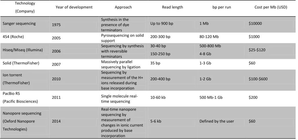
Since its emergence in the market in 2005, the technology behind the first next-generation sequencing (NGS) devices has evolved substantially, together with the broadening of its applications. Several NGS technologies emerged, based on different sequencing chemistries, such as the 454 sequencing technology, the Illumina technology, the Solid technology and the Ion torrent technology, providing a wide range of amplification strategies and having different number of Mb sequenced per run, of read length and of cost per run (Table 1). These technologies overcame the limitation of Sanger sequencing (developed in the 1970s)(18), that was the need to
amplify the number of target DNA fragments before sequencing, which was done usually by cloning into specific bacterial hosts (19).
The very first NGS technology developed was 454 sequencing technology, that was released by 454 Life Sciences in 2005 (20), a company that was later acquired by Roche (Basel, Switzerland). The development of this technology was ground-breaking since it was based on a new DNA amplification method called the emulsion PCR (19) that consists in the shearing of the genome into DNA fragments which are then ligated to streptavidin beads. The beads are then captured into separate emulsion droplets, which act as individual amplification reactors (19). Each droplet is then transferred into a well of a microtiter plate and sequenced by pyrosequencing technology. The pyrosequencing technology is based on the measurement of the release of inorganic pyrophosphate by chemiluminescence upon incorporation of labeled dNTPs in the nascent DNA chain (21). This technology produces reads of around 300 bp and when it was introduced in the market, was very costly (Table 1). In addition, it has the disadvantage of producing a high number of sequencing errors.
Shortly after the release of the first 454 apparatus, a new NGS technology was introduced in the market by Illumina (Hiseq, San Diego, USA). Briefly, the Illumina technology consists in shearing the genome into small single-stranded DNA fragments (the libraries). These are then attached to a flowcell and sequencing is achieved through solid-phase bridge amplification (19). During this process, one end of a single DNA molecule is attached to the flowcell using an adapter; the molecules subsequently bend over and hybridize to complementary adapters, thereby forming the template for the synthesis of their complementary strands (19). The templates are sequenced in a massively parallel fashion using the basic principles of Sanger sequencing (18). This technology produces short sequence reads (50-250 bp) (19) (Table 1) but due to its sequencing chemistry, it produces less sequencing errors than 454 technology. Furthermore, the cost of each run decreased dramatically due to the low cost of the reagents used in the sequencing reaction, which promoted the use of this technology, particularly for sequencing bacterial genomes. The development of a benchtop sequencer by Illumina, MiSeq, in 2011 has further increased the use of this NGS technology for the control of outbreaks and molecular epidemiology of pathogens,
General Introduction
Chapter I | 9 namely staphylococcal species. In fact, for the two major staphylococcal pathogens S.
aureus and S. epidermidis, 4369 and 106 whole-genome sequences have been
deposited in the NCBI database by August 2015 (www.ncbi.nlm.nih.gov/), respectively, illustrating the growing interest in this high throughput technology.
Several different NGS technologies were introduced in the market shortly after the release of Hiseq by Illumina (Table 1). Briefly, in the Solid technology (Applied Biosystems, ThermoFisher, Waltham, Massachusetts, United States) a library of DNA fragments linked to clonal beads is prepared by emulsion PCR. Then adapter primers hybridize to the template beads and a set of four fluorescently labeled di-base probes are added to the mixture, that compete for ligation to the sequencing primer. The sequencing is achieved after multiple cycles of ligation, detection and cleavage. On the other hand, the Ion Torrent technology (Life Technologies, ThermoFisher, Waltham, Massachusetts, United States) is based on the release of pyrophosphate and a positively charged hydrogen ion, when incorporation of dNTPs occurs into a nascent DNA strand. Briefly, the DNA polymerase is immobilized on a semiconductor chip that is then flooded with many copies of single-stranded DNA template and each of the four dNTPs, sequentially. If a certain dNTP is incorporated, the hydrogen ion is released and the pH of the solution changes, which is detected by the chip. Both of these technologies have been used to study bacterial genomes (19), but their low throughput power and elevated cost have not made them first choices to study small genomes.
Table 1. Summary of the most used sequencing technologies and their main characteristics. Adapted from (19, 22, 23). Technology
(Company) Year of development Approach Read length bp per run Cost per Mb (USD)
Sanger sequencing 1975 Synthesis in the presence of dye terminators
Up to 900 bp 1 Mb $10000
454 (Roche) 2005 Pyrosequencing on solid
support 200-300 bp 80-120 Mb $1000
Hiseq/Miseq (Illumina) 2006 Sequencing by synthesis with reversible terminators
30-40 bp 150-250 bp
500-800 Mb
4-8 Gb $25-$120
Solid (ThermoFisher) 2007 Massively parallel
sequencing by ligation 35 bp 1-3 Gb $60
Ion torrent
(ThermoFisher) 2010
Sequencing by
measurement of the H+ ions released during base incorporation
200-400 bp 1-2 Gb $100-$600
PacBio RS
(Pacific Biosciences) 2011
Single molecule
real-time sequencing 10-60 kb 500 Mb-1 Gb $200 Nanopore sequencing (Oxford Nanopore Technologies) 2014 Real-time nanopore sequencing by measurement of changes in ionic current produced by base incorporation
General Introduction
Chapter I | 11 The principles and chemistry of each NGS technology are different but all of these technologies produce short reads. Therefore, the reads obtained produce highly fragmented assemblies and a closed genome is hard to achieve. To overcome this problem, third generation sequencing technologies have been introduced in the market, which produce longer reads. PacBio sequencing (Pacific Biosciences, San Francisco, USA) is a high throughput method that has been recently released, that produces sequencing reads by reading in real-time a continuous sequence from the molecular template. Briefly, a specialized cell, called the SMRT cell is used, that contains a single DNA polymerase molecule immobilized in each well. The libraries are prepared by shearing the DNA into double-stranded fragments (2-5 kb) and ligating hairpin adapters to each end of the fragments (http://www.pacificbiosciences.com/), thus creating a single-stranded end. The sequencing reads are obtained in real-time, by monitoring the incorporation of fluorescent-dyed nucleotides in the nascent DNA chain. This method allows obtaining long sequence reads (3-9 kb) (Table 1); the drawback is that it is prone to sequencing errors and it is still very costly (Table 1). More recently, a third generation sequencing technology developed by Oxford Nanopore Technologies (Oxford, UK) has been introduced in the market (Table 1). This technology is incorporated in a small machine called MinION. Briefly, the technology is based in the measurement of changes in the electronic current as single molecules of DNA are passed through the DNA polymerase, immobilized in a biological nanopore. By using a hairpin adapter, each molecule is read twice and the resulting reads are long. Although it is suitable to assemble closed genomes, the error rate has been reported to be high (24). This technology is promising though, because of the low cost of sequencing a genome in comparison with PacBio sequencing (23)(Table 1).
1.2.3.2. Whole-genome sequencing analysis
The limiting step of whole-genome sequencing (WGS) has been the low availability of user-friendly software to analyze the huge amounts of data produced by this technology. However, the recent application of WGS to the molecular epidemiology of bacteria, including Staphylococcus, is trending towards an increase, with the emergence of improved analysis methods and user-friendly software. The fast development of accessible informatics tools to analyze clinically relevant targets has allowed the application of WGS in clinical laboratories.
1.2.3.2.1. Closed reference genome-based analysis
The first reports that applied WGS to the study of Staphylococcus epidemiology used a method called single-nucleotide polymorphisms (SNP) analysis and was developed for S. aureus (25). This method involves aligning the reads produced by sequencing the genome of the isolate, with a fully sequenced and annotated reference genome. A phylogenetic reconstruction with this data is performed with appropriate algorithms, the end result being a SNP tree (25). This method has proven to be very useful to detect bacterial lineages, as well as SNP variation in the part of the genome that is shared between all isolates tested and the reference strain (25-28). However, interpretation of the results is not yet optimal, since the variation of the number and genetic location of SNPs among clonal lineages and even inside the same clonal lineage is not uniform. Therefore, an appropriate cut-off value for clonal relatedness in case of outbreaks is difficult to define and depends on the type of collection analyzed. Thus interpretation of the SNP tree should be done only locally, using appropriate epidemiological information (29). The recent advances in phylogeny reconstruction with a Bayesian inference, such as BEAST (30), that takes into consideration also the epidemiological data, are promising and have proven to be suitable to use in surveillance and epidemiological studies (31, 32).
General Introduction
Chapter I | 13
1.2.3.2.2. De novo assembly
An appropriate closed reference genome is not always available. Therefore, assembling the short reads into long contigs, in a reference-free manner, stimulated the interest of researchers and several algorithms have been developed for this purpose, such as VELVET (33). The so-called assembly de novo has been widely used in several epidemiological studies. The drawback of this method is the end result, which consists of hundreds of contigs with no overlapping genomic regions that represent different segments of the genome (22). To overcome this difficulty, alignment of the contigs, produced for each strain by VELVET, is achieved with powerful iteractive alignment algorithms, such as Mauve (34). Mauve can align a reasonable number of genomes, partitioned in hundreds of contigs, while accounting for rearrangements (22).
Another useful approach to deal with the high number of genomic contigs of a given isolate is to perform a hybrid assembly, which consists in combining the reads obtained with Illumina (or other NGS technologies) with the reads obtained with PacBio sequencing (35) or nanopore sequencing reads (23) . In this approach, the hybrid assembly method allows for correction of sequencing errors produced with third generation sequencing technologies, since it incorporates the short reads obtained by NGS technologies (35). Powerful assemblers that allow error corrections, have been developed to work with reads from different technologies, such as SPAdes (36) and CELERA (37), among others.
Finally, a commonly used strategy to analyze the high number of contigs produced by the de novo assembly is to perform a gene-by-gene analysis, or whole-genome MLST (38). This “super MLST” utilizes the assembly de novo data and consists in extracting alleles of genes present in the predicted core genome of all strains (38, 39). Following the rationale for MLST data, an allele number is assigned to each gene and an allelic profile is attributed to each strain. This strategy was first implemented in an open-source database system called the BIGSDB (bacterial isolate genome sequence database)(38). A similar approach has been recently described and incorporated in an automated program, called the SeqSphere (Ridom GmbH,
Germany). This method has proven to be valuable to infer epidemiological relatedness between isolates in outbreak situations (39). Another method consists on predicting the core genome of the strains, by selecting the genes that are present in all strains (22). An alignment can then be produced with the concatenated gene sequences of the predicted core genome, by using algorithms that have been widely used before for single gene alignments, such as CLUSTALW (40) and MUSCLE (41). Phylogenetic trees are then performed using the resulting alignment. This strategy has been recently used to study the core genomes of S. aureus and S. epidermidis (42).
1.2.3.2.3. Downstream analysis
The contigs obtained in the assembly de novo can be used for many objectives. Several tools have been developed to study draft genomes, particularly of bacterial origin. For instance, different algorithms and databases have been recently developed to rapidly detect antibiotic resistance genes, with the interest of aiding clinicians in the course of treatment of infectious diseases (43). Other databases and web-based servers have been developed, which identify virulence genes, plasmid genes, prophages and insertion sequences, among many other functional categories; examples are the antibiotic resistance genes online database ResFinder, https://www .cge.cbs.dtu.dk/services/ResFinder/, the insertion sequence online database ISFinder, https://www-is.biotoul.fr,among others. The great majority of these platforms work with the BLAST algorithm that identifies homologues of the genes in the contigs by comparing these with the ones deposited in a database.
One useful and most used approach is functional annotation of genomes or genomic regions. In these cases, the annotation is usually confined to the protein-coding sequences (CDS) present in contigs containing regions of interest of the genome. Briefly, the annotation process is usually based in programs that use ab initio algorithms trained on gene models from related species (for instance, AUGUSTUS (44)) or gene alignments (using for example tblastx) and databases (such as KEGG) that complement the predicted gene models (45). However, as the evidence available for some species is mostly incomplete and sometimes contradictory, this is a difficult task
General Introduction
Chapter I | 15 that often benefits from manual curation. A complete genome annotation represents a considerable effort and requires bioinformatic proficiency; furthermore, it strongly depends on the quality of the genome assembly. Only near-complete genomes interrupted only by small gaps will yield satisfactory results (45).
1.3. The opportunistic pathogen Staphylococcus epidermidis
1.3.1. Staphylococcus epidermidis as a commensal
Staphylococcus epidermidis is mainly a commensal of the human skin, being
one of the first bacteria that colonize the skin of newborns after birth (46). Its main niches are the humid areas of the human skin, such as the anterior nares, the axillae, the inginal and perineal areas, the toe webs and also the conjunctiva (2), where it forms populations of cells, which are often composed of multiple strains of this species (47). S. epidermidis can be also found colonizing the skin of non-human mammals, but this colonization is thought to be transient and of human origin (48, 49). Nevertheless, relatively high frequencies of carriage of S. epidermidis have been described, particularly in production animals in Europe (40%)(50).
While colonizing, S. epidermidis cells are believed to adhere to the human skin (51). In fact, S. epidermidis genome is well equipped with genes for adhesion, such as adhesins (aae and sdrG, among many others, see below) (52). The role of S.
epidermidis as part of the protection barrier provided by the skin is well documented.
In fact, some studies suggest that S. epidermidis has an antagonistic effect against more virulent species, like S. aureus. One example of this is the inhibition of S. aureus by S. epidermidis through the excretion of the serine protease Esp; specifically, it has been found that purified Esp inhibits biofilm formation by S. aureus and destroys pre-existing biofilms (53). Furthermore, it was shown that Esp enhances the susceptibility of S. aureus biofilms to immune system components. Other exoproteins produced by
S. epidermidis that have antimicrobial activity against S. aureus are the PSMs
epidermidis strains carrying specific alleles of the quorum-sensing system agr (one of
the most important virulence regulons in staphylococci) inhibit the colonization of the skin of healthy volunteers by S. aureus strains carrying specific agr alleles, which suggests that cross-interference between agr alleles affects negatively the colonization of these bacteria of the same niche (56). Furthermore, S. epidermidis strains with the ability to produce bacteriocins have been also identified (57). In particular, epidermicidin produced by a S. epidermidis strain recovered from a human skin swab, was shown to have an inhibitory activity against S. aureus, other CNS species and enterococci (58) and a thiopeptide produced by S. epidermidis of avian origin, was reported to have activity against several different gram-positive pathogens (including
S. aureus, enterococci, Clostridium difficile and others) (59). Finally, several studies
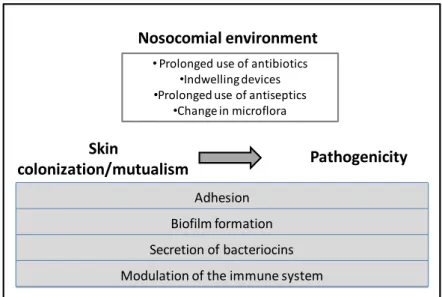
have showed that S. epidermidis can interact with the human immune system. Specifically, the main component of S. epidermidis biofilms, PIA (polysaccharide intercellular adhesin, see below), is able to stimulate the immune system through binding to toll-like receptor 2 (TLR2) present in human cells (60) (Figure 2). PIA was found to be able to induce the production of pro-inflammatory cytokines via TLR2. S.
epidermidis can also interact with antimicrobial peptides (AMPs). AMPs are small
peptides that have antimicrobial activity and are part of the innate immune system. A study has showed that a PSM produced by S. epidermidis can boost the production of AMPs by the host, leading to an increased killing capacity of human neutrophils against pathogenic bacteria such as group A streptococci (55). Altogether, these data suggest that the presence of S. epidermidis in the human skin contributes to its homeostasis and prevents colonization by pathogenic species.