Article
J. Braz. Chem. Soc., Vol. 22, No. 5, 919-928, 2011. Printed in Brazil - ©2011 Sociedade Brasileira de Química
0103 - 5053 $6.00+0.00
A
*e-mail: [email protected]
Monitoring of
β
-Blockers Ozone Degradation via Electrospray Ionization Mass
Spectrometry
Cristina Quispe,a,d Fabiane M. Nachtigall,b Maria Francesca R. Fonseca,b Rosana M.
Alberici,b Luis Astudillo,c Jorge Villaseñor,a Marcos N. Eberlinb and Leonardo S. Santos*,d
aLaboratory of Physical Chemistry, Instituto de Química de Recursos Naturales, Talca University, PO Box 747, Talca, Chile
bThoMSon Mass Spectrometry Laboratory, Institute of Chemistry, University of Campinas, 13083-970 Campinas-SP, Brazil
cLaboratory of Organic Synthesis and dLaboratory of Asymmetric Synthesis, Talca University, PO Box 747, Talca, Chile
As estruturas dos produtos de degradação mediada por ozônio de diferentes compostos farmacêuticos foram estudadas. Uma completa degradação do nadolol foi observada nas condições empregadas após 100 min. Os produtos de degradação obtidos nas soluções aquosas foram caracterizados por espectrometria de massas com ionização por electrospray (ESI-MS e ESI-MS/MS). O mecanismo proposto para a degradação consiste inicialmente do ataque de ozônio ao grupo amino da anilina formando nitro compostos, seguida de uma série de processos oxidativos. Monitoramento por ESI-MS(/MS) mostrou que a degradação de atenolol (ATE) e acebutolol (ACE) por ozônio ocorrem via um mecanismo similar ao do nadolol.
The structures of intermediate products of ozone degradation of different pharmaceutical compounds have been studied. Under the conditions employed, complete ozone degradation of nadolol was achieved after 100 min. The degradation products obtained in aqueous solution were characterized by electrospray ionization mass (and tandem mass) spectrometry (ESI-MS and ESI-MS/MS). The proposed mechanism for degradation, ozone attacks at the aniline amino group giving rise to nitro compounds and further degradation occurs via a series of oxidative processes. Continuous online monitoring by ESI-MS(/MS) with high accuracy mass measurements showed that ozone degradation of atenolol (ATE) and acebutolol (ACE) occurs via mechanisms similar to that of nadolol.
Keywords: ozonation, degradation, ESI-MS, pharmaceutical compounds, reaction mechanism
Introduction
Concerns about the prejudicial effects to life of accumulation of pharmaceuticals in the environment has greatly increased in recent years.1,2 But such accumulation is governed mainly by the environmental bio- and photo-transformation3 and the speed and nature of the natural degradation of these potentially harmful contaminants.4,5 The photolytic degradation of chemicals in the environment may occur via direct sunlight absorption or by reactions with strong oxidizing species such as hydroxyl radicals and singlet oxygen.
Pharmaceuticals enter the aquatic environment after its ingestion by humans and other animals and subsequent excretion, either in their intact forms or as metabolites.6 Wastewater treatment is applied worldwide but pharmaceuticals may not be eficiently removed,3,7 and these pharmaceutical residues and degradation products can be downloaded directly into aquatic environments. Several studies have stated that the long-term risk to humans from any single pharmaceutical at sub-µg L−1 levels is negligible, but the toxicological implications of chronic exposure to trace contaminants is still unclear.8,9
common oxidizing reagents such as H2O2 or chlorine, is more eficient in pollutant degradation and less harmful to most living organisms.10 In water, ozone can oxidize contaminants via direct selective reactions11 such as ozone addition to double bonds or the initiation of chain radical reactions by the production of free hydroxy radicals (equations 1- 4). Hydroxy radicals are generated from ozone decomposition, which is catalyzed by the presence of hydroxyl anions or by traces of other substances, such as transition metal cations.
O3 + H2O→ 2 HO•+ O2 (1)
O3 + OH−→ O2•– + HO2• (2)
O3 + HO•→ O2 + HO2• (3)
O3 + HO2•→ 2 O2+ HO• (4)
The decomposition of ozone in aqueous solutions is known to proceed via the chain reactions shown in equations 1-4.12 Ozone therefore indirectly attacks organic compounds since such reactions are faster than direct O3 attack. In previous studies,13,14 we described the O3 interaction with pharmaceutical compound nadolol (anti-hypertensive). In this study, we report the characterization via electrospray ionization mass spectrometry (ESI-MS)15,16 of the oxidation intermediates and products of degradation of nadolol generated during O3 oxidation. Furthermore, the O3 degradation of two other β-blockers compounds atenolol and acebutolol was also studied.
ESI-MS (and its tandem version ESI-MS/MS) has been used extensively to monitor the composition and degradation of organic compounds in the environment.17,18 ESI-MS(/MS) with its unique characteristics has become a major technique to elucidate reaction mechanism especially in aqueous solutions via the detection and identiication of reactants, products, and intermediates,19,20 even the short-lived ones that occur at very low concentrations.21,22
Gas chromatography-mass spectrometry (GC-MS) has often been the technique of choice for reaction screening and product elucidation.23 However, GC-MS is an ofline technique that demands extraction and sometimes derivatization pre-steps, transient, most polar, or relatively unstable compounds may be missed. We have then performed ESI-MS(/MS) monitoring of the O3 advanced oxidation of nadolol, atenolol and acebutolol in water to search for all intermediates and products of this environmentally important process. These products are the ones most likely to be found in treated wastewaters. We also used high performance liquid chromatography (HPLC), ultraviolet spectroscopy (UV), and total organic carbon (TOC) analyses to collect additional
data on substrate degradation rates and mineralization and to investigate the presence of residual organic compounds in the aqueous solutions.24
Experimental
Chemicals
Nadolol (NAD), atenolol (ATE) and acebutolol (ACE) were purchased from Sigma. All the solvents were of HPLC grade and purchased from Merck and used without puriication. Doubly distilled water was used to prepare all the solutions in the experiments. Ozone was generated using a laboratory ozone generation (OZOCAV) ozonizer fed with air/oxygen.
Degradation procedures
The ozonation reactions were performed in a batch glass reactor thermostated at 25 °C. The reactor was charged with 100 mL of the drug aqueous solutions (1.6 × 10⁻⁴ mol L−1) and fed with 1.2 × 10⁻⁴ mol L−1 of ozone at a constant low of 46 ± 1 mL min−1. The ozone concentration was monitored measuring the absorbance at 254 nm, using the molar extinction coeficient of ozone of 2900 L mol-1 cm-1. Aliquots (1.0 mL) were taken at several reaction times and promptly analyzed by ESI-MS(/MS) and HPLC. All samples were protected from light and refrigerated at 4 °C prior to analyses.
Analytic methods
Drugs concentrations were monitored using a Perkin Elmer Series 200 chromatograph HPLC system equipped with a UV-Vis detector. A Merck-Chromolith Performance RP-18 column (4.6 mm × 100 mm) was used with H2O:MeOH (70:30, v/v) mobile phase at a low rate of 0.6 mL min−1. Water was previously acidiied to pH 3 using aqueous solution of HCl. Organic acids were quantiied with a transgenomic ORH-801 column (6.5 mm × 300 mm) using 5.0 × 10⁻³ mol L−1 sulphuric acid as mobile phase at a low rate of 0.8 mL min−1.
Ultraviolet (UV) absorbance measurements were performed using a HP 8453 (Agilent) spectrophotometer equipped with a quartz cell with a 1.0 cm path length. Absorbances were measured with baseline correction, scan rate of 200-600 nm, and integrate time of 0.5 s and intervals of 1 nm.
ESI-MS(/MS) data were collected using a high resolution hybrid quadrupole (Q) and orthogonal time-of-light (TOF) mass spectrometer (Q-Tof, Micromass UK) with constant nebulizer temperature of 50 °C. The ESI source and the mass spectrometer were operated in the positive ion mode, and the cone and extractor potentials were set to 40 and 10 V, respectively, with a scan range of
m/z 50-1000. Samples were infused into the ESI source at low rates of ca 15 µL min−1 via a microsyringe pump. ESI-MS/MS experiments were carried out by selection of a speciic ion in Q1 and by performing its collision-induced dissociation (CID) with argon in the collision chamber. The product ions MS analysis was accomplished with the high-resolution (ca. 5.000) and high accuracy (ca. 10 ppm) orthogonal TOF analyzer.
Results and Discussion
Degradation of nadolol
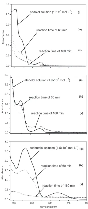
Our investigation began with HPLC monitoring for the O3 degradation of nadolol (NAD) in an aqueous solution at a concentration of 1.6 × 10⁻⁴ mol L−1 at pH 3 or 9, with O
3 concentration at 1.2 × 10⁻⁴ mol L−1 (Figure 1). Regardless the pH (acidic or basic), more than 90% of NAD was found to be degraded after 60 min at similar rates. TOC analyses were then performed to evaluate the mineralization extent, which was found to be only 20% according to TOC removal. This result suggests that NAD was completely consumed after 60 min of O3 degradation, but desirable mineralization to CO2, H2O and NH3 was far for completion. UV monitoring of O3 degradation (Figure 2) also indicated complete NAD consumption, but little information is provided of the nature of the degradation products. The UV spectra of Figure 2i show three characteristic absorption bands of nadolol at 203, 217 and 277 nm. After
O3 treatment, the UV spectra show that intensities of these absorption bands were greatly reduced, suggesting nadolol was degraded, but not totally mineralized with persistent organic intermediates.
Figure 1. HPLC monitoring of the ozonation of nadolol at pH 3 () and pH 9 ().
Figure 2. UV spectra of (i) an aqueous solution of nadolol at 1.6 × 10⁻⁴ mol L−1, (ii) an aqueous solution of atenolol at 1.9 × 10⁻⁴ mol L−1,
(iii) an aqueous solution of acebutolol at 1.5 × 10⁻⁴ mol L−1 and aliquots
ESI-MS(/MS) monitoring
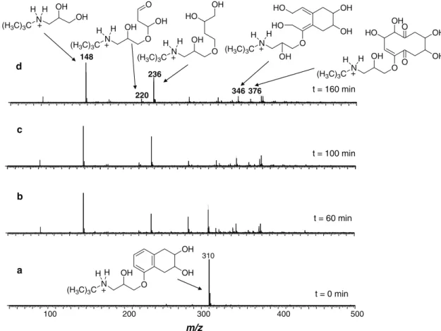
Figure 3 displays representative ESI(+) mass spectra of nadolol aqueous solution (pH 9) monitored after 60, 100 and 160 min from ozone treatment. Similar spectra (not shown) were acquired when using solutions at pH 3. The spectra at zero irradiation time (Figure 3a) detected, as expected, an intense ion of m/z 310 corresponding to protonated NAD. The ESI-MS/MS (Figure 4a) for collision induced dissociation of protonated NAD of m/z 310 shows a series of major and structurally diagnostic fragment ions arising from the loss of isobutene (m/z 254), H2O plus isobutene (m/z 236), 2 × H2O plus isobutene (m/z 218) and NH3 plus 2 × H2O and isobutene (m/z 201).
The degradation process was monitored by intervals of 20 min during 160 min of reaction. Illustrative spectra are displayed in Figure 3. The degradation of nadolol of m/z 310 was clearly perceived as well as the formation of organic intermediates. After 60 min of ozonation (Figure 3b), three additional and increasingly abundant ions of m/z 92, 148 and 236 were detected. The ion of m/z 148 (Figure 3d), which indicates the loss of a subunit of 161 Da (from nadolol of 309 Da), was characterized as the protonated form of dihydroxylamine 2 (Figure 5). Further degradation
of 2 afforded hydroxylamine [3 + H]+ by loss of isobutene as noted through the ion of m/z 92, as depicted in Figure 3d.
The ion of m/z 236 (Figure 3d) was likely formed by aromatic ring opening, which produced a series of degradation products detected as the ions of m/z 378, 342 and 376. Furthermore, ESI-MS was able to gently and eficiently ish directly from the solution to the gas phase for MS analysis also an increasingly abundant ion of m/z 236. This ion was characterized as aminotriol 9
(Figure 5). Figure 4c shows the ESI-MS/MS spectra of intermediate [9 + H]+ that dissociated by loss of isobutene (C4H8) producing the fragment ion of m/z 180. That loss is indicative of tert-butyl group in the molecule, as observed in Figure 4. Another interesting fragmentation pathway was observed for [9 + H]+ by loss of C
4H8O2, which structure could be realized as 2-hydroxytetrahydrofuran.
Two major mechanisms can be considered for O3 treatment. The irst one assumes O3 addition to double bonds, whereas the second considers indirect reaction in water via hydroxyl radicals (equations 1-4). Formation of lower and higher masses intermediates was observed. Considering the lower mass compounds, Figure 5 presents a proposed route of degradation based on the ESI-MS(/MS) data, whereas Figure 6 presents proposed pathways
Figure 4. ESI-MS/MS of (a) protonated nadolol [1 + H]+ of m/z 310; (b) the ion of m/z 148 attributed to [2 + H]+; and (c) the ion of m/z 236 attributed to
[9 + H]+.
leading to the higher mass products. HPLC analyses with standards corroborated unequivocally the structure of several structures proposed for the intermediates detected and characterized by ESI-MS(/MS). Among them are oxalic acid, formic acid and glyoxylic acid, which are known as recalcitrant compounds very resistant to further degradations.13 The resistance for this class of compounds is due to the lack of C–H bonds, and this may explaining why oxalic acid was less susceptible to OH• radicals attack which are produced mainly at high pH from O3. Thus, although the parent NAD was totally consumed by O3 treatment at
different pH in a similar reaction rates, the intermediates observed in solution at pH 3 or 9 were different.
Degradation of atenolol
Figure 5. Mechanism proposed for nadolol (NAD) degradation by ozone based on ESI-MS(/MS) data (lower mass products).
[1 + H]+ m/z 310
[2 + H]+ m/z 148
[3 + H]+ m/z 92
[4 + H]+ m/z 90
[7 + H]+ m/z 235
[10 + H]+ m/z 220 [9 + H]+
m/z 236
Organic acids
CO2 + H2O
ring-opening
[O]
[O]
[O] [O]
[O] [O ]
[O] O3 / HO
.
O3 / HO.
O H N (H3C)3C
OH OH OH OH H N (H3C)3C
OH
O H N (H3C)3C
OH HO
OH
OH H2N
OH
OH
OH
HO O O
H
O
O H2N
OH OH O O OH H N (H3C)3C
.
[11− H]− m/z 243
[8 − H]− m/z 229 [5− H]−
m/z 159 [1− H]−
m/z 308
ring-opening
O3 / HO HO [O]
O H N (H3C)3C
OH
OH
OH
HO COOH
OH HOOC COOH
COOH OH HOOC COOH COOH O HO
.
.
HO O O HO O O [O] OH H OH O [O]CO2 + H2O
Figure 6. Mechanism proposed for nadolol (NAD) degradation by ozone based on ESI-MS(/MS) data (higher mass products).
[1 + H]+
m/z 310
[12 + H]+
m/z 358 O3
O N (H3C)3C
OH H OH OH H + O N
(H3C)3C OH H OH OH H + O O O
[13 + H]+
m/z 342
[14 + H]+
m/z 344 ring-opening
O3 / HO
O3 / HO
.
.
O N (H3C)3C
OH H OH OH H HO OH O N (H3C)3C
OH H OH OH H O + O
.
[15 + H]+
m/z 392 [18 + H]+
m/z 376 [9 + H]+
m/z 236
[16 + H]+
m/z 394
[17 + H]+
m/z 378* O N (H3C)3C
OH H OH OH H + HO OH O O O O N (H3C)3C
OH H OH OH H HO OH O OH OH + O N (H3C)3C
OH H OH OH H HO OH OH OH + O N (H3C)3C
OH H H HO OH + OH OH OH HO OH O N (H3C)3C
H H
O
O
+
of mineralization. These results suggested, as for NAD, that the mineralization leading to CO2, H2O and NH3 was mostly incomplete.
Figure 7 displays representative ESI(+)-MS of atenolol aqueous solutions according with time at pH 3. Similar spectra (not shown) were acquired at pH 9. The ESI-MS at zero irradiation time (Figure 7a) shows, as expected, predominantly the ion of m/z 267 for protonated ATE. After 160 min of ozonation (Figure 7b), ATE (m/z 267) was totally consumed and three intense ions of m/z 134, 206 and 222 were observed. It was reported that photocatalytic TiO2 degradation
25 and biological advanced oxidation26
of atenolol afforded comparable degradation products as characterized by ESI-MS. Furthermore, the degradation species reported through TiO2 and biodegradation suggested a HO• radical Fenton-like pathway showing
higher mass products than observed by us. However, based on the ESI-MS(/MS) data, a different mechanism for O3 treatment of atenolol is proposed as depicted in Figure 8.
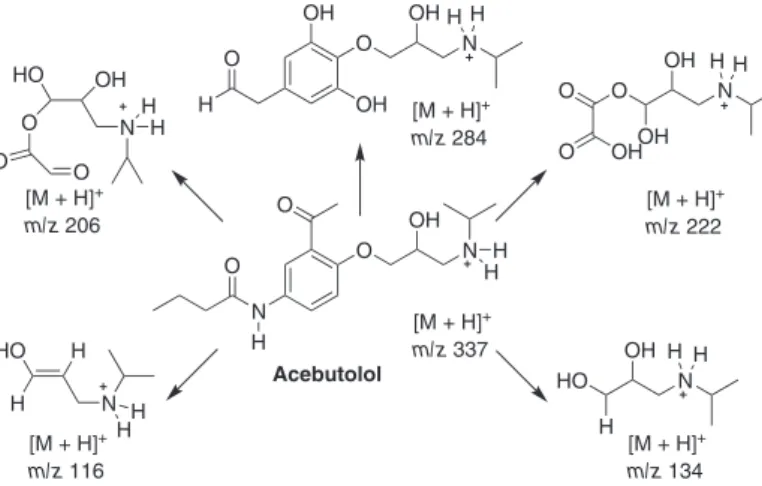
Degradation of acebutolol
Figure 2 shows the UV spectra for the O3 treatment of acebutolol (1.5 × 10⁻⁴ mol L−1). The two characteristic absorption bands of acebutolol are those at 231 and 320 nm. As observed previously for nadolol and atenolol, complete consumption of the parent molecule is achieved but 15% of TOC removal was observed.
Figure 9 displays two representative ESI(+)-MS of acebutolol solutions for O3 treatment. The ESI-MS at zero irradiation time (Figure 9a) detects, as expected, the ion of m/z 337 corresponding to protonated acebutolol.
Figure 7. ESI(+) Mass spectra acquired for aliquots of the reaction mixture after degradation of atenolol with ozone system for (a) 0 min and (b) 160 min.
Figure 8. Mechanism proposed for atenolol degradation by ozone based on ESI-MS(/MS) data.
H2N
O O N
OH
H
O O N
OH
H
OH
OH
HO
O N
O
OH OH
O N
OH O
O
HO OH
N H
O
H3N O
OH O
O H
HO
H OH
N H HO
H H
N O N
OH HO H H
H H
O
H H
H
H
HH O
H
HH [M + H]+
m/z 267
[M + H]+
m/z 158 [M + H]+
m/z 222
[M + H]+
m/z 164 [M + H]+
m/z 134 [M + H]+
m/z 116 [M + H]+
m/z 206 [M + H]+
m/z 224
[M + H]+
m/z 284
Figure 9. ESI(+) Mass spectra acquired for aliquots of the reaction mixture after O3 degradation of acebutolol for (a) 0 min and (b) 160 min.
Figure 10. Mechanism proposed for acebutolol degradation by ozone based on ESI-MS(/MS) data.
O O N
OH
H
OH
OH
OH OH
O N
OH O
HO
H OH
N H HO
H H
N
H H
O
H H
HH
H [M + H]+
m/z 337
[M + H]+
m/z 222
[M + H]+
m/z 134
[M + H]+
m/z 116
[M + H]+
m/z 206
[M + H]+
m/z 284
Acebutolol
N
O O N
OH O
H
H H O
HO OH
N H
O O
H
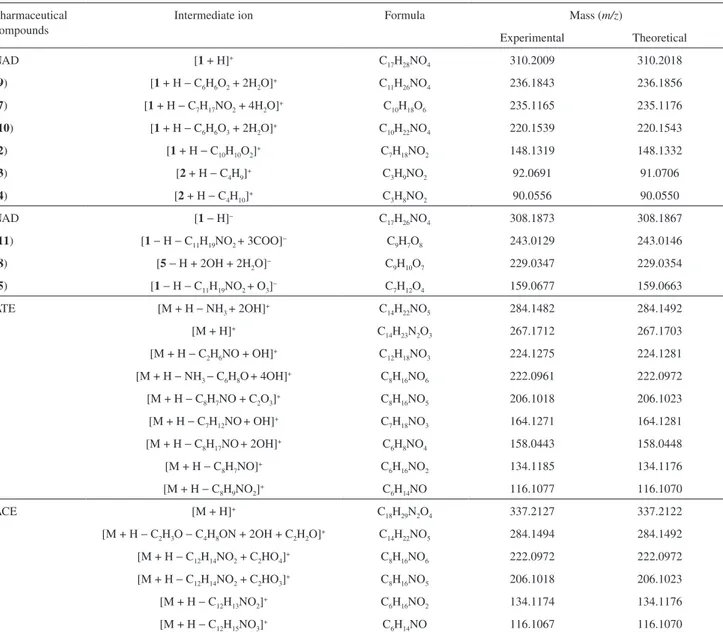
After ozonation for 160 min (Figure 9b), acebutolol was completely consumed and four ions of m/z 134, 206, 222 and 284 were observed. These ions were characterized by ESI-MS/MS experiments and based on the MS data Figure 10 proposes a mechanism for its O3 treatment, assuming similar degradation pathways as proposed previously for atenolol and nadolol. Table 1 summarizes the accurate mass data of product ions for all species identiied during the degradation processes of NAD, ATE and ACE.
Conclusions
A s e x e m p l i fi e d h e r e i n f o r t h r e e c o m m o n pharmaceuticals, ESI mass spectrometry (and its tandem version) is a powerful technique able to provide detailed information about organic intermediates connecting
Acknowledgments
Financial support from the Proyecto Bicentenario de Ciencia y Tecnología PSD-16 and the Brazilian agencies FAPESP and CNPq is gratefully acknowledged. L. S. S. thanks FONDECYT (1085308) for inancial support.
References
1. Daughton, C. G.; Ternes, T. A.; Environ. Health Perspect. 1999,
107, 907.
2. Ternes, T. A.; Water Res. 1998, 32, 3245.
3. Kolpin, D. W.; Skopec, M.; Meyer, M. T.; Furlong E. T.; Zaugg, S. D.; Sci. Total Environ.2004, 328, 119.
4. Boyd, G. R.; Reemtsma, H.; Grimm, D. A.; Mitra, S.; Sci. Total Environ.2003, 311, 135.
Table 1. Accurate mass measurement of intermediate ions of nadolol (NAD), atenolol (ATE) and acebutolol (ACE) as determined by ESI-MS(/MS)
Pharmaceutical compounds
Intermediate ion Formula Mass (m/z)
Experimental Theoretical
NAD [1 + H]+ C
17H28NO4 310.2009 310.2018
(9) [1 + H − C6H6O2 + 2H2O]+ C
11H26NO4 236.1843 236.1856
(7) [1 + H − C7H17NO2 + 4H2O]+ C10H18O6 235.1165 235.1176
(10) [1 + H − C6H6O3 + 2H2O]+ C
10H22NO4 220.1539 220.1543
(2) [1 + H − C10H10O2]+ C7H18NO2 148.1319 148.1332
(3) [2 + H − C4H9]+ C3H9NO2 92.0691 91.0706
(4) [2 + H − C4H10]+ C3H8NO2 90.0556 90.0550
NAD [1− H]− C
17H26NO4 308.1873 308.1867
(11) [1− H − C11H19NO2 + 3COO]− C9H7O8 243.0129 243.0146
(8) [5− H + 2OH + 2H2O]− C9H10O7 229.0347 229.0354
(5) [1− H − C11H19NO2 + O3]− C
7H12O4 159.0677 159.0663
ATE [M + H − NH3 + 2OH]+ C14H22NO5 284.1482 284.1492
[M + H]+ C
14H23N2O3 267.1712 267.1703
[M + H − C2H6NO + OH]+ C12H18NO3 224.1275 224.1281
[M + H − NH3 − C6H8O+ 4OH]+ C8H16NO6 222.0961 222.0972
[M + H − C8H7NO + C2O3]+ C8H16NO5 206.1018 206.1023
[M + H − C7H12NO+ OH]+ C7H18NO3 164.1271 164.1281
[M + H − C8H17NO+ 2OH]+ C
6H8NO4 158.0443 158.0448
[M + H − C8H7NO]+ C6H16NO2 134.1185 134.1176
[M + H − C8H9NO2]+ C
6H14NO 116.1077 116.1070
ACE [M + H]+ C
18H29N2O4 337.2127 337.2122
[M + H − C2H3O − C4H8ON + 2OH + C2H2O]+ C14H22NO5 284.1494 284.1492
[M + H − C12H14NO2 + C2HO4]+ C8H16NO6 222.0972 222.0972
[M + H − C12H14NO2 + C2HO3]+ C8H16NO5 206.1018 206.1023
[M + H − C12H13NO2]+ C
6H16NO2 134.1174 134.1176
[M + H − C12H15NO3]+ C6H14NO 116.1067 116.1070
5. Glassmeyer, S. T.; Furlong, E. T.; Kolpin, D. W.; Cahill, J. D.; Zaugg, S. D.; Werner, S. L.; Meyer, M. T.; Kryak, D. D.;
Environ. Sci. Technol.2005, 39, 5157.
6. Halling-Sørensen, B.; Nors Nielsen, S.; Lanszky, P. F.; Ingerslev, F.; Holten Lützhaft, H. C.; Jørgensen, S. E.;
Chemosphere1998, 36, 357.
7. Halling-Sorensen, B.; Lykkeberg, A.; Ingerslev, F.; Blackwell, P; Tjornelund, J.; Chemosphere2003, 50, 1331.
8. Snyder, S. A.; Westerhoff, P.; Yoon, Y.; Sedlak, D. L.; Environ. Eng. Sci. 2003, 20, 449.
9. Jones, O. A.; Lester, J. N.; Voulvoulis, N.; Trends Biotechnol. 2005, 23, 163.
10. Von Gunten, U.; Water Res.2003, 37, 1443.
11. Staehelin S.; Hoigne J.; Environ. Sci. Technol. 1982, 16, 676. 12. Kasprzyk-Hordern, B.; Ziolek, M.; Nawrocki, J.; Appl. Catal.,
13. Quispe, C.; Villaseñor, J.; Pecchi G.; Reyes P.; J. Chil. Chem. Soc.2006, 51, 1049.
14. Hernando, M. D.; Gomez M. J.; Aguera A.; Fernandez-Alba, A. R.; Trends Anal. Chem.2007, 26, 581.
15. Eberlin, M. N.; Eur. J. Mass Spectrom.2007, 13, 19. 16. Santos, L. S.; Knaack, L.; Metzger, J. O.; Int. J. Mass Spectrom.
2005, 246, 84.
17. Santos L. S.; Eur. J. Org. Chem.2008, 235.
18. Dalmazio, I.; Santos, L. S.; Lopes, R. P.; Eberlin, M. N.; Augusti, R.; Environ. Sci. Technol. 2005, 39, 5982.
19. Domingos, J. B.; Longhinotti, E.; Brandao, T. A. S.; Bunton, C. A.; Santos, L. S.; Eberlin, M. N.; Nome, F.; J. Org. Chem. 2004, 69, 6024.
20. Gozzo, F. C.; Santos, L. S.; Augusti, R.; Consorti, C. S.; Dupont, J.; Eberlin, M. N.; Chem. - Eur. J.2004, 10, 6187.
21. Santos, L. S.; In Reactive Intermediates: MS Investigations in Solution; Santos, L. S., ed.; Wiley-VCH: Weinheim, 2010, ch. 5.
22. Chen, P.; Angew. Chem., Int. Ed.2003, 42, 2832.
23. Vogna, D.; Marotta, R.; Andreozzi, R.; Napolitano, A.; d’Ischia, M.; Chemosphere2004, 54, 497.
24. Moura, F. C. C.; Araujo, M. H.; Dalmazio, I.; Alves, T. M. A.; Santos, L. S.; Eberlin, M. N.; Augusti, R.; Lago, R. M.; Rapid Commun. Mass Spectrom.2006, 20, 1859.
25. Yang, H.; An, T.; Li, G.; Song, W.; Cooper, W. J.; Luo, H.; Guo, X.; J. Hazard. Mat. 2009, 179, 834.
26. Marco-Urrea, E.; Radjenovic, J.; Caminal, G.; Petrovic, M.; Vicent, T.; Barcelo, D.; Water Res. 2010, 44, 521.
Submitted: June 3, 2010
Published online: February 3, 2011
![Figure 4. ESI-MS/MS of (a) protonated nadolol [1 + H] + of m/z 310; (b) the ion of m/z 148 attributed to [2 + H] + ; and (c) the ion of m/z 236 attributed to [9 + H] + .](https://thumb-eu.123doks.com/thumbv2/123dok_br/18995484.462061/5.892.200.722.349.1059/figure-esi-ms-ms-protonated-nadolol-attributed-attributed.webp)
![Figure 5. Mechanism proposed for nadolol (NAD) degradation by ozone based on ESI-MS(/MS) data (lower mass products).[1 + H]+m/z 310[2 + H]+m/z 148[3 + H]+m/z 92[4 + H]+m/z 90[7 + H]+m/z 235[10 + H]+m/z 220[9 + H]+m/z 236OrganicacidsCO2 + H2Oring-opening[O]](https://thumb-eu.123doks.com/thumbv2/123dok_br/18995484.462061/6.892.220.645.114.545/figure-mechanism-proposed-nadolol-degradation-products-organicacidsco-opening.webp)