molecules
ISSN 1420-3049www.mdpi.com/journal/molecules Communication
Stereochemistry of 16
α
-Hydroxyfriedelin and
3-Oxo-16-methylfriedel-16-ene Established by 2D NMR Spectroscopy
Lucienir Pains Duarte 1,*, Roqueline Rodrigues Silva de Miranda 2, Salomão Bento Vasconcelos Rodrigues 1, Grácia Divina de Fátima Silva 1, Sidney Augusto Vieira Filho 3 and Vagner
Fernandes Knupp 4
1
NEPLAM – Departamento de Química, ICEx, Universidade Federal de Minas Gerais, Av. Antônio Carlos, 6627, Pampulha, CEP 31270-901, Belo Horizonte, Minas Gerais, Brazil
2
NEProNat - Departamento de Química – FACESA, Universidade Federal dos Vales do Jequitinhonha e Mucuri, Campus II - Rodovia MG 367 – Km 583, nº 5000, CEP 39100-000, Diamantina, Minas Gerais, Brazil
3
DEFAR, Escola de Farmácia, Universidade Federal de Ouro Preto, Rua Costa Sena, 171, CEP 35400-000, Ouro Preto, Minas Gerais, Brazil
4
Centro Tecnológico de Minas Gerais, Av. José Cândido da Silveira, 2000, CEP 31170-000, Belo Horizonte, Minas Gerais, Brazil
* Author to whom correspondence should be addressed; E-mail: lupadu@netuno.lcc.ufmg.br; Fax: +55 31-3409-5700.
Received: 20 November 2008; in revised form: 10 January 2009 / Accepted: 21 January 2009 / Published: 4 February 2009
Abstract: Friedelin (1), 3β-friedelinol (2), 28-hydroxyfriedelin (3), 16α-hydroxyfriedelin (4), 30-hydroxyfriedelin (5) and 16α,28-dihydroxyfriedelin (6) were isolated through fractionation of the hexane extract obtained from branches of Salacia elliptica. After a week in CDCl3 solution, 16α-hydroxyfriedelin (4) reacted turning into
3-oxo-16-methylfriedel-16-ene (7). This is the first report of a dehydration followed by a Nametkin rearrangement of a pentacyclic triterpene in CDCl3 solution occurring in the NMR tube.
These seven pentacyclic triterpenes was identified through NMR spectroscopy and the stereochemistry of compound 4 and 7 was established by 2D NMR (NOESY) spectroscopy and mass spectrometry (GC-MS). It is also the first time that all the 13C-NMR and 2D NMR spectral data are reported for compounds 4 and 7.
Keywords: Salacia elliptica; Celastraceae; 16α-Hydroxyfriedelin; 3-Oxo-16-methylfriedel-16-ene.
Introduction
The genus Salacia (Celastraceae) has a great variety of species spread throughout Brazil and other regions of South America [1]. Different bioactive compounds like salacinol [2], kotalonol [2], sesquiterpene alkaloids [3], quinonemethide triterpenes [3] and pentacyclic triterpenes (PCTT) [4] have already been isolated from Salacia sp.
From the hexane extract of Salacia elliptica branches, the following PCTT: friedelin (1), 3β -friedelinol (2), 28-hydroxyfriedelin (canophyllol, 3), 16α-hydroxyfriedelin (4), 30-hydroxyfriedelin (5) and 16α,28-dihydroxyfriedelin (celasdin-B, 6) (Figure 1) were isolated and identified by TLC comparisons with reference standards and NMR spectroscopy. Compounds 1, 2, 3, 5 and 6 have been isolated from species of the Celastraceae family [5-7]. And, this is the first report of the presence of compound 4 in Celastraceae and the isolation of compound 4, 5 and 6 from specie of the genus Salacia.
Figure 1. Pentacyclic triterpenes isolated from Salacia elliptica.
R1
R2 R3 R4
Friedelin (1)
3β-Friedelinol (2)
28-Hydroxyfriedelin (3)
16α-Hydroxyfriedelin (4)
30-Hydroxyfriedelin (5)
16α,28-Dihydroxyfriedelin (6)
Pentacyclic triterpene R1 R2 R3 R4
=O H H H
OH H H H
=O H OH H
=O OH H H
=O H H OH
=O OH OH H
compound 4. After the acquisition of 1D NMR data, the CDCl3 sample solution was maintained inside
the tube for a week, until the 2D NMR experiments were performed. The 2D spectral data obtained showed that compound 4 was not the same. The preliminary analysis indicated that compound 4 had been fully converted to 3-oxo-16-methylfriedel-16-ene (7). This process can be due to a dehydration accompanied by methyl migration of C-17 to C-16, which is in agreement with the Nametkin rearrangement [11, 12].
The triterpene 7 had already been produced by the reaction of the C-16-epimer of 4 with MsCl, but, in this case, besides compound 7, the products 3-oxo-methylfriedel-17(22)-ene and 3-oxo-16-methylfriedel-15-ene were also obtained [13].
The literature reports occurrence of olefinic and allylic hydrogen rearrangements in the presence of CDCl3, but those reactions were purposely carried out under acidic conditions to study the behavior of
compounds [14-16]. In the case at hand the transformation of compound 4 into 7 is undoubtedly due to traces of DCl, which is always present in commercial CDCl3, unless the solvent is passed through
basic alumina (acidity I) immediately before use.
In order to accomplish our initial aim, i.e., establish the complete chemical shifts assignments of 4, the NMR experiments were repeated, but using pyridine-D5 as solvent, and, the results showed that no
rearrangement was observed.
This work describes for the first time the isolation of 16α-hydroxyfriedelin (4), 30-hydroxifriedelin (5) and 16α,28-dihydroxyfriedelin (celasdin-B, 6) from Salacia sp.; the dehydration of a PCTT (compound 4) accompanied by structural rearrangement that occurred in CDCl3, normally used as a
solvent in NMR experiments, and also, the complete 2D NMR spectral data of the compound 4 and 7.
Results and Discussion
The identification of 1, 2, 3, 5 and 6 was initially developed through TLC using patterns of PCTT compounds which are commonly isolated from species of the Celastraceae family, and followed by the comparison of the NMR spectral features with literature data [5, 17]. The structural elucidation, including the stereochemistry of compounds 4 and 7, was based on the chemical shifts assignments obtained from 1D (1H, 13C and DEPT-135) and 2D (HMQC, HMBC, COSY and NOESY) NMR spectral data and mass spectrometry (GC-MS).
The 1H-NMR spectrum of 4 presented a double doublet at δH 4.25 (J=7.0 and 10.4 Hz), typical of
hydrogen bounded to an oxygenated carbon, suggesting the presence of hydroxyl group in the structure. It also presented a signal at δH 2.34, characteristic of a hydrogen bonded to a carbon adjacent
to a carbonyl group. The 13C-NMR of 4 presented a signal at δC 212.12, attributed to a carbonyl,
confirming the presence of a ketone group, and also a signal at δC 75.70, which was attributed to a
carbon bonded to a hydroxyl [17]. The 13C-NMR spectral data of 4 was compared to the data of 16β -hydroxyfriedelin [17] since, no 13C-NMR data has been reported so far for 16α-hydroxyfriedelin,. This led us to establish the structure of 4 as being 16α-hydroxyfriedelin.
Compound 4, dissolved in CDCl3, was submitted to 2D NMR experiments aiming to confirm its
showed new signals at δC 122.57 and δC 129.50, assignable to olefinic carbons. This modified
compound was numbered 7. Comparison of the spectral data of 4 and 7 suggested that 4 had undergone dehydration, probably due to the residual acidity of CDCl3, and acquired a double bound
accompanied by methyl migration from C-17 to C-16. This process is the expected outcome of the so-called Nametkin rearrangement [11, 12]. To confirm the effect of the acidity of CDCl3 in this specific
reaction, another 1D NMR experiment was realized with a sample of 4 dissolved in pyridine-D5.
Similarly to the anterior experiment, the pyridine-D5 solution of 4 was also kept inside the NMR tube
for a week and then a 2D NMR analysis was carried out. The NMR spectral data obtained using pyridine-D5 as solvent did not show any structural modifications of compound 4. The results suggest
that indeed the acidity of CDCl3 was sufficient to promote the dehydration of compound 4. From the
HSQC and HMBC spectra of 4 was it possible to correlate each hydrogen signal with its corresponding carbon signal. Through preliminary analysis the chemical shifts assignments of C-16 (δC 75.70, δH 4.25) and C-2 (δC 41.90, δH 2.34 and δH 2.44) were identified.
In the HMBC spectra correlations of the signal at δC 212.12 (C-3) with the signals at δH 2.34, 2.44
(H-2), 1.62, 1.86 (H-1), δH 0.95 (H-23) and at δH 2.20 (H-4) were observed. This last one presented
correlations with the signals at δC 7.55 (C-23), 15.08 (C-24), 42.37 (C-5) and at δC 59.77 (C-10). This
last signal correlated with δH 2.34, 2.44 (H-2), 2.20 (H-4) and δH 1.51 (H-8). The signal of C-8 (δC
50.82) presented correlation with the signals at δH 0.84 (H-25), 0.93 (H-26), 1.32 (H-11), 1.66 and δH
2.08 (H-15). The signal at δH 4.25 (H-16) correlated with the signals at δC 27.76 (C-22), 31.03 (C-28),
37.75 (C-17), 40.25 (C-14 and C-15) and δC 46.66 (C-18). This signal was correlated to the signals at δH 1.35 (H-19), 1.34 (H-27) and δH 1.37 (H-28). The signal of H-28 correlated with the δC 27.76
(C-22) and δC 37.75 (C-17). The signals at δH 1.05 and δH 1.09 presented correlations with the signal at δC
28.62 (C-20), 34.28 (C-19) and δC 34.97 (C-21). These two signals could only be attributed to H-29
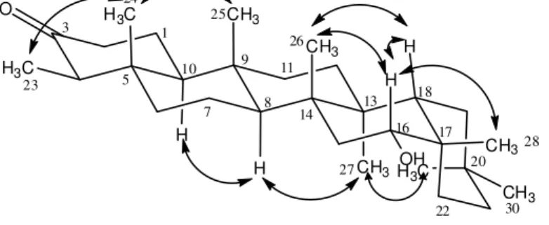
and H-30. Both signals have close chemical shifts values and, for this reason, it becomes difficult to distinguish these two methyl groups through the HMBC spectrum. However, they could be distinguished and the stereochemistry of 4 established from the NOESY spectrum, since it was possible to observe NOEs between H-16 axial and H-15 equatorial, H-18, H-26 and H-28. NOEs were also observed between H-23 and H-24; and between H-24 and H-25. This last signal presented NOE with the signal of H-26 and this one correlated with H-18. It was observed correlation between H-27 and H-29 which presented correlation with H-21 equatorial. The signal of H-27 was correlated with the signal of H-8, which presented correlation with the signal of H-10. Some correlations, observed in the NOESY spectrum of compound 4, are shown in Figure 2.
Figure 2. Some correlations observed in the NOESY spectrum of 16α-hydroxyfriedelin(4).
O
H3C
H3C CH3
CH3
CH3
CH3
H3C
The 1D NMR spectral data of 16β-hydroxyfriedelin [17] are compared with those of compound 4
and the complete 1D/2D NMR data for compounds 4 and 7 are presented in Table 1.
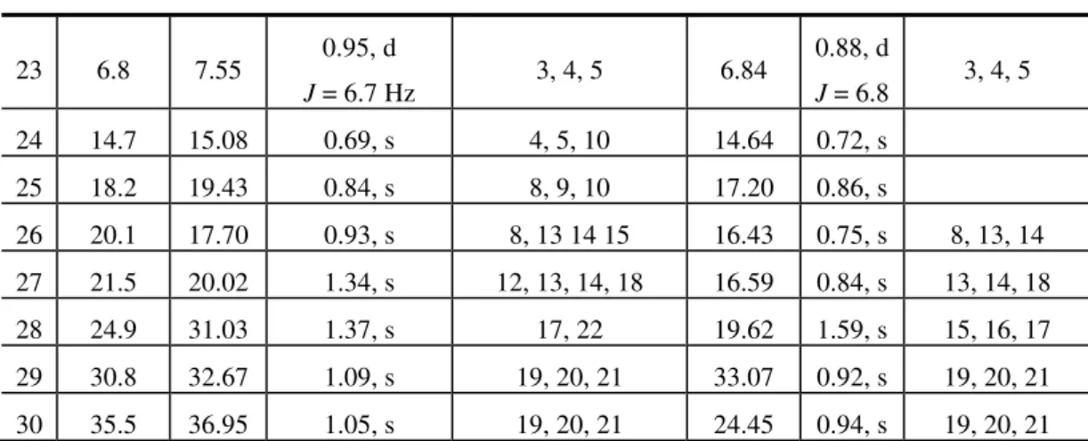
Table 1. 1H (400 MHz) and 13C (100 MHz) NMR spectral data of 16α -hydroxyfriedelin (4) (δ values, Py-d5) and 3-oxo-16-methylfriedel-16-ene (7) (δ
values, CDCl3) * (Literature data of 16 -hydroxyfriedelin) [17].
No δC (lit) * δC (4) δH (4) HMBC δC (7) δH (7) HMBC
1 22.3 22.77 1.62 ax
1.86 eq 22.32
1.68 ax 1.98 eq 2 41.6 41.90 2.34 eq
2.44 ax 41.55
2.39 ax 2.45 eq
3 212.5 212.12 - 1, 2, 4, 23 213.21 - 4, 23 4 58.3 58.28 2.20, m 23, 24, 5, 10 58.25 2.27, m
5 42.3 42.37 - 42.07 - 4, 10
6 41.4 41.54 1.54, m 41.09 1.23 eq 1.74 ax 7 18.6 18.95 1.64, m 18.23 1.39 eq 1.48 ax
8 53.5 50.82 1.51, m 12, 15, 25, 26 50.18 1.38, m 6, 10, 11, 25, 26
9 37.6 37.97 - 37.53 -
10 59.7 59.77 1.56, m 2, 4, 8 59.25 1.55, m 8 11 35.8 35.64 1.32, m 35.50 1.27 ax
1.51 eq 12 30.8 30.42 1.29 ax
1.40 eq 28.14 1.35, m
13 39.3 39.95 - 37.29 - 8, 12, 26, 27
14 40.1 40.25 - 37.65 -
15 44.4 40.25 1.66 ax, m
2.08 eq, m 43.07
1.52 eq 1.58 ax 16 75.6 75.70 4.25
J = 7.0; 10.4 Hz 14, 15, 17, 18, 22, 28 122.57 - 28
17 32.1 37.75 - 129.50 - 28
18 44.8 46.66 1.63, m 19, 27, 28 40.43 1.87, m 12, 19 19 35.8 34.28 1.35, m 37.69 1.03 ax
1.29 eq
20 28.0 28.62 - 30.00 -
21 32.1 34.97 1.62, m 38.33 1.19 eq 1.36 ax 22 36.0 27.76 1.97, m
2.05, m 24.60
Table 1. Cont.
23 6.8 7.55 0.95, d
J = 6.7 Hz 3, 4, 5 6.84
0.88, d
J = 6.8 3, 4, 5 24 14.7 15.08 0.69, s 4, 5, 10 14.64 0.72, s
25 18.2 19.43 0.84, s 8, 9, 10 17.20 0.86, s
26 20.1 17.70 0.93, s 8, 13 14 15 16.43 0.75, s 8, 13, 14 27 21.5 20.02 1.34, s 12, 13, 14, 18 16.59 0.84, s 13, 14, 18 28 24.9 31.03 1.37, s 17, 22 19.62 1.59, s 15, 16, 17 29 30.8 32.67 1.09, s 19, 20, 21 33.07 0.92, s 19, 20, 21 30 35.5 36.95 1.05, s 19, 20, 21 24.45 0.94, s 19, 20, 21
The 1H-NMR spectrum of 7 showed multiple signals in the region between δH 0.70 and δH 2.50. As
mentioned, a lack of signals at the region of the H-C-O hydrogen was observed. The 13C-NMR spectrum presented a signal at δC 213.21, which was assigned to a carbonyl group, and two
non-hydrogenated carbon signals at δC 122.57 and δC 129.50 that were attributed to olefinic carbons. All
chemical shifts of hydrogens and carbons of compound 7 were assigned through the HMBC spectra. The signal at δC 213.21 (C-3) correlated with the signals at δH 2.27 (H-4) and δH 0.88 (H-23). This last
one showed correlation with the signal at δC 42.07 (C-5), which presented correlation with δH 2.27
(H-4) and δH 1.55 (H-10). This last one correlated with the signal at δC 50.18 (C-8). The signal of C-8
correlated with the signal at δH 1.74 (H-6), 1.55 (H-10), 1.51 and δH 1.27 (H-11), and also with the two
methyl signals at δH 0.75 and 0.86, attributed to H-26 and H-25. The signal at δH 0.75 presented
correlation with the signal of C-13 (δC 37.29), then this signal could only be associated to H-26, and
consequently, the signal at δH 0.86 was attributed to H-25. The signal of C-13 (δC 37.29) correlated
with δH 1.38 (H-8), 1.35 (H-12) and δH 0.84 (H-27) and this last one was also correlated with δC 40.43
(C-18). The signal of C-18 correlated with the signals at δH 1.35 (H-12), 1.03 and δH 1.29 (H-19). Also
were observed correlations between the signals of olefinic carbon at δC 122.57 (C-16) and δC 129.50
(C-17) with the signal of methyl hydrogen at δH 1.59 attributed to H-28, because it is the only methyl
group able to correlate with carbons C-16 and C-17. The signal of H-28 presented yet a correlation with the signal at δC 43.07 (C-15).
Figure 3. Some correlations observed in NOESY spectrum of 3-oxo-16-methylfriedel-16-ene (7).
1 3
5
7 8
9
10 11
13 14
16 17
18 20
22 23
24
25
26
27
28
30
O
H3C
H3C CH3
CH3
CH3 H
H
H
CH3
CH3
The analysis of the NOESY spectrum permitted us to determine the stereochemistry of compound
7. It was possible to observe NOEs between H-30, H-18 and H-19 equatorial. These correlations indicated that the E ring has a chair conformation. NOEs were also observed between H-18 and H-26, between 25, 24 and 26, and also between 27 and 11 axial, 19 axial, 12 equatorial, H-15 axial and H-8. It was possible to observe correlations between H-23/H-24 and H-10/H-4, and this last one presented correlation with H-2 axial. The H-28 hydrogen presented a NOE with H-22 equatorial. Some correlations observed in the NOESY spectrum of compound 7 are shown in Figure 3. The complete 1D/2D NMR spectral data of 3-oxo-16-methylfriedel-16-ene (7) are presented in Table 1. The mass spectrum of 4 did not show the molecular ion at m/z 442, but rather showed peaks at m/z 411 (M-OH,-CH2) and m/z 273, confirming it to be a friedelane derivative [18]. On the other hand, the
mass spectrum of compound 7 showed the molecular ion at m/z 424, corroborating the molecular formula C30H48O.
By the data obtained through NMR and CG/MS was possible to confirm that the structure of PCTT
4 was modified by a process of dehydration accompanied by methyl rearrangement induced by the acidity CDCl3 that is normally used as NMR solvent, producing compound 7. To the best of our
knowledge, this is the first report of the Nemetkin rearrangement of a pentacyclic triterpene dissolved in CDCl3, inside an NMR tube, and also the isolation of compounds 1 to 6 from Salacia elliptica.
Experimental
General
Melting points (uncorrected) were determined on a Mettler FP 80 HT.The IR spectra were obtained on a Perkin Elmer, Spectrum One (SN 74759) spectrophotometer. Plates of silica gel G-60 were previously activated at 100°C/30min, and developed with an acidic soln. of vanillin in perchloric acid [19] after the TLC processes. Column chromatography (CC) processes were developed using silica gel (Merck, 230-400 mesh). GC-MS analysis was carried out on a Hewlett Packard HP5890 instrument, equipped with a HP 7673 injector, HP-1 (50 m x 0.25 mm i.d. x 0.2 mm film) column and helium as mobile phase. Operating conditions: injector temperature at 300 °C, 2 μL of sample solution (10
μg/100 μL); splitless of 30s followed by split 1:40 (30 psi stream pressure). The initial oven temperature was 200 ºC/3min, followed by 10°C/min until 300ºC and with 40 min holding time. Interface: quadrupole mass spectrometer model HP 5971, electron impact ionization, 70 eV potential.
NMR spectra
NMR spectra were recorded on a Bruker DRX400-AVANCE spectrometer operating at 400 and 100 MHz at 27 0C equipped with a direct detection 5 mm 1H/13C dual probe and a 5 mm inverse probe with z-gradient coil. The solvent was CDCl3 or pyridine-D5. Compound 4 (about 10 mg) was dissolved
in 0.7 mL of CDCl3 or pyridine-D5, and transferred to a 5 mm o.d. tube. The chemical shifts are
[nJ (C, H), n = 2 and 3], 1H homonuclear correlation spectroscopy (COSY) and homonuclear 2D-NOESY (mixing time = 441 ms) experiment were used to confirm the assignments of all carbons and hydrogens of the compounds.
Plant Material and Compound Isolation
Leaves and branches of Salacia elliptica were collected in the Mata Samuel de Paula, Nova Lima region, Minas Gerais, Brazil, in August of 2005. They were separated, dried at room temperature (r.t.) and powdered in a mill. The branches (1158 g) were successively submitted to exhaustive extraction at r.t. with solvents of different polarity. Each solvent was removed under vacuum furnishing the hexane (6.11 g), ethyl acetate (8.16 g) and finally ethanol (144.5 g) extracts.
During the hexane removal, the formation of a white solid was observed. The solid material (0.98 g) was separated by filtration. This material was fractionated by silica gel CC eluted with dichloromethane, ethyl acetate and ethanol pure or in mixture of gradient polarity, obtaining 69 fractions. Fraction 4-5 was analyzed by TLC and GC together with PCTT standards and identified as friedelin (1). By these comparative analyses also was possible to identify fraction 6-7 as a mixture of 1
and 3β-friedelinol (2). Fraction 10-15 gave a white solid material (mp. 279.5-281.8 °C). Its 1H- and
13
C-NMR data were compared with the literature data [17] and identified as 28-hydroxyfriedelin (canophyllol, 3). Fraction 18 (200 mg) was submitted to TLC, which showed the presence of only three components. It was then separated by CC using CHCl3, ethyl acetate and ethanol as eluents
furnishing 16α-dihydroxyfriedelin (4, 18 mg, m.p. 238.7-244.9 oC) and compound 5 (102.5 mg, m.p. 270.6-278.7 °C), identified as 30-hydroxyfriedelin. After solvent evaporation, fraction 46-47 presented as a white solid (20.2 mg, m.p. 170-172.5 0C). By comparison of its NMR spectral data with the literature [5] this solid material was identified as 16α,28-dihydroxyfriedelin (6). For the NMR analysis, a sample of 4 (10 mg) was dissolved in CDCl3 (0.7 mL) and placed within the NMR tube.
After the acquisition data of 1D NMR, the solution was kept inside the tube during a week, and then submitted to 2D NMR experiments. After CDCl3 evaporation the solid was recovered and named as 7
(melting point 240-242 oC).
Acknowledgements
The authors thank the Fundação de Amparo a Pesquisa de Minas Gerais (FAPEMIG) for financial support (Process CEX-1004/05) and the Anglogold Ashanti Ltd for the permission to collect Salacia elliptica in the Mata Samuel de Paula, Nova Lima, MG, Brazil.
References
1. Wolf, B.M.; Weisbrode, S.E. Safety evaluation of an extract from Salacia oblonga. Food Chem. Toxicol. 2003, 4, 867-870.
the ayurvedic traditional medicine Salacia reticulata in Sri Lanka and India. Tetrahedron Lett.
1997, 38, 8367-8370.
3. Jeller, A.H.; Silva, D.H.S.; Lião. L.M.; Bolzani, V.S.; Furlan; M. Antioxidant phenolic and quinonemethide triterpenes from Cheiloclinium cognatum. Phytochemistry 2004, 65, 1977-1982. 4. Deepa, M.A.; Bai, V.N.; Antibacterial activity of Salacia beddomei. Fitoterapia 2004, 75,
589-591.
5. Kuo, Y.H.; Kuo, L.M.Y. Antitumour and anti-AIDS triterpenes from Celastrus hindsii. Phytochemistry 1997, 44, 1275-1281.
6. Oliveira, M. L. G.; Duarte, L. P.; Silva, G. D. F.; Vieira Filho, S.A.; Knupp, V.F.; Alves; F.G.P. 3-Oxo-12α-hydroxyfriedelane from Maytenus gonoclada: structure elucidation by (1)H and (13)C chemical shifts assignments and 2D-NMR spectroscopy. Magn. Reson. Chem. 2007, 45, 895-898. 7. Oliveira, D.M.; Silva, G.D.F.; Duarte, L.P.; Vieira Filho, S.A. Chemical constituents isolated from roots of Maytenus acanthophylla Reissek (Celastraceae). Bioch. Syst. Ecol., 2006, 34, 661-665.
8. Rizvi, S.H.; Shoeb, A.; Kapil, R.S.; Popli, S.P. Antidesmanol - New Pentacyclic Triterpenoid From Antidesma menasu Miq Ex Tul. Experientia 1980, 36, 146-147.
9. lves, J.S.; Castro, J.C.M.; Freire, M.O.; da-Cunha; E.V.M.; Barbosa-Filho, J.M.; Silva, M.S. Complete assignment of the 1H and 13C NMR spectra of four triterpenes of the ursane, artane, lupane and friedelane groups. Magn. Reson.Chem. 2000, 38, 201-206.
10. Vieira, H.S.; Takahashi, J.A.; Gunatilaka, A.A.L.; Boaventura, M.A.D. 1H and 13C NMR signal assignments of a novel Baeyer-Villiger originated diterpene lactone. Magn. Reson. Chem. 2006, 44, 146-150.
11. Fernandez, A.H.; Cerero, S.M.; Jimenez, F.M. About the Timing of Wagner-Meerwein and Nametkin Rearrangements, 6,Hydride Shift, Proton Elimination and Cation Trapping in 2-Norbornyl Carbocations Tetrahedron 1998, 54, 4607-4614.
12. Starling, S.M.; Vonwiller, S.C.; Reek, J.N.H. Effect of Ortho Substituents on the Direction of 1,2-Migrations in the Rearrangement of 2-exo-Arylfenchyl Alcohols. J. Org. Chem. 1998, 63, 2262-2272.
13. Kikuchi, T.; Niwa, M.; Yokoi, T.; Kadota, S. Studies on the Neutral Constituents of Pachysandra terminalis SIEB. et ZUCC. VIII. Methyl Migration in the Dehydration Reaction of Pachysonol and Pachysandiol-B Derivatives. Chem. Pharm. Bull. 1981, 29, 1819-1826.
14. Hao; X.; Xuehui, L.; Yuxin, C.; Min, Z.; Jiaxiang, S. NMR study on the reaction of rearrangement of 17β-hydroxy-7α-methyl-19-nor-17-α-pregn-5(10)-en-20-yn-3-one. Bopuxue Zazhi 2000, 17, 17-22.
15. Lebedeva, T.L.; Vointseva, I.I.; Gilman, L.M.; Petrovskii; P.V.; Larina, T.A.; Topchiev, A.V. Solvent-induced allylic rearrangements in poly(trichlorobutadiene) chains. Russ. Chem.Bull.
1997, 46, 732-738.
16. Mitsuo, K.; Zhang, L.C.; Kabuto, C.; Sakurai, H. Synthesis and Reactions of Neutral Hypercoordinate Allylsilicon Compounds Having a Tropolonato Ligand Organometallics 1996, 15, 5335-5341.
18. Courtney, J.L.; Shannon, J.S. Studies in mass spectrometry triterpenoids: structure assignment to some friedelane derivatives. Tetrahedron Lett. 1963, 1, 13-20.
19. Matos, F.J.A. Introdução a Fitoquímica Experimental. Editora UFC: Fortaleza, Brazil, 1988; p. 126
Sample Availability: Samples of compounds 1, 2, 3 and 5 are available from the authors.