Viability of autogenous bone grafts obtained by using bone
collectors: histological and microbiological study
Viabilidade dos enxertos autógenos obtidos com a utilização de
coletores para osso: estudo histológico e microbiológico
Alberto Blay* Samy Tunchel*
Wilson Roberto Sendyk**
ABSTRACT:The use of autogenous bone grafts is considered to be the best choice for reconstructive surgery. In the pe-riodontal literature, the utilization of osseous coagulum was suggested by the end of the sixties. The purpose of this study is to consider the use of bone collectors (bone traps) as an alternative method for obtaining material to fill small bone imperfections, such as fenestrations and dehiscences. Thirty samples were obtained from bone drilling during fixture installation in patients (13 men and 17 women, with an average age of 54 years) requiring treatment at the De-partment of Periodontology and Implant Dentistry, University of Santo Amaro. These samples were fixed in 10% neu-tral formaldehyde for 24 hours and subjected to histological preparation, in order to evaluate the presence of viable os-teoblasts. In addition, the material was placed in a fluid thioglycolate medium and incubated for 24 hours at 36 ± 1°C in aerobiosis and anaerobiosis. Bacterial growth evaluation was made by using six different culture media (MacCon-key agar, blood agar base, mannitol salt agar, Anaerokit LTD medium, Anaerokit LTD – bile medium, Anaerinsol). The results show that, if proper care is taken to prevent saliva contamination during the surgical procedure, this method of collecting autogenous bone may be useful in situations where small amounts of bone are required.
DESCRIPTORS:Dental implantation; Bone transplantation; Bone regeneration.
RESUMO:A utilização de enxertos autógenos é considerada a melhor opção nos tratamentos cirúrgicos de reconstru-ção óssea. Na literatura periodontal, a utilizareconstru-ção de coágulo ósseo foi sugerida no final da década de 60. O objetivo des-te estudo é considerar a utilização de coletores para osso como um método aldes-ternativo de se obdes-ter osso autógeno para preenchimento de defeitos ósseos como fenestrações e deiscências. Trinta amostras foram obtidas no processo de per-furação do tecido ósseo, durante a instalação de implantes em pacientes (13 homens e 17 mulheres, com média etária de 54 anos) que foram submetidos a tratamento na Disciplina de Periodontia e Implantodontia da Universidade de Santo Amaro. Essas amostras foram fixadas em solução de formol neutro a 10% por 24 horas para serem analisadas histologicamente com o intuito de avaliar a presença de osteoblastos viáveis. Além das amostras fixadas, também fo-ram obtidos espécimens que fofo-ram incubados em aerobiose e em anaerobiose, em meio de tioglicolato por 24 h a 36 ± 1°C. A avaliação do crescimento bacteriano foi feita através de seis meios seletivos de cultura (ágar MacConkey, ágar-sangue, ágar manitol, meio Anaerokit LTD, meio Anaerokit LTD – bile e Anaerinsol). Os resultados mostraram que, se forem tomados certos cuidados para prevenir a contaminação com saliva durante o procedimento cirúrgico, este método de coletar osso autógeno pode ser útil em situações em que pequenas quantidades de osso são necessá-rias.
DESCRITORES:Implante dentário; Transplante ósseo; Regeneração óssea.
INTRODUCTION
The loss of one or more dental elements not only harms the patient psychologically, but also causes an atrophic condition of the tooth supporting tis-sues. The highest rate of alveolar bone reabsorpti-on occurs in the period between 6 mreabsorpti-onths and 2 years after a tooth extraction7
, and this fact is
ex-*DDS, Assistant Professor; **PhD, Chairman – Department of Implantology and Periodontology, School of Dentistry, University of Santo Amaro.
tremely important, since it will affect the position and the angle of an implant1,16
.
This may result in situations in which the im-plant, to be placed in a favorable functional and aesthetical position, contains parts of its surface clear of bone coverage, originating bone imperfecti-ons such as dehiscences and fenestratiimperfecti-ons3,4,6,7,10
ex-posure of parts of the implants, may impair bone integration within a short period, as well as cause related infection problems18
.
Minimization of these problems requires pre-operatory diagnostics in order to evaluate the qua-lity and quantity of the bone host. Intra-oral bone mapping as well as X-ray techniques are manda-tory for evaluating the conditions of the bone tis-sue that will receive the implants8,13,21
. However, even after careful preliminary planning, small bone imperfections might occur, leading us to use some guided bone regeneration technique.
Considering autogenous bone grafts as the best choice for reconstruction surgery procedures15
, we adopted a technique based on the use of bone col-lectors for obtaining autogenous bone material, which allowed us to fill small bone defects, such as fenestrations and dehiscences, without having to involve a second (intra-oral or extra-oral) surgical area for obtaining autogenous bone.
The purpose of this study is to evaluate the mi-crobiological and histological viability of a bone tis-sue which, to date, has been disregarded in bone-integrated implant surgeries.
MATERIALS AND METHODS
Clinical procedure
Patient selection
Thirty patients (13 men and 17 women, aged between 38 and 70, with an average age of 54 ye-ars), in good general health condition, were inclu-ded in this study. They were all patients of the De-partment of Implantology and Periodontology, University of Santo Amaro, Brazil. Patient selecti-on criteria for this study were established so as to include patients with loss of one or more dental
elements and with moderate alveolar atrophy. The-se characteristics were diagnoThe-sed in pre-operatory X-ray examinations, such as panoramic radio-graphy and computed tomoradio-graphy scans. An ethi-cal committee evaluated and accepted the guideli-nes of the study. Before being included in the research all patients gave their informed consent.
Surgical procedure
All surgical procedures were performed under local anesthesia (with mepivacaine with 2% adre-naline - Scandicaine®
, Specialites Septodont, Sa-int-Maur-des-Fosses, France). Patients received antibiotic prophylaxis during 7 days (amoxine, Smithkline Beecham Brasil, Rio de Janeiro, Brazil, 1 g, 2 hours before surgery and 500 mg every 8 hours after surgery), and anti-inflammatory medi-cation (sodium diclofenac, Novartis Biociências, São Paulo, Brazil) 50 mg every 12 hours, during 4 days.
Implants were installed following the protocol proposed by Branemark5
and Schulte19
. Samples were obtained during the process of alveolar bone drilling, using two types of special bone collectors that can be described as autoclavable intermediate pieces with a collection chamber where the bone filters are located; these filters can be autoclavable or disposable (Osseous Coagulum Trap - Quality Aspirators; Duncaville, Texas, USA/Frios Bone Collector - Friadent, Mannheim, Germany). One of the extremities of this intermediate piece is con-nected to a high power suction pump and in the ot-her extremity a surgical aspirator is inserted (Figu-res 1 and 2).
Precautions were taken in order to avoid conta-mination by saliva, which consisted in the manda-tory utilization of two aspiration tips, one of them
FIGURE 1 -Bone collectors used in the research. FIGURE 2 - Parts of the bone collectors before being
only for saliva aspiration and the other for the col-lector, the latter being kept at a distance of 5 to 10 millimeters from the area being prepared with the drills.
At 10-minute intervals, the collector’s filter (Fi-gure 3) was removed in order to avoid excessive dehydration of bone particles due to the action of the aspiration unit.
The collected tissue was placed in a glass vial containing sterile saline solution (Biossintética, São Paulo, Brazil) at room temperature and cove-red with a cap in order to minimize the risk of con-tamination. After fixture installation, the excess of saline solution was removed from the vial and the collected material was used to fill the bone defects (Figures 4 and 5).
At that moment, a small sample of the collected material was sent to microbiological and histologi-cal analysis. The flaps were closed with Donath
type surgical thread and simple individual sutu-res.
Mouth rinsing with a 0.12% solution of chlore-xidine digluconate, twice a day, during two weeks after the surgery was prescribed.
Sutures were removed 7 days after surgery, and this patient was monitored at monthly appoint-ments up to the moment of implant reopening sur-gery.
Second surgical stage
Reopening was performed 6 to 8 months after installation of the implants. Periapical X-rays were made at this treatment stage (Figure 6).
The areas were reopened by means of full-thick-ness flaps (Figure 7).
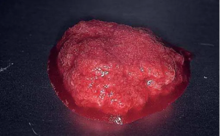
Biopsies were performed in the dehiscence area to provide small bone fragments for histological analysis. All cases showed bone growth over the
FIGURE 3 -Bone particles obtained from the filters. FIGURE 4 -Buccal dehicensce showing exposure of part
of the implant at the time of installation.
FIGURE 5 -The defect filled with autogenous bone from
the collector.
FIGURE 6
cover screw, so we could take the biopsies without jeopardizing the bone around the implant.
Microbiological analysis
The samples were transported in a thioglycolate medium and incubated for 24 hours at a tempera-ture of 36±1ºC under aerobic and anaerobic con-ditions, in order to evaluate the presence of bacte-rial contamination. Evaluation of the colonies of the bacteria found was made using six different se-lective culture media (MacConkey agar, blood agar, mannitol agar, Anaerokit LTD medium, Ana-erokit LTD - bile medium, Anaerinsol – Probac, São Paulo, Brazil).
Histological analysis
Samples at fixture installation
The samples collected at the time of implant installation in order to be processed histologically
were fixed during 24 hours in a neutral formal-dehyde solution at 10%. Subsequently, they were decalcified in a vial containing 10% EDTA during 4 weeks. The EDTA solution was changed every week in order to remove the calcium from the bone fragments through chelation.
After decalcification, the samples were embed-ded in paraffin, sliced with a microtome and stai-ned with fuchsine, after which they were ready for microscopical analysis.
RESULTS
Histological analysis
Samples at fixture installation
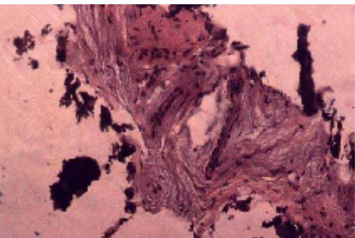
Histological evaluation of the samples with an optical microscope showed that, even in a particu-late state, the bone structure was well preserved (Figure 8), containing large numbers of osteocytes within the calcified matrix and suggesting that the viability of the bone tissue was maintained.
Samples at second stage surgery
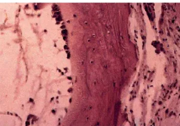
All samples showed complete bone formation over the dehiscence defect. Samples at the second stage period showed a histologic bone structure si-milar to that of secondary bone. The Haversian system, typical osteocytes and a sharply defined layer of osteoblasts covering the mature bone frag-ment can be seen in Figure 9.
Some of the cells are surrounded by a newly for-med bone matrix, indicating early stages of oste-ocyte maturation.
A basophilic line suggesting apositional growth, as well as the presence of osteo-progenitor or
oste-FIGURE 7 -Reopening at second stage surgery. FIGURE 8 -Histological appearance of the bone obtained
from the collector (acid fuchsin, 160 X)
FIGURE 9 -At 6 months, haversian remodeling started
ogenic cells (non-differentiated mesenchymal cells) can be observed in Figure 10.
Microbiological analysis
Under aerobic and anaerobic conditions, the presence of bacteria was clearly visible in the 30 samples submitted to microbiological analysis, when clouding was observed in the vials contai-ning the samples and the transport medium thi-oglycolate, after an incubation period of 24 hours at a temperature of 36±1ºC.
The bacteria found in the samples were ascerta-ined by including the samples, after an incubation period, in a thioglycolate medium, in six different selective culture media. Of these, three were selec-tive media under aerobic conditions, and the other three were selective media under anaerobic condi-tions.
The blood agar selective medium, being an enri-ched medium, determined the presence ofaandb
hemolytic streptococci, under aerobic conditions. The mannitol agar selective medium determined the presence of S. aureusandStaphylococcus ne-gative coagulate under aerobic conditions.
The MacConkey agar selective medium determi-ned the presence of Pseudomonaespp. and Citro-bacterspp. The inclusion of these bacilli in another selective medium called IAL (Rugai medium, Pro-bac, São Paulo, Brazil, modified by Pessoa and Sil-va19) determined which of them fermented lactose
and which of them did not. The identified lactose fermenting bacilli wereEnterobacterand Citrobac-ter.
The selective media used for anaerobic conditi-ons were Anaerokit and Anaerinsol, which
deter-mines the presence of Gram-negative bacilli, which produce black pigment. No other types of bacteria were found in this analysis.
In terms of genus, the percentages of the morp-hological types mentioned above are shown in Ta-ble 1.
It is important to point out that the microbiolo-gical evaluation was qualitative and we did not count the number of bacterial colonies. It is known that most of the bacteria found in the samples are usually present in the normal microbiologic envi-ronment of the oral and/or oral-pharyngeal cavity.
DISCUSSION
Atrophy of the alveolar bone always presents difficulties for carrying out procedures which in-volve the placement of implants, and this problem has prompted the development of a number of techniques in order to obtain bone in sufficient quantity and quality. Utilization of autogenous bone in regeneration procedures, being a techni-que of proven success, is motivating a large num-ber of researchers to continue developing procedu-res destined to overcome situations of bone atrophy2,11.
Utilization of autogenous bone is justified by the fact that this material has a high osteogenic potential in comparison with other types of materi-als. In addition, it has cells with bone induction ca-pacity and does not produce immunological reacti-on17
.
For these reasons, we decided to investigate the method of obtaining autogenous bone tissue by means of collectors, a technique which is being used empirically by a large number of professio-nals and which, although apparently simple, re-quires a number of precautions in order to yield satisfactory results.
The use of bone collectors must be considered as an extremely conservative technique, since it
TABLE 1 -Percentage of bacteria found in the 30
sam-ples.
Genera Percentage
Streptococcusalpha hemolytic 66.6
Staphylococcus aureus 43.3
Streptococcusbeta hemolytic 36.6
Staphylococcusnegative coagulate 26.6
Pseudomonas spp. 26.6
Citrobacter spp. 16.6
FIGURE 10 - Morphology of the bone after the healing
eliminates the need of obtaining autogenous bone material from a second surgical area which always involves complex procedures. Furthermore, the bone material obtained by means of collectors is already in a particulate state, thus reducing opera-tion time and the probability of contaminaopera-tion, since in this case there is no need to use bone crushers.
In our research project, all cases were carried out without membranes, and we noted that a bone graft made with a collector was efficient without having to use barriers. We think that in this way we have eliminated another risk factor, which is the use of membranes12,14,22
.
As to the presence of microorganisms found du-ring the collection process, we would like to point out that we found no Gram-negative anaerobic mi-croorganisms, which are present in several forms of periodontal diseases and are known to produce enzymes capable of causing bone reabsorption. Furthermore, we must emphasize that the bacteria found in the samples did not show sufficient pat-hogenicity or virulence to harm the graft or the im-plant in any way. This is probably due to the syste-mic action of the broad spectrum antibiotics administrated to patients, or to the low virulence of the microorganisms found.
Therefore, we emphasize that it is mandatory to use two surgical aspirators, one of them only for saliva and another directly applied to the drilling site, collecting only cut bone and saline solution,
thus reducing the risk of excessive bacterial conta-mination. We chose to close the suction unit of the bone collector at the moment we finished drilling, so as to avoid an excessive dehydration of the col-lected bone material, which could impair the viabi-lity of the bone inducing cells. At that moment the bone was removed from the collector and placed into a saline solution at room temperature.
From the results obtained we could conclude that, clinically as well as hystologically, the collec-tion method was capable of preserving cells with bone induction capacity. Another positive factor is the utilization of autogenous bone tissue of mem-branous and not of endocondral origin, a material known to be more efficient because it has lower re-absorption levels23
.
CONCLUSIONS
The surgery technique demonstrated here for obtaining particulate intra-oral autogenous bone material proved to be simple, efficient and safe.
During the bone collection procedure it is extre-mely important to avoid saliva contamination, which could cause failure of the graft and increase the risk of failure of the implant and of infection.
In spite of all precautions taken to prevent the presence of bacteria, this presence has been detec-ted in all samples; the technique must therefore be improved in an attempt to eliminate any risk of contamination.
REFERENCES
1. Adell R, Lekholm U, Branemark PI. A 15-year-old study of osseointegrated implants in the treatment of the edentu-lous jaw. Int J Oral Surg 1981;6:387-96.
2. Adell R, Lekholm U, Grondahl K, Branemark PI, Lindstrom J, Jacobson M. Reconstruction of severely resorbed eden-tulous maxillae using osseointegrated fixtures in immedia-te autogenous bone grafts. Int J Oral Maxillofac Implants 1990;5:233-46.
3. Albrektsson T. Osseointegrated titanium implants. Requi-rements for ensuring a long-lasting, direct bone anchorage in man. Acta Orthop Scand 1981;52:155-70.
4. Becker W, Becker BE. Guided tissue regeneration for im-plants placed into extraction sockets and for implant de-hiscences: surgical techniques and case report. Int J Perio-dontics Restorative Dent 1990;10:376-91.
5. Branemark PI, Zarb GA, Albrektsson T. Tissue-integrated prostheses: osseointegration in clinical dentistry. Chicago: Quintessence; 1985.
6. Branemark PI. Intra-osseous anchorage of dental prosthe-ses. I. Experimental studies. Scand J Plast Reconstr Surg 1969;3:81-100.
7. Buser D, Dula K, Belser U, Hirt HP, Berthold H. Localized ridge augmentation using guided bone regeneration. I. Surgical procedure in the maxilla. Int J Periodontics Res-torative Dent 1993;13:29-45.
8. Buser D, Schoroeder A, Sutter F, Lang NP. The new con-cept of ITI hollow-cylinder and hollow-screw implants. Part 2: Clinical aspects, indications, and early clinical re-sults. Int J Oral Maxillofac Implants 1988;3:173-81. 9. Carlsson GE, Persson G. Morphologic changes of the
man-dible after extraction and wearing of dentures. A longitudi-nal, clinical and X-ray cephalometric study covering 5 ye-ars. Odontol Revy 1967;18:27-54.
10. Dahlin C, Sennerby L, Kekholm U, Linde A, Nyman S. Ge-neration of new bone around titanium implants using a membrane technique: an experimental study in rabbits. Int J Oral Maxillofac Implants 1989;4:19-25.
12. Jovanovic SA, Schenk RK, Orsini M, Kenney B. Supra-crestal bone formation around dental implants: an experi-mental dog study. Int J Oral Maxillofac Implants 1995;10:23-31.
13. Kassebaum DK, Nummikoski PV, Triplett RG, Langlais RP. Cross-sectional radiography for implant site assessment. Oral Surg Oral Med Oral Pathol 1990;70:674-82.
14. Leghissa GC, Botticelli AR. Resistance to bacterial aggres-sion involving exposed nonresorbable membranes in the oral cavity. Int J Oral Maxillofac Implants 1996;11:210-5. 15. Lundgren S, Moy P, Johanson C, Nilsson H. Augmentation of the maxillary sinus floor with particulated mandible: a histologic and histomorphometric study. Int J Oral Maxil-lofac Implants 1996;11:760-6.
16. Mecall RA, Rosenfeld AL. Influence of residual ridge resorp-tion patterns on implant fixture placement and tooth posi-tion. Int J Periodontics Restorative Dent 1991;11:8-23. 17. Moy PK, Lundgren S, Holmes RE. Maxillary sinus
augmen-tation: histomorphometric analysis of graft materials for maxillary sinus floor augmentation. J Oral Maxillofac Surg 1993;51:857-62.
18. Newman M, Flemmic T. Periodontal consideration of im-plants and implant associated microbiota. J Dent Educat 1988;52:737-44.
18. Pessoa GV, da Silva EA. A new medium for the rapid pre-sumptive identification of enterobacteriae, aeromonas and vibrios. Ann Microbiol 1974;125A(3):341-7.
20. Schulte W, Heimke G. Das tübinger sofort-implantat. Qu-intessenz 1976;27:17-23.
21. Schwarz MS, Rothman SL, Rhodes ML, Chafetz N. Compu-ted tomography: Part 1. Preoperative assessment of the mandible for endosseous implant surgery. Int J Oral Maxil-lofac Implants 1987;2:137-41.
22. Simion M, Scarano A, Gionso L, Piattelli A. Guided bone re-generation using resorbable and nonresorbable membra-nes: a comparative histologic study in humans. Int J Oral Maxillofac Implants 1995;10:23-30.
23. Sindet-Pedersen S, Enemark H. Reconstrution of alveolar clefts with mandibular or iliac crest bone grafts: a compa-rative study. J Oral Maxillofac Surg 1990;48:554-8.