Short Report
Printed in Brazil - ©2017 Sociedade Brasileira de Química0103 - 5053 $6.00+0.00*e-mail: [email protected]
Conyza canadensis
: Green Extraction Method of Bioactive Compounds and
Evaluation of Their Antifungal Activity
Rafael S. Porto,a Susanne Ratha and Sonia C. N. Queiroz*,b
aDepartamento de Química Analítica, Instituto de Química, Universidade de Campinas, CP 6154,
13084-971 Campinas-SP, Brazil
bEMBRAPA Meio Ambiente, CP 69, 13820-000 Jaguariúna-SP, Brazil
Synthetic fungicides are a common treatment for post-harvest diseases in fruits, although demand is increasing for alternatives such as biopesticides. The aim of this work was to investigate the presence of naturally occurring antifungal compounds in Brazilian specimens of the weed
Conyza canadensis. Two compounds [(4Z)-lachnophyllum lactone and (4Z,8Z)-matricaria lactone] were isolated and their antifungal activities were evaluated against eight postharvest disease fungi.
Aspergillus niger, Cladosporium sp., and Penicillium digitatum proved susceptible to the treatment (minimum inhibitory concentrations varying from 32 to 64 µg mL-1). These fungi are common
postharvest pathogens of fruits and vegetables, causing rots and secreting mycotoxins. A green and sustainable extraction method employing pressurized hot water was developed and optimized (100 °C, 4 × 1 min cycles). Yields of 1.46 and 0.24 mg g-1 were obtained for (4Z)-lachnophyllum
lactone and (4Z,8Z)-matricaria lactone, respectively. The extract could be applied directly to fruits and vegetables in postharvest treatments.
Keywords: Conyza canadensis, acetylene, horseweed, antifungal, PHWE
Introduction
Postharvest diseases are the main factors responsible for fruit losses along the production chain.1,2 These diseases
are usually caused by fungal pathogens that are commonly controlled by the application of synthetic fungicides.3
However, natural products, classified as biopesticides by the U.S. Environmental Protection Agency,4 are emerging
as a promising alternative for crop protection.5 This is due
to increasing concerns regarding the environmental impacts of synthetic pesticides, as well as stricter controls on their use and the appearance of fungicide-resistant strains.1,6
Given the importance of developing new biopesticides and investigating their spectra of action, a recent survey showed promising results with the allelopathic plant Conyza canadensis (L.) Cronquist, which belongs to the Asteraceae family and is popularly known as horseweed.7
This plant is a potential source of bioactive compounds8
and several studies have reported the fungitoxic effects of its essential oils,9-11 although the compounds
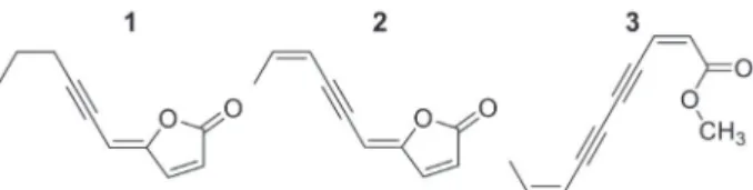
responsible for such activity were not identified. Queiroz et al.7 found three fungitoxic compounds in
dichloromethane extracts of C. canadensis specimens collected in the USA: (4Z)-lachnophyllum lactone (1), (4Z,8Z)-matricaria lactone (2), and (2Z,8Z)-matricaria acid methyl ester (3), whose chemical structures are shown in Figure 1. Compounds 1 and 2 showed antifungal activity against three phytopathogenic fungi: Colletotrichum acutatum, Colletotrichum fragariae and Colletotrichum gloeosporioides. The activity of compound 3 against these fungi was reported previously.12
The aim of this study was to investigate the presence of compounds 1, 2 and 3 in Brazilian specimens of C. canadensis and to determine their antifungal activity against eight common postharvest disease fungi. Additionally, a new extraction method was evaluated as an alternative to solvent extraction with dichloromethane. This green procedure employed pressurized hot water
extraction (PHWE) and provided crude aqueous extracts that could be used directly in postharvest treatments.
Experimental
Instrumentation
Flash chromatography was performed using an Isolera Four system (Biotage, Uppsala, Sweden) equipped with a SNAP cartridge (KP-Sil 100 g, 39 × 157 mm, 45-50 µm) and a UV detection system (254 and 280 nm). 1H and 13C nuclear magnetic resonance (NMR)
spectra, as well as 1H-13C heteronuclear single quantum
coherence (HSQC) spectra, were recorded using either an AVANCE 400 MHz spectrometer or an AVANCE 500 MHz spectrometer (Bruker, Rheinstetten, Germany). The fractions and the purified compounds were analyzed by GC-MS using a 7890A gas chromatograph (Agilent, Santa Clara, CA) coupled to a 5975C inert XL MSD single quadrupole detector operated with 70 eV electron ionization. An Agilent HP-5 ms capillary column (30 m × 0.25 mm, 0.25 µm) was used. The identities of the compounds were confirmed by analysis using a GC-2010 Plus gas chromatograph (Shimadzu, Kyoto, Japan) coupled to a TQ8040a triple quadrupole detector (70 eV electron ionization) and equipped with a Restek Rtx-5MS capillary column (30 m × 0.25 mm, 0.25 µm). Pressurized hot water extractions were performed using a Dionex ASE 350 accelerated solvent extractor (Thermo Scientific, Waltham, MA) with 34 mL stainless steel sample cells and glass fiber thimbles. Quantitation of bioactive compounds in the extracts was performed by gas chromatography with flame ionization detector (GC-FID) using a 7890A gas chromatograph (Agilent, Santa Clara, CA) equipped with an HP-5 capillary column (30 m × 0.32 mm, 0.25 µm). Details of the chromatography and NMR operational conditions are provided in the Supplementary Information section.
Plant material
The aerial parts of C. canadensis were collected in Jaguariúna, São Paulo, Brazil (22º43’13’’S and 46º01’10’’W) in August 2013. A voucher specimen was deposited under the number 179015 at the University of Campinas Herbarium (São Paulo, Brazil).
Isolation of bioactive compounds
A 250 g sample of fresh aerial parts of C. canadensis was air-dried in a fume hood at 40 °C for one week. After
grinding in a cutting mill, the material was submitted to maceration extraction with 500 mL of dichloromethane over three days, with solvent renewal each day, yielding 13.08 g of raw extract.
The dichloromethane extract was dissolved in methanol:water (90:10, v/v) and subjected to partitioning with 200 mL of hexane (3 times). Water was added to the remaining methanolic fraction to achieve 70:30 (v/v) methanol:water, followed by partitioning with 200 mL of chloroform (3 times). The chloroform fractions were combined and dried over magnesium sulfate, and the solvent was evaporated at 40 °C using a rotary evaporator. This procedure yielded 3.31 g of chloroform extractable.
The chloroform extract was separated by flash chromatography. Based on gas chromatography-mass spectrometry (GC-MS) analysis, six similar fractions were combined, yielding 338 mg of compound 1. In the same way, six other fractions were combined, yielding 53 mg of compound 2.
Isolates
(4Z)-Lachnophyllum lactone (1)
Brownish oil (338 mg); molecular formula C10H10O2;
MS/MS (EI, 70 eV) m/z (%) 162.0 [M+] (precursor
ion), 147.0 (23) [M − CH3]+, 133.0 (25) [M − C2H5]+,
119.1 (31) [M − C3H7]+, 105.1 (14) [M − C2H5 − CO]+,
91.1 (47) [M − C3H7 − CO]+, 82.0 (100) [C4H2O2]+. 1H and 13C NMR data (Supplementary Information section) were
in agreement with published values,13 except for the
attributions of C-8 and C-9, as discussed.
(4Z,8Z)-Matricaria lactone (2)
Brownish oil (53 mg); molecular formula: C10H8O2;
MS/MS (EI, 70 eV) m/z (%): 160.1 [M+] (precursor ion),
145.0 (7) [M − CH3]+, 131.0 (100) [M − C2H5]+, 103.0 (37)
[M − C2H5 − CO]+, 82.0 (18) [C4H2O2]+, 78.1 (58)
[M − C4H2O2]+. 1H and 13C NMR data (Supplementary
Information section) were in agreement with published values.14,15
The purities of compounds 1 and 2 were confirmed by GC-MS, using area normalization, giving purities exceeding 99% (m/m) for both compounds. The isolated and purified compounds were used as standards for the quantitation.
Standard solutions
a refrigerator at 4 °C for a maximum of 30 days. Working standard solutions were prepared daily by dilutions of the stock solutions with ethyl acetate.
Optimization of PHWE
The PHWE procedure was optimized considering the following parameters: temperature (70, 90, 100, 110 and 130 °C), time per cycle (1, 5, 10, 20 and 30 min), and number of cycles (up to four consecutive cycles). Each parameter was evaluated separately, in a univariate approach, using extractions of 5 g of ground plant material (aerial parts). Each extraction was carried out in duplicate. An average of 40 mL of aqueous extract was obtained in each procedure.
Quantitation of the bioactive compounds in the PHWE extracts
Purified extracts were prepared by partitioning 20 mL of the aqueous PHWE extracts with ethyl acetate (3 × 15 mL) in conical tubes, with vortex agitation. The fractions were combined and the solvent was removed under vacuum in a rotary concentrator (50 °C, 60 min, 1500 rpm).
Quantitation of compounds 1 and 2 was performed by GC-FID, using coumarin (15 µg mL-1) as internal standard.
The analytical curves were obtained using six concentration levels in the range 5-50 µg mL-1. The precision of the
extraction procedure was assessed by analyzing the purified extracts obtained from five independent extractions carried out on the same day (intra-day) and three more extractions carried out on a second day (inter-day).
Disk diffusion assays against postharvest fungi
Isolates of Colletotrichum gloeosporioides, Alternaria alternata, Lasiodiplodia theobromae, B o t r y o s p h a e r i a d o t h i d e a, A s p e rg i l l u s n i g e r, Cladosporium sp., Fusarium palidoroseum and Penicillium digitatum were obtained from the Culture Collection of Microorganisms of Agricultural and Environmental Relevance (EMBRAPA Environment, Jaguariúna, São Paulo, Brazil). The species were isolated from lesions of different fruits (grapes, mangos, oranges and melons) (Table 2). The fungi were grown on potato dextrose agar (PDA) medium (HiMedia, Mumbai, India) in 9 cm Petri dishes, with incubation in a growth chamber at 28 ± 2 °C. Filter paper disks (Ø = 6 mm) containing 10 µL of a solution (5 mg mL-1) of either compound 1 or 2 in
ethyl acetate were applied to the surfaces of the PDA plates previously inoculated with a test microorganism (mycelial
plug or surface inoculation). Pure ethyl acetate was used as the control. The plates were incubated at 28 ± 2 °C for between 2 and 20 days, depending on the fungi growth rates. The inhibition zone diameter (including the disk diameter) was measured at the point of marked decrease in fungal density.
Minimum inhibitory concentration (MIC)
The minimum inhibitory concentration (MIC) assays against Aspergillus niger, Cladosporium sp. and Penicillium digitatum were adapted from the CLSI M38 method for antifungal susceptibility testing of filamentous fungi.16 Isolated colonies were suspended and diluted to
yield final inocula of 106 CFU mL-1. Tests were performed
using sterile 96-well microplates, with addition to each well of 95 µL of culture medium (RPMI 1640), 5 µL of inoculum, and 100 µL of a solution of either compound 1
or 2 in the culture medium (total volume of 200 µL). The final concentrations in the wells ranged from 4 to 64 µg mL-1. Nistatin (64 µg mL-1) was used as the negative
control and sodium chloride solution (0.85% m/v) was the positive control. The plates were incubated for 48 h at 35 ± 2 °C. Fungal growth was evaluated visually, and the MIC was defined as the lowest concentration of the compound that inhibited growth. All experiments were performed in quintuplicate.
Results and Discussion
In a first trial, C. canadensis specimens collected in Brazil were extracted using dichloromethane, chloroform, and hexane, as described by Queiroz et al.,7 in order to
determine whether the compounds isolated from North American specimens were also present in specimens growing in Brazil. Using GC-MS analysis, it was possible to confirm that compounds 1 and 2 were present in both hexanic and chloroformic fractions. Compound 3 was also detected in both fractions, but in very small quantities, rendering its isolation impossible. The low concentrations of compound 3 in the extracts differed from the earlier findings of Queiroz et al.,7 who isolated it in larger amounts
from North American specimens. This was not unexpected, because the composition of C. canadensis essential oil can vary according to the collection site and the growth phase of the plant.17,18
The chloroform fraction, with fewer concomitant compounds, was submitted to preparative flash chromatography in order to isolate the analytes of interest. The NMR spectra of the isolated compounds 1 and 2
carbons of compound 1. For this compound, 1H-13C HSQC
analysis (Figure S7, Supplementary Information section) revealed that the carbon at d 21.8 (C-8) was correlated to the hydrogen at d 2.43 (H-8), and that the carbon at d 22.1 (C-9) was correlated to the hydrogen at d 1.62 (H-9), rather than the opposite, as reported by Rivera et al.13 These data
are consistent with reports in the literature for the chemical shifts of carbons and hydrogens near triple bonds.19 For
compound 2, the 1H-13C HSQC analysis confirmed the
attributions available in the literature.14 To the best of our
knowledge, these are the first 2D NMR experiments carried out for compounds 1 and 2.
Optimization of the extraction using PHWE
Temperature, extraction time, and the number of cycles are the main factors affecting PHWE efficiency and selectivity.20,21 Temperature affects the polarity of
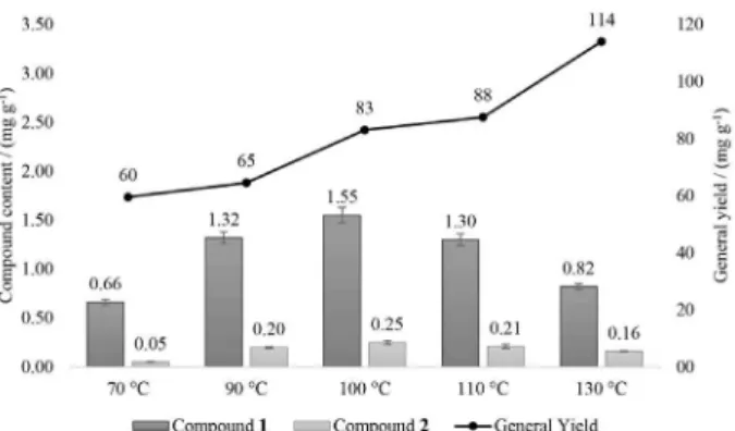
the extraction solvent and its capacity to penetrate into the matrix, altering its surface tension and viscosity. Furthermore, selection of a suitable temperature is essential to avoid degradation of thermolabile compounds and to prevent hydrolysis and oxidation reactions.20-22 Extractions
at different temperatures (70, 90, 100, 110 and 130 °C), maintaining a constant extraction time of 3 cycles of 20 min each, showed that the general extraction yield (the mass of purified extract per gram of plant material) increased significantly with temperature (Figure 2). The general extraction yield was 60 mg g-1 at 70 °C,
and increased nearly two-fold at 130 °C, to 114 mg g-1.
Nevertheless, the extraction at 100 °C gave better yields of compounds 1 (1.55 mg g-1) and 2 (0.25 mg g-1), compared
to the other extraction temperatures evaluated (Figure 2).
Lower yields of these compounds were obtained at temperatures of 70 and 90 °C, due to less significant reductions of the surface tension and viscosity of water, which hindered its ability to penetrate the matrix and
extract the analytes. Above 100 °C, although degradation products were not observed in the chromatograms, it is possible that some degradation of the analytes occurred and that the products were not extracted by partitioning with ethyl acetate.
In samples consisting of plant material, the limiting extraction steps are those involving diffusion processes and solubilization of the compounds.21 It is therefore expected
that a longer extraction should provide higher efficiency. However, it is also possible that prolonged heating could lead to degradation of the compounds of interest.20
Extractions performed using different times per cycle (1, 5, 10, 20 and 30 min), employing 3 extraction cycles at a fixed temperature of 100 °C, showed that the general extraction yield increased with the extraction time. The general extraction yield ranged from 64 mg g-1 (1 min per cycle) to
82 mg g-1 (20 min per cycle), an increase of approximately
29% in efficiency. However, extraction using 1 min cycles was more efficient for both analytes, yielding 1.91 and 0.31 mg g-1 of compound 1 and compound 2, respectively
(Figure 3). Here again, it can be supposed that prolonged heating in longer extractions caused degradation of the analytes prior to their elution from the extraction cell. However, no degradation products were found in the chromatograms to confirm this hypothesis.
In static mode, the efficiency of PHWE depends on the partition coefficient and the solubility of the compounds at the selected temperature. However, extractions may be incomplete due to the limited volume of solvent in the extraction cell. One way to minimize this problem is to use a greater number of extraction cycles.20 On the other hand,
the use of many extraction cycles results in a more time- and solvent-consuming procedure. Therefore, the percentages of compounds 1 and 2 were evaluated in each extraction cycle, using a total of 4 cycles (Figure 4).
For compound 1, the second cycle shows the highest yield, while the fourth (final) cycle was, on average, only
Figure 3. Compound yields and general yields for the purified extracts
(ethyl acetate) during optimization of the extraction time per cycle.
Figure 2. Compound yields and general yields for the purified extracts
responsible for 6.8% of the total amount of the substance present in the purified extract. For compound 2, the first and second cycles showed comparable average yields (44.3 and 41.4%, respectively), while the fourth cycle was responsible for 3.2%, on average, of the total amount of the substance in the purified extract. Since the extractions of these two compounds in the fourth cycle were low, compared to the other cycles, the use of four extraction cycles was considered sufficient. Additional cycles would increase the aqueous extract volume and would require greater amounts of ethyl acetate for partitioning, which would be disadvantageous.
Once the extraction had been optimized (100 °C, 4 cycles of 1 min each), the repeatability of the procedure was evaluated by means of the inter-day and intra-day precisions, with the results expressed as the relative standard deviations (RSD) (Table 1). The RSD values were below 6.7%, which was considered acceptable for the desired application.
These results reinforced the viability and efficiency of PHWE for the extraction of bioactive compounds from C. canadensis. The ability to obtain an aqueous extract of these compounds represents an advance in the direct
application of plant extracts in postharvest treatments, because aqueous extracts are less hazardous to use than organic (chlorinated) solvent extracts.
Disk diffusion assays against postharvest fungi
Compounds 1 and 2 showed similar antifungal activities. As shown in Table 2, both inhibited growth of Aspergillus niger (CMAA 1132) and Cladosporium sp. (CMAA 1136). For the later, however, compound 1
was more active than compound 2. Additionally, only compound 1 significantly inhibited the growth of Penicillium digitatum (CMAA 1190). This assay was used as a screening method to determine which fungi would be submitted to the MIC assays.
Minimum inhibitory concentration (MIC)
The purpose of this test was to determine the minimum amount of each compound that caused inhibition of fungal growth. Given this information, subsequent tests involving the application of aqueous extracts of C. canadensis to fruits could be carried out using appropriate concentrations of the compounds. Compound 1 was most active against the fungus Penicillium digitatum (32 mg L-1),
while compound 2 showed greater activity against Aspergillus niger (32 mg L-1). The MIC results are listed
in Table 2.
Figure 4. Compound contents in the purified extracts (ethyl acetate) during optimization of the number of extraction cycles.
Table 1. Precision of the extraction procedure, expressed as the relative standard deviation (RSD)
Compound RSD / %
Intra-day (n = 5) Inter-day (n = 8)
1 6.3 5.7
2 5.8 6.7
Table 2. Fungal species evaluated and their corresponding codes, the fruits from which they were isolated, inhibition scores for each compound (cpd.),
and minimum inhibitory concentrations (MIC)
Code Fungus Fruit Inhibition MIC / (µg mL
-1)
Cpd. 1 Cpd. 2 Cpd. 1 Cpd. 2
CMAA 1129 Colletotrichum gloeosporioides mango − − n.a. n.a.
CMAA 1130 Alternaria alternata mango − − n.a. n.a.
CMAA 1131 Lasiodiplodia theobromae mango − − n.a. n.a.
CMAA 1133 Botryosphaeria dothidea mango − − n.a. n.a.
CMAA 1132 Aspergillus niger grape + + 64 32
CMAA 1135 Alternaria alternata grape − − n.a. n.a.
CMAA 1136 Cladosporium sp. grape ++ + 64 64
CMAA 1134 Fusarium pallidoroseum melon − − n.a. n.a.
CMAA 1137 Alternaria alternata melon − − n.a. n.a.
CMAA 1190 Penicillium digitatum orange + − 32 64
The results of this test were promising, because the susceptible fungi are common postharvest pathogens of fruits and vegetables.23 Aspergillus niger causes black
mold and other rots in citrus fruits, grapes, figs, peaches, mangoes, apples and strawberries. Moreover, in Brazil it is responsible (amongst other fungi) for the contamination of grapes and wines with ochratoxin A,24 a mycotoxin with
nephrotoxic, teratogenic, hepatotoxic and immunotoxic effects in animals and possibly humans, which is classified as a possible carcinogen.25,26 The Cladosporium genus
causes rots in capsicum, berries, tomatoes and eggplants.23
Lastly, Penicillium digitatum is the causative agent of green mold in citrus fruits and is responsible for 60 to 80% of losses during the storage period.27
Conclusions
The allelopathic weed Conyza canadensis proved to be a promising source of antifungal compounds with potential use in the treatment of postharvest diseases of fruits and vegetables. The compounds 4Z-lachnophyllum lactone and 4Z,8Z-matricaria lactone, both isolated and identified by mass spectrometry and NMR analysis in this work, demonstrated significant antifungal activity against Aspergillus niger, Penicillium digitatum and another fungus of the Cladosporium genus. Aside the traditional extraction method employing organic solvents, it was possible to extract these compounds from the aerial parts of Conyza canadensis by employing pressurized hot water as solvent. This green extraction method was optimized and had its reproducibility assessed, proving to be a viable alternative to obtain the referred antifungal compounds with an adequate yield. Thus, it could be possible to apply the crude aqueous extracts on harvested fruits and vegetables to prevent or delay the appearance of postharvest diseases, prolonging their shelf life and reducing production losses.
Supplementary Information
NMR spectra (1H, 13C and HSQC) and attributions for
compounds 1 and 2, together with the chromatography and NMR operational conditions, are available free of charge at http://jbcs.sbq.org.br as a PDF file.
Acknowledgments
We thank Prof Daniel Terao for his assistance with the selection of the postharvest disease fungi species, Prof Marta Camargo de Assis for her help with plant collection and voucher deposition, and Rodrigo Castanha
and Dr Suikinai Nobre dos Santos for technical assistance. We acknowledge the financial support provided by CNPq (grant number 482675/2012-8) and FAPESP (grant numbers 2012/017670 and 2013/09543-7).
References
1. Sivakumar, D.; Bautista-Baños, S.; Crop Prot. 2014, 64, 27. 2. Prusky, D.; Food Secur. 2011, 3, 463.
3. Liu, J.; Sui, Y.; Wisniewski, M.; Droby, S.; Liu, Y.; Int. J. Food Microbiol. 2013, 167, 153.
4. http://www.epa.gov/ingredients-used-pesticide-products/what-are-biopesticides, accessed in July 2016.
5. Dayan, F. E.; Cantrell, C. L.; Duke, S. O.; Bioorg. Med. Chem. 2009, 17, 4022.
6. Nunes, C. A.; Eur. J. Plant Pathol. 2011, 133, 181.
7. Queiroz, S. C.; Cantrell, C. L.; Duke, S. O.; Wedge, D. E.; Nandula, V. K.; Moraes, R. M.; Cerdeira, A. L.; J. Agric. Food Chem. 2012, 60, 5893.
8. Duke, S. O.; Dayan, F. E.; Rimando, A. M.; Schrader, K. K.; Aliotta, G.; Oliva, A.; Romagni, J. G.; Weed Sci. 2002, 50, 138. 9. Curini, M.; Bianchi, A.; Epifano, F.; Bruni, R.; Chem. Nat.
Compd. 2003, 39, 191.
10. Franzener, G.; Martinez-Franzener, A. S.; Stangarlin, J. R.; Czepak, M. P.; Schwan-estrada, K. R. F.; Eugênia, M.; Cruz, S.; Semina: Cienc. Agrar. 2007, 28, 29.
11. Veres, K.; Csupor-Löffler, B.; Lázár, A.; Hohmann, J.; Sci. World J. 2012, 2012, ID 489646.
12. Meepagala, K. M.; Sturtz, G.; Wise, D.; Wedge, D. E.; Pest Manag. Sci. 2002, 58, 1043.
13. Rivera, P.; Arancibia, L.; Castillo, M.; J. Nat. Prod. 1989, 52, 433.
14. Csupor-Loffler, B.; Hajdu, Z.; Zupko, I.; Molnar, J.; Forgo, P.; Vasas, A.; Kele, Z.; Hohmann, J.; Planta Med. 2011, 77, 1183. 15. Bär, B.; Schultze, W.; Planta Med. 1996, 62, 329.
16. The Clinical and Laboratory Standards Institute (CLSI); M38-A: Reference Method for Broth Dilution Antifungal Susceptibility
Testing of Filamentous Fungi; Approved Standard; CLSI:
Wayne, USA, 2002.
17. Tzakou, O.; Vagias, C.; Gani, A.; Yannitsaros, A.; Flavour Fragrance J. 2005, 20, 425.
18. Lis, A.; Piggott, J. R.; Góra, J.; Flavour Fragrance J. 2003, 18, 364.
19. Pretsch, E.; Bühlmann, P.; Badertscher, M.; Structure Determination of Organic Compounds-Tables of Spectral Data,
4th ed.; Springer: Berlin, Germany, 2009.
20. Teo, C. C.; Tan, S. N.; Yong, J. W.; Hew, C. S.; Ong, E. S.; J. Chromatogr. A 2010, 1217, 2484.
21. Mustafa, A.; Turner, C.; Anal. Chim. Acta 2011, 703, 8. 22. Heng, M. Y.; Tan, S. N.; Yong, J. W. H.; Ong, E. S.; TrAC, Trends
23. Coates, L.; Johnson, G. In Plant Pathogens and Plant Diseases, 1st ed.; Brown, J. F.; Ogle, H. J., eds.; Rockvale Publications: Armidale, Australia, 1997, ch. 33.
24. Varga, J.; Houbraken, J.; Samson, R. A.; Frisvad, J. C. In
Mycotoxins in Fruits and Vegetables; Barkai-Golan, R.; Paster,
N., eds.; Academic Press: Amsterdam, 2008, ch. 9.
25. International Agency for Research on Cancer (IARC);
IARC Monographs on the Evaluation of Carcinogenic Risks
to Humans; Some Naturally Occurring Substances: Food
Items and Constituents, Heterocyclic Aromatic Amines and
Mycotoxins, IARC: Lyon, France, 1993.
26. Pozo-Bayon, M. A.; Monagas, M.; Bartolome, B.; Moreno-Arribas, M. V.; Crit. Rev. Food Sci. Nutr. 2012, 52, 31. 27. Ballester, A. R.; Lafuente, M. T.; Forment, J.; Gadea, J.; de Vos,
R. C.; Bovy, A. G.; Gonzalez-Candelas, L.; Mol. Plant Pathol. 2011, 12, 879.
Submitted: May 27, 2016
Published online: August 8, 2016