RBCCV 44205-1641 DOI 10.5935/1678-9741.20150020
Effects of ischemia and omeprazole preconditioning
on functional recovery of isolated rat heart
Efeitos da isquemia e pré-condicionamento com omeprazol na recuperação funcional do coração isolado
de rato
Nevena Jeremic
1, Mr Pharm; Anica Petkovic
2; Ivan Srejovic
2, MD; Vladimir Zivkovic
2, MD, PhD;
Dragan Djuric
3, MD, PhD; Vladimir Jakovljevic
2, MD, PhD
1Department of Pharmaceutical chemistry, Faculty of Medical Sciences, University of Kragujevac, Serbia.
2Department of Physiology, Faculty of Medical Sciences, University of Kragujevac, Serbia.
3Institute of Medical Physiology “Richard Burian”, School of Medicine, University of Belgrade, Serbia.
Work carried out at Department of Physiology, Faculty of Medical Sciences, University of Kragujevac, Serbia.
No inancial support.
Correspondence address: Vladimir Jakovljevic
Svetozara Markovica 69, P.O.Box 124, 34000 Kragujevac E-mail: [email protected]
Article received on September 9th, 2014 Article accepted on March 9th, 2015 Abstract
Objective: The aim of this study was to compare protective effects of ischemic and potential protective effects of pharma-cological preconditioning with omeprazole on isolated rat heart subjected to ischemia/reperfusion.
Methods: The hearts of male Wistar albino rats were excised and perfused on a Langendorff apparatus. In control group (CG) after stabilization period, hearts were subjected to global ischemia (perfusion was totally stopped) for 20 minutes and 30 minutes of reperfusion. Hearts of group II (IPC) were submit-ted to ischemic preconditioning lasting 5 minutes before 20 min-utes of ischemia and 30 minmin-utes of reperfusion. In third group
(OPC) hearts irst underwent preconditioning lasting 5 minutes
with 100µM omeprazole, and then submitted 20 minutes of isch-emia and 30 minutes of reperfusion.
Results: Administration of omeprazole before ischemia in-duction had protective effect on myocardium function recovery especially regarding to values of systolic left ventricular pressu-re and dp/dt max. Also our indings are that values of coronary
low did not change between OPC and IPC groups in last point
of reperfusion.
Conclusion: Based on our results it seems that ischemic
pre-conditioning could be used as irst window of protection after
ischemic injury especially because all investigated parameters showed continuous trend of recovery of myocardial function.
On the other hand, preconditioning with omeprazole induced sudden trend of recovery with positive myocardium protection, although less effective than results obtained with ischemic pre-conditioning not withstand, we must consider that omeprazole may be used in many clinical circumstances where direct coro-nary clamping for ischemic preconditioning is not possible.
Descriptors: Coronary circulation. Ischemic Precondition-ing, Myocardial. Omeprazole.
Resumo
Objetivo: O objetivo deste estudo foi comparar os efeitos protetores de efeitos protetores isquêmicos e potenciais de pré-condicionamento farmacológico com omeprazol no coração iso-lado de rato submetido à isquemia/reperfusão.
pré-condiciona-INTRODUCTION
Myocardial preconditioning represents exposure of myo-cardium to sublethal stimulus in order to protect it from a subsequent normal lethal stress[1]. Myocardium can be
pre-conditioned by two basic techniques such as ischemic and pharmacological preconditioning. Ischemic preconditioning (ICP) is a concept introduced by Murry et al. in 1986 by us-ing canine models. He showed that sus-ingle or multiple brief periods of myocardial ischemia that produce reversible myo-cyte injury can limit the size of the infarct and the degree of reperfusion injury after a subsequent and more prolonged period of myocardial ischemia[2].
These protective effects of ICP on heart can be conse-quence of reduction in reactive oxygen species generation, delay in ATP depletion, reduction of: infarct size, apoptosis and neutrophil accumulation, as well as improvement of en-dothelial function and reduction of intracellular Ca++
over-load[3-5]. Two different time frames have been reported for
pre-conditioning, early or ’’classical preconditioning’’ phase and late or ’’second window’’ phase. Duration of irst phase, which involves the activation of different membrane recep-tors, is from several seconds and to 3h and for second phase from 12-72 h which represents changes in gene expression leading to production cardioprotective stress proteins[4].
Ischemia is characterized by an absolute or relative de-crease in the blood supply of tissue or organ due to blockage of blood vessels. Blood vessel can be occluded by thrombus, atherosclerotic plaque, vasoconstriction or inlammation. During myocardial ischemia absence of oxygen and metabol-ic substrates to cardiomyocite can cause functional, structural and metabolic diseases. As a consequence, cell switches me-tabolism to anaerobic, resulting in accumulation of lactate and generation of acidosis. Hypoxic conditions lead to diminished intracellular concentrations of ATP (adenosine triphosphate)
and CP (creatine phosphate) which results in decreased activ-ity of ATP reliant ion pumps including Na+/K+ ATP-ase pump
and exacerbation of contractile function. Inactivation of Na+/
K+ ATP-ase contributes to intracellular Na+ overloading.
Low-er intracellular pH induces the Na+/H+ exchanger to extrude H+
and results in intracellular accumulation of Na+, which leads
to activation of the 2Na+/Ca2+ exchanger in order to extrude
Na+ and accumulate intracellular Ca2+[6-9]. All these facts and
generation of reactive oxygen species (ROS) can lead to cell death induced by ischemic episodes[10].
Furthermore, reperfusion is restoration of blood low after an ischemic episode and it may result in paradoxical cardiomyocite dysfunction caused by ROS, intracellular and mitochondrial Ca++ overload and accumulation of inlamma
-tory cells. This phenomenon is called “reperfusion injury”, where prompt changes in intracellular ions and normalization of pH can occur cell death and greater damage than it can be induced by pre-reperfusion ischemia[6,8,11].
Besides ischemic preconditioning, which represents an adaptive response triggered by a brief ischemia applied before a prolonged coronary occlusion, the same response can be in-duced with pharmacological agents[3-5]. Proton pump inhibitors
(PPI) have been one of most important advances in the ield of gastroenterology in past 15 years. These medications showed signiicant progress in acid-related diseases over other acid reducing medications. Most commonly used PPI are omepra-zole, lansoprazole and pantoprazole[12,13]. Omeprazole was irst
introduced into clinical practice and it is commonly used for treatment of gastroesophageal relux and erosive esophagitis in children. The main mechanism of action of these drugs is suppression of acid secretion by binding to H+/K+ ATP-ase
known as “proton pump” or “acid pump”[14,15]. Proton pump
is enzymatic pump expressed in different tissues like parietal cells where hydrochloride acid (HCl) is secreted. The main physiological effect of this pump is H+ exchange for K+ ions.
Abbreviations, acronyms & symbols
CF Coronary low
DLVP Diastolic left ventricular pressure
HCl Hydrochloride acid
HR Heart rate
ICP Ischemic preconditioning
PPI Proton pump inhibitors
ROS Reactive oxygen species
SLVP Systolic left ventricular pressure
mento com duração de 5 minutos com 100 µM de omeprazol, e, então, submetidos a 20 minutos de isquemia e 30 minutos de reperfusão.
Resultados: A administração de omeprazol antes da indução da isquemia teve efeito protetor sobre a recuperação funcional do miocárdio especialmente em relação aos valores de pressão
sistólica ventricular esquerda e dp/dt max. Também os nossos
achados são de que os valores de luxo coronário não se altera -ram entre os grupos OPC e IPC no último ponto de reperfusão.
Conclusão: Com base nos nossos resultados, o pré-condicio-namento isquêmico poderia ser usado como primeira janela de proteção após a lesão isquêmica, especialmente porque todos os parâmetros analisados apresentam tendência contínua de recu-peração da função do miocárdio. Por outro lado, o pré-condicio-namento induzido com omeprazol apresenta tendência repen-tina de recuperação com proteção miocárdio positiva, embora menos efetiva da obtida com o pré-condicionamento isquêmico. Devemos considerar que o omeprazol pode ser usado em muitas circunstâncias clínicas em que o pinçamento coronariano direto para pré-condicionamento isquêmico não é possível.
Due to existence of proton pump in myocardial tissue, which was irst proven by Nagashima et al.[16], mechanical and
electrical properties can be changed by using PPI[6]. Recently
proton pump inhibitors had showed protective effects in treat-ment of myocardial ischemia in patients with coronary artery disease and gastroesophageal relux[17].
Regarding all above presented data, the aim of this study was to compare protective effects of ischemic and potential pro-tective effects of pharmacological preconditioning with omepra-zole on isolated rat heart subjected to ischemia/reperfusion.
METHODS
Preparation of isolated rat hearts
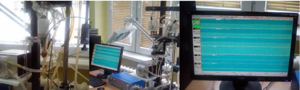
The hearts of male Wistar albino rats (n=36, 12 in each exper-imental group, body mass 180–200 g) were excised and perfused on a Langendorff apparatus (Experimetria Ltd,1062 Budapest, Hungary). After a short-term ketamine/xylasin narcosis, animals were killed by cervical dislocation (Schedule 1 of the Animals/ Scientiic Procedures, Act 1986, UK), and premedicated with heparin as an anticoagulant. After emergency thoracotomy and rapid cardiac arrest by superfusion with ice-cold isotonic saline, rapidly excised, the aortas were cannulated and retrogradely per-fused under a constant perfusion pressure (CPP).
The composition of the non-recirculating Krebs-Hense-leit perfusate was as follows (mM): NaCl 118, KCI 4.7, CaCI2x2H2O 2.5, MgSO4x7H2O 1.7, NaHCO3 25, KH2PO4 1.2, glucose 11, pyruvate 2, equilibrated with 95 % O2 plus 5% CO2 and warmed to 37 oC (pH 7.4). Immediately after
the restoration of normal heart rhythm, through the created entrance to the left atrium of the heart and damaged mitral valve, the sensor (transducer BS473-0184, Experimetria Ltd, Budapest, Hungary) was inserted into the left ventricle for continuous monitoring of cardiac function (Figure 1).
Physiological assay and experimental protocol
All study groups underwent 30 min perfusion at CPP of 70 cm H2O. In control group (CG) after stabilization period,
hearts were subjected to global ischemia (perfusion was totally stopped) for 20 minutes and 30 minutes of reperfusion. Twelve hearts of group II (IPC) were submitted to ischemic precondi-tioning lasting 5 minutes before 20 minutes of ischemia and 30 minutes of reperfusion. In third group (OPC) hearts irst underwent preconditioning lasting 5 minutes with 100µM omeprazole, then submitted 20 minutes of ischemia and 30 minutes of reperfusion. In control group after 20 minutes of global ischemia during period of reperfusion (30 minutes) all cardiodynamic parameters and coronary low were measured in intervals of 5 minute (RP1-RP7). In IPC group, after short period of ischemia (5 minutes) during period of reperfusion (10 minutes), all cardiodynamic parameters and coronary low were measured in intervals of 1 minute (PR1-PR10), while during second period of ischemia (20 minutes)/reperfusion (30 minutes) cardiodynamic parameters and coronary low were measured in intervals of 5 minute (RP1-RP7).
In OPC group (after 5 minutes preconditioning with omeprazole), during period of reperfusion all cardiodynamic parameters and coronary low were measured during second period of ischemia (20 minutes)/reperfusion (30 minutes) in intervals of 5 minute (RP1-RP7). When the low was con-sidered stable (three measurements of the same values), cor-onary low was recorded. Only the properly performed ex-periments were included in the study (i.e., the groups of the hearts in which the CPP/CF relationship was studied twice in the absence of any drug).
After placing the sensor in the left ventricle, the follow-ing parameters of myocardial function have been continuous-ly registered:
1. Maximum rate of pressure development in the left ventricle (dp/dt max)
2. Minimum rate of pressure development in the left ventricle (dp/dt min)
3. Systolic left ventricular pressure (SLVP) 4. Diastolic left ventricular pressure (DLVP) 5. Heart rate (HR)
Coronary low (CF) was measured lowmetrically.
Drugs
All drugs were purchased from Sigma–Aldrich Chemie GmbH, Germany.
Statistical analysis
For statistical analysis we examined three measured points, irst point was stabilization, second was the irst min-ute of reperfusion and third was the 30 minmin-ute of reperfusion. Values are expressed as mean ± SE. Statistical analysis was performed by ANOVA test. P values lower than 0.05 were considered to be signiicant.
The experimental protocol was approved by the Facul-ty of Medical Sciences Ethics Committee for the welfare of experimental animals, University of Kragujevac, number
01-12149 and by Ministry of Agriculture, Forestry and Wa-ter Management, Authority for VeWa-terinary of Serbia number 323-07-09426/2013-05.
RESULTS
Maximum Rate of Left Ventricular Pressure Develop-ment (dp/dt max)
There were no signiicant differences among groups in the values of point of stabilisation and irst minute of reperfusion.
In control and IPC group there were no signiicant dif-ference between periods of stabilisation an irst minute of reperfusion, however there were high statistical signiicant increase (P<0.01**) of values of dp/dt max between these
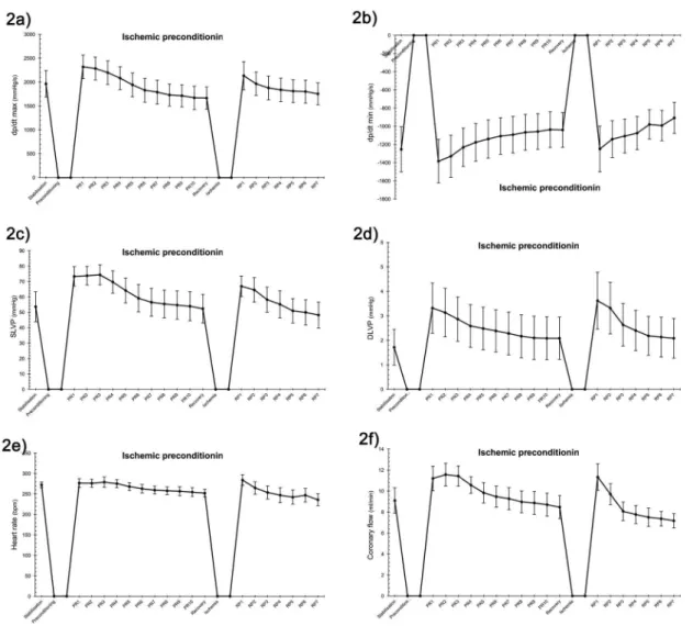
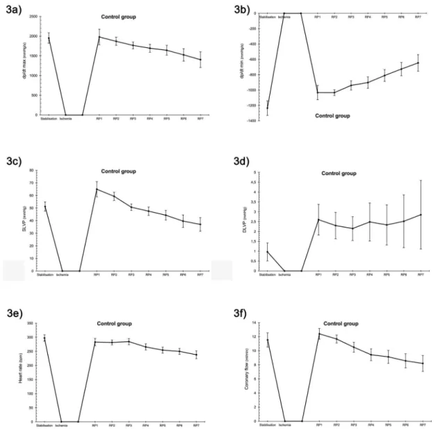
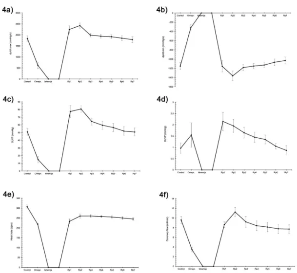
points in OPC group. Trend of values in period of reperfu-sion was the same in all investigated groups. Furthermore, there were statistical signiicant changes between control group and IPC group in values of the last point of reperfusion (P<0.05*) but in comparison with mentioned groups, OPC group has statistical different values (P<0.01**) which were very similar with values before omeprazole administration (Figures 2a, 3a and 4a).
Minimum Rate of Left Ventricular Pressure Develop-ment (dp/dt min)
There were no signiicant differences among groups in the values of point of stabilisation and irst minute of reperfusion.
In control, IPC and OPC groups there were no sig-niicant difference between periods of stabilisation an irst minute of reperfusion. Trend of values in period of reperfusion was the same in OPC, IPC and control group without any statistical difference. Furthermore, there were changes between control group and OPC group in values of the last point of reperfusion (P<0.01**) in compari-son between control group with IPC group values were statistical different (P<0.05*) at the end; in comparison OPC group with IPC group values were statistical differ-ent (P<0.01**) but in OPC group values were similar with values before omeprazole administration (Figures 2b, 3b and 4b).
Systolic Blood Pressure in the Left Ventricle (SLVP) There were no signiicant differences among groups in the values of point of stabilisation and irst minute of reperfusion.
In control and IPC group there were no signiicant differ-ence between periods of stabilisation an irst minute of reper-fusion, however there were high statistical signiicant increase (P<0.01**) of values of SLVP between these points in OPC group. Trend of values in period of reperfusion was the same in OPC and control group on the other hand trend in IPC group was statistical signiicantly lower than in mentioned groups (P˂0.05*). Furthermore, there were statistical signiicant changes between control group and IPC group in values of the last point of reperfusion (P<0.05*) but in comparison with
Fig. 4 - The inluence of preconditioning with omeprazole (5 minutes) on cardiodynamic parameters of the isolated rat heart during subsequent ischemia (20 minutes)/reperfusion (30 minutes): 4a) dp/dtmax, 4b) dp/dtmin, 4c) SLVP, 4d) DLVP, 4e) HR, 4f) CF.
mentioned groups, OPC group has statistical different values (P<0.01**) which were very similar with values before ome-prazole administration (Figures 2c, 3c and 4c).
Diastolic Blood Pressure in the Left Ventricle (DLVP) There were no signiicant differences among groups in the values of point of stabilisation and irst minute of reperfusion.
control group values of DLVP in period of reperfusion increased compared with period before ischemia. In IPC and OPC groups values of DLVP in period of reperfusion decreased compared with period before preconditioning. Furthermore, there were statistical significant changes between control group and IPC group. Compared with values in control group, IPC and OPC groups values were statistical different in last point of reperfusion (P<0.05*) (Figures 2d, 3d and 4d).
Heart Rate (HR)
There were no signiicant differences between IPC group and control group and also between OPC and control group, but there were signiicant difference between IPC and OPC at the point of stabilisation.
In control and IPC group there were no signiicant dif-ference between periods of stabilisation at the irst minute of reperfusion, however there were high statistical signiicant drop (P<0.01**) of values of HR between these points in OPC group. Trend of values in period of reperfusion was the same in IPC and control group on the other hand trend in OPC group was the same as in mentioned groups but with statistical signiicant lower decrease. Furthermore, there were no changes between control group and OPC group in values of the last point of reperfusion but in comparison with IPC group those values were statistically different (P<0.05*) (Figures 2e, 3e and 4e)
Coronary Flow (CF)
There were no signiicant differences among groups in the values of point of stabilisation and irst minute of reperfusion.
In control and IPC group there were no signiicant dif-ference between periods of stabilisation and irst minute of reperfusion however there were high statistical signiicant increase (P<0.01**) of values of CF between these points in
OPC group. Trend of values in period of reperfusion was the same in OPC, IPC and control group without any statistical difference. Furthermore, there were no changes among con-trol, IPC and OPC group in last point of reperfusion (Figures 2f, 3f and 4f).
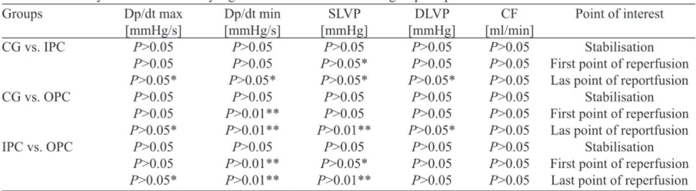
Summary table shows all cardiodynamic parameters compared between the groups (Table 1).
DISCUSSION
Acute myocardial infarction is leading cause of morbidi-ty and mortalimorbidi-ty worldwide each year. Consequence of acute myocardial infarction, a diminished blood supply to the heart exceeds a critical threshold and overwhelms myocardial cel-lular repair mechanisms designed to maintain normal operat-ing function and homeostasis[18]. The treatment of acute
myo-cardial infarction has been prospering in last few decades and new methods such as preconditioning, post-conditioning and pharmacological agents have been examined to protect the heart[19,20].
Preconditioning (PC) involves reduction of necrotic tis-sue mass, improvement of cardiac contractile performance after ischemia and reperfusion, and reduction of arrhyth-mias. Although the ischemic preconditioning is not entirely clariied, recently parts of the signal transduction cascade of ischemic preconditioning have been identiied. According to different species, the organism increases the production of several chemical mediators that trigger the cardio protection process[21]. To avoid the problems which can cause ischemic
preconditioning in clinical use, administration of pharma-cological agents could be ideal solution. Pharmapharma-cological agents such as a cardio-selective β1-blocker and the adenos-ine triphosphate-sensitive potassium channel openers have been shown the ability to protect the heart but none has been widely accepted[19].
Table 1. Summary table of statistically signiicant difference between groups in points of interest. Groups
CG vs. IPC
CG vs. OPC
IPC vs. OPC
Dp/dt max [mmHg/s] P>0.05 P>0.05 P>0.05* P>0.05 P>0.05 P>0.05* P>0.05 P>0.05 P>0.05* Dp/dt min [mmHg/s] P>0.05 P>0.05 P>0.05* P>0.05 P>0.01** P>0.01** P>0.05 P>0.01** P>0.01** SLVP [mmHg] P>0.05 P>0.05* P>0.05* P>0.05 P>0.05 P>0.01** P>0.05 P>0.05* P>0.01** DLVP [mmHg] P>0.05 P>0.05 P>0.05* P>0.05 P>0.05 P>0.05* P>0.05 P>0.05 P>0.05 CF [ml/min] P>0.05 P>0.05 P>0.05 P>0.05 P>0.05 P>0.05 P>0.05 P>0.05 P>0.05
Point of interest
Stabilisation First point of reperfusion Las point of reportfusion
Stabilisation First point of reperfusion Las point of reportfusion
Stabilisation First point of reperfusion Last point of reperfusion *Statistically signiicant
**High statistically signiicant
Model of isolated rat heart is one the most convenient ex-perimental tool for preclinical investigations of mammalian heart, and also very reliable for connection between animal and human studies. Generally, viewed morphology of the rat heart is very similar with human one[22]. Namely, structure of
the left ventricular, wall thickness and properties of the pap-illary muscles are almost the same as in the human heart[22].
In addition, examination of nodal cells showed that they are very similar to human T cells, and begin with the functioning during the early embryogenesis[23]. Moreover, both
ventricu-lar and atrial cardiomyocites are showed to possess high per-cent of histological similarity[24]. In this sense, we can assume
that there are signiicant analogy between cardiac (patho) physiological events in rat and human heart.
In that sense, data collected from these experimental studies could be of great interest in improving knowledge about ischemic and especially pharmacological form of pre-conditioning.
The present study aimed to compare potential protective effects of ischemic and pharmacological preconditioning with omeprazole on isolated rat heart subjected to ischemia/ reperfusion. Considering the fact that myocardial tissues have H+/K+ - ATPase[19], we examined the effects of one of
clinically most used H+/K+-ATPase inhibitor (omeprazole) in
isolated rat heart.
Proton pump inhibitors may be a particularly important in patients with intrinsic cardiac disease however their safety has not been well studied. Omeprazole was irst introduced into clinical practice and it is commonly used for treatment of gastroesophageal relux and erosive esophagitis but some studies pioneering demonstrated the protective effect of ome-prazole on myocardial contractility in isolated rat hearts[25].
Considering the fact that H+/K+-ATPase exists in
myocar-dial tissues it could be expected that speciic proton pump inhibitors might change the mechanical and electrical proper-ties of the myocardium and might cause intracellular acidii-cation via decreasing the extracellular H+ transport and
mem-brane depolarization through intracellular K+ import[6,16].
Gomes et al.[25] have shown that administration of
ome-prazole before ischemia induction had protective effect on myocardium function recovery and our results were very similar especially regarding to values of SLVP and dp/ dt max. These results are coherent with indings in studies where only ischemic preconditioning was induced[2].
On the other hand, in case of coronary low (CF), in the present study there were no difference between OPC and IPC groups in last point of reperfusion (Figures 2a and 4a), which was very close to indings of Gomes et al.[25].
It has been shown, that ischemic preconditioning can re-duce the magnitude of ischemia/reperfusion injury via activa-tion of K+ adenosine triphosphate (ATP)-sensitive (K(ATP))
channels[26]. Concerning this fact, Kersten et al.[27] found that
left-ventricular pressure and coronary low, respectively,
were recovered to a greater extent after inducing ischemic preconditioning. In our study, results are very similar (Figures 2a, 2b, and 2f).
A study evaluating animal model of frog by Gautam et al.[28], showed interesting results. They found that PPI in
minimal used dose did not change heart rate, but when they increased doses twice, they noticed bradycardia. However, Gomes et al., on rat model did not show any effect on heart rate[25]. In this investigation, we found high statistical
signif-icant drop of values of HR between period of stabilisation and irst minute of reperfusion in OPC group (Figure 4e).
Birnbaum et al.[29] showed that induction of ischemic
pre-conditioning on isolated rabbit heart leads to slower heart rate then in control groupas we also demonstrated (Figures 2e and 3e).
In our study, omeprazole decreased tension and complete inhibition of cardiac contractility (Figure 4d) and these re-sults correlate with studies of other authors who investigat-ed another PPI on similar models[6,30,31]. In OPC group we
remarked that values of every parameter was signiicantly increased in irst minute of reperfusion compared to point of stabilisation (Figures 4a to 4f).
Based on indings by Murry et al.[2], we investigated
ischemic preconditioning using similar procedureand we also concluded that in IPC group there was recovery of all measured parameters.
Furthermore, based on our results it seems that ischemic preconditioning could be used as irst window of protection after ischemic injury especially due to all investigated pa-rameters showed continuous trend of recovery of myocardial function. On the other hand, after administration of ome-prazole we noticed sudden trend of recovery with positive myocardium protection, although less effective than results obtained with ischemic preconditioning not withstand we must consider that omeprazole may be used in many clinical circumstances where direct coronary clamping for ischemic preconditioning is not possible.
CONCLUSION
ACKNOWLEDGMENTS
This study is supported by Grant no. 175043 from the
Ministry of Science and Technical Development of the Re-public of Serbia.
Authors’ roles & responsibilities
NJ Analysis and/or interpretation of data; statistical analysis; study design; operations and/or experiments conduct; writing of the manuscript or critical review of its content
AP Analysis and/or interpretation of data; statistical analysis; i-nal approval of the manuscript; operations and/or experiments conduct
IS Conception and design; operations and/or experiments con-duct; writing of the manuscript or critical review of its content VZ Final approval of the manuscript; study design; operations
and/or experiments conduct; writing of the manuscript or crit-ical review of its content
DD Final approval of the manuscript; study design VJ Final approval of the manuscript; study design
REFERENCES
1. Luh SP, Yang PC. Organ preconditioning: the past, current status, and related lung studies. J Zhejiang Univ Sci B. 2006;7(5):331-41.
2. Murry CE, Jennings RB, Reimer KA. Preconditioning with ischemia: a delay of lethal cell injury in ischemic myocardium. Circulation. 1986;74(5):1124-36.
3. Zhao ZQ, Corvera JS, Halkos ME, Kerendi F, Wang NP, Guyton RA, et al. Inhibition of myocardial injury by ischemic postconditioning during reperfusion: comparison with ischemic preconditioning. Am J Physiol Heart Circ Physiol. 2003;285(2):H579-88.
4. Sanada S, Komuro I, Kitakaze M. Pathophysiology of myocardial reperfusion injury: preconditioning, postconditioning, and translational aspects of protective measures. Am J Physiol Heart Circ Physiol. 2011;301(5):H1723-41.
5. Yellon DM, Hausenloy DJ. Myocardial reperfusion injury. N Engl J Med. 2007;357(11):1121-35.
6. Bacaksiz A, Teker ME, Buyukpinarbasili N, Inan O, Tasal A, Sonmez O, et al. Does pantoprazole protect against reperfusion injury following myocardial ischemia in rats? Eur Rev Med Pharmacol Sci. 2013;17(2):269-75.
7. Gomes OM, Magalhães Mde M, Abrantes RD, Kallás E. Pantoprazole provides myocardial protection similar to ischemic preconditioning: experimental study of isolated hearts of rats. Rev Bras Cir Cardiovasc. 2011;26(3):433-9.
8. Hausenloy DJ, Yellon DM. Myocardial ischemia-reperfusion injury: a neglected therapeutic target. J Clin Invest. 2013;123(1):92-100.
9. Jennings RB, Reimer KA. The cell biology of acute myocardial ischemia. Annu Rev Med. 1991;42:225-46.
10. Sommerschild HT, Kirkebøen KA. Preconditioning - endogenous defense mechanisms of the heart. Acta Anaesthesiol Scand. 2002;46(2):123-37.
11. Verma S, Fedak PW, Weisel RD, Butany J, Rao V, Maitland A, et al. Fundamentals of reperfusion injury for the clinical cardiologist. Circulation. 2002;105(20):2332-6.
12. Richardson P, Hawkey CJ, Stack WA. Proton pump inhibitors. Pharmacology and rationale for use in gastrointestinal disorders. Drugs. 1998;56(3):307-35.
13. Fock KM, Ang TL, Bee LC, Lee EJ. Proton pump inhibitors: do differences in pharmacokinetics translate into differences in clinical outcomes? Clin Pharmacokinet. 2008;47(1):1-6.
14. Monzani A, Oderda G. Delayed-release oral suspension of omeprazole for the treatment of erosive esophagitis and
gastroesophageal relux disease in pediatric patients: a review.
Clin Exp Gastroenterol. 2010;3:17-25.
15. Lindberg P, Nordberg P, Alminger T, Brändström A, Wallmark B. The mechanism of action of the gastric acid secretion inhibitor omeprazole. J Med Chem. 1986;29(8):1327-9.
16. Nagashima R, Odashiro K, Morita S. Evidence for the existence
of myocardial H+-K+ATP and its electrophysiological effects. Jpn
Heart J. 1994; 35(suppl):473-4.
17. Dobrzycki S, Baniukiewicz A, Korecki J, Bachórzewska-Gajewska H, Prokopczuk P, Musial WJ, et al. Does
gastro-esophageal relux provoke the myocardial ischemia in patients
with CAD? Int J Cardiol. 2005;104(1):67-72.
18. Xia A, Xue Z, Li Y, Wang W, Xia J, Wei T, et al. Cardioprotective effect of betulinic acid on myocardial ischemia reperfusion injury in rats. Evid Based Complement Alternat Med. 2014;2014 [Acessed on: Set 1, 2014]. Available from: http://www.hindawi. com/journals/ecam/2014/573745/
19. Li J, Iorga A, Sharma S, Youn JY, Partow-Navid R, Umar S, et al. Intralipid, a clinically safe compound, protects the heart against
ischemia-reperfusion injury more eficiently than cyclosporine-A.
Anesthesiology. 2012;117(4):836-46.
20. Ghyasi R, Sepehri G, Mohammadi M, Badalzadeh R, Ghyasi A. Effect of mebudipine on oxidative stress and lipid peroxidation in myocardial ischemic-reperfusion injury in male rat. J Res Med Sci. 2012;17(12):1150-5.
21. Schulz R, Cohen VM , Behrends M , Downey MJ , Heusch D. Signal transduction of ischemic preconditioning. Cardiovasc Res. 2001;52(2):181-98.
23. Domenech-Mateu JM, Boya-Vegué J. An ultrastructural study of sinuatrial node cells in the embryonic rat heart. J Anat. 1975;119(Pt 1):77-83.
24. Anversa P, Loud AV, Vitali-Mazza L. Morphometry and autoradiography of early hyperthropic changes in the ventricular myocardium of adult rat: an electron microscopic study. Lab Invest. 1976;35(5):475-83.
25. Gomes OM, Magalhães MM, Abrantes RD. Myocardium functional recovery protection by omeprazole after ischemia-reperfusion in isolated rat hearts. Rev Bras Cir Cardiovasc. 2010;25(3):388-92.
26. Schillinger W, Teucher N, Sossalla S, Kettlewell S, Werner C, Raddatz D, et al. Negative inotropy of the gastric proton pump inhibitor pantoprazole in myocardium from humans and rabbits: evaluation of mechanisms. Circulation. 2007;116(1):57-66.
27. Kersten JR, Orth KG, Pagel PS, Mei DA, Gross GJ, Warltier
DC. Role of adenosine in isolurane-induced cardioprotection.
Anesthesiology. 1997;86(5):1128-39.
28. Gautam CS, Utreja A, Goel D, Sandhu G, Gogia N. Negative chronotropic effect of proton pump inhibitors on frog-heart preparation. Indian J Gastroenterol. 2009;28(4):147-9.
29. Birnbaum Y, Hale SL, Kloner RA. Ischemic preconditioning at a distance: reduction of myocardial infarct size by partial reduction of blood supply combined with rapid stimulation of the gastrocnemius muscle in the rabbit. Circulation. 1997;96(5):1641-6.
30. Novalija E, Fujita S, Kampine JP, Stowe DF. Sevolurane mimics ischemic preconditioning effects on coronary low
and nitric oxide release in isolated hearts. Anesthesiology. 1999;91(3):701-12.